Introduction
Head and neck squamous cell carcinoma (HNSCC)
accounts for >500,000 newly diagnosed cases annually worldwide
(1). The primary risk factors are
tobacco use and alcohol consumption, which appear to exert a
synergistic effect (2). Patient
prognosis is largely determined by the extent of the tumor, as well
as the presence of lymph node and distant metastases (1,2).
Furthermore, a unique subset of HNSCCs, particularly oropharyngeal
tumors, are associated with human papillomavirus (HPV) infection,
which has been demonstrated to be of significant prognostic value
(3,4).
HPV-positive tumors are characterized by increased expression of
p16 due to functional inactivation of retinoblastoma protein by the
viral oncoprotein E7. Therefore detection of p16 by
immunohistochemistry (IHC) is considered to be a robust marker of
HPV infection in these tumors (5,6).
In locally advanced HNSCC, concomitant
chemoradiation therapy (CRT) has emerged as a useful treatment
modality for organ preservation, treatment of unresectable disease
and as an adjuvant therapy in resected high-risk disease (7). However, CRT is associated with
significant short- and long-term treatment-associated morbidities
and complications. Furthermore, histologically similar tumors
receiving identical therapies may exhibit varying clinical
outcomes, and a proportion of patients will not respond to CRT
(3–5,7).
Therefore, identification of markers that predict the prognosis of
advanced HNSCC treated by CRT will be significant for the
improvement of clinical treatment decisions.
HNSCC is a heterogeneous disease with respect to its
complex molecular abnormalities and tumor-associated proteome. Over
20 recurrent chromosomal alterations have been identified in
invasive HNSCC, and gains at 3q, 11q and 12q, as well as losses at
5q, 6q, 8p, 21q and 22q have been associated with poor prognosis
(8–10). A number of tumor-associated markers
have been identified for HNSCC carcinogenesis, and have been
considered as potential reasons for various responses to therapy.
They may also have value as prognostic markers in patients with
HNSCC. Epidermal growth factor receptor (EGFR) and survivin are two
candidates that have been investigated as promising prognostic
biomarkers for HNSCC (11–17).
EGFR, a transmembrane glycoprotein receptor, is
encoded by the human epidermal growth factor receptor
1/c-ERBB1 proto-oncogene located on chromosome 7p12. Ligand
binding causes activation of the intracellular tyrosine kinase
domain, resulting in signal transduction involving proliferation,
cell division and differentiation (18). EGFR is overexpressed in the majority
of epithelial malignancies and a prognostic impact of EGFR
overexpression has been identified in a number of types of human
cancer, including ovarian, bladder, cervical, gastric, esophageal,
breast, endometrial and colorectal cancer (19). Overexpression of EGFR has additionally
been observed in >80% of HNSCC cases and occurs early during
head-neck carcinogenesis (20,21).
However, conflicting results have been reported from certain
laboratories. Meta-analyses have discussed the effects of various
clinicopathological parameters and evaluation methods for the
prognostic value of EGFR (11,12).
Survivin, a member of the apoptosis inhibitor family
located on chromosome 17q25.3, serves a significant role in cell
cycle progression and is expressed in fetal tissues, as well as in
a wide range of cancer tissues; however, it is not expressed in
normal adult differentiated tissues (22,23).
Notably, survivin is detected as a cytoplasmic and nuclear protein
in cancer patients. Nuclear survivin is suspected to control cell
division, whereas cytoplasmic/mitochondrial survivin is considered
to be cytoprotective (13).
Consequently, intracellular localization of survivin in tumor cells
has been analyzed as a prognostic marker in a number of
patient-based studies, achieving differing results (13).
Gain of 3q is the most frequently detected
chromosomal alteration in HNSCC and has additionally been reported
in numerous other tumor entities, including prostate, esophageal
squamous cell, cervical squamous cell and non-small cell lung
cancer (NSCLC) (24–27). Furthermore, an amplification of the
chromosomal region 3q was identified as the most frequent genomic
alteration in oropharyngeal squamous cell carcinoma (OSCC),
independent of HPV status, and was significantly associated with
advanced tumor stage in the total patient group, as well as in the
non-HPV-associated subgroup (28).
Notably, studies have identified SEC62 as the most commonly
overexpressed gene within this 3q region, based on increased
messenger RNA and protein levels in prostate and thyroid cancer, as
well as NSCLC (24,29–31).
SEC62, a gene that encodes an endoplasmic reticulum (ER)
transmembrane protein, has an essential function in cell migration
(29) and ER stress tolerance
(30). Furthermore, Sec62
overexpression has been demonstrated to be correlated with the
presence of lymph node metastases (increased Sec62 levels in
node-positive tumors compared with node-negative tumors) and tumor
cell dedifferentiation (increased Sec62 levels in
poorly-differentiated grade 3 tumors compared with grade 2 tumors)
in NSCLC patients (31). Furthermore,
it is reported to be associated with significantly poorer overall
survival (OS), particularly in NSCLC with squamous differentiation
(32).
In the present study, Sec62 expression was analyzed
for the first time in HNSCC by IHC, and the results were correlated
with the clinicopathological data of a cohort of 35 patients with
advanced HNSCC treated by CRT. In addition, the immunohistological
expression of EGFR, survivin and p16 as biomarkers of interest in
HNSCC were investigated and included in a multiple regression
analysis.
Materials and methods
Patient characteristics
The records of 35 patients treated at Saarland
University Hospital (Homburg, Germany) between August 1996 and May
2006 were analyzed retrospectively. All patients exhibited newly
diagnosed squamous cell carcinoma of the head and neck region.
Informed consent was obtained from the patients and the study
design was approved by the local ethics committee (reference
#218/10). The primary tumors were located as follows: Oropharynx,
24 cases (69%); hypopharynx, 6 cases (17%); larynx, 3 cases (9%);
and nasopharynx, 2 cases (6%). The median age at diagnosis was 55
years (range, 43–74 years), and the mean Karnofsky performance
index amounted to 8 (6–10). The cohort included 33 male patients
(94%) and 2 female patients (6%). Smoking history data was not
available. All tumors were classified as stage IV according to the
International Union Against Cancer system (33), and distant metastases had been
excluded.
All patients underwent simultaneous definitive CRT
without prior surgery. A total dose of 72 Gy in single fractions of
1.2 Gy was administered 10 times/week and twice daily in a
hyperfractionated manner (inter-fraction interval, ≥6 h). A linear
accelerator was used to apply 6 MV photons and electrons.
Chemotherapy was administered to all patients, and consisted of two
cycles of cisplatin (20 mg/m2 for 5 days, for a total
dose of 60 mg/m2) and fluorouracil (5-FU; continuous
infusion of 600 mg/m2/day on days 1–5) in 34 patients,
or mitomycin and 5-FU in the remaining patient (continuous infusion
of 600 mg/m2/day 5-FU on days 1–5; single intravenous
bolus injection of 10 mg/m2 mitomycin C on days 5 and
36). Follow-up time was defined from the date of diagnosis to the
date of the final visit or date of mortality. The mean follow-up
time was 52.2 months, with a minimum of 1.6 and a maximum of 203
months (median, 24 months). The clinicopathological characteristics
of the patient group are presented in Table I.
 | Table I.Clinicopathological features of head
and neck squamous cell carcinoma patients (n=35) and influence on
survival time. |
Table I.
Clinicopathological features of head
and neck squamous cell carcinoma patients (n=35) and influence on
survival time.
|
| Patients | P-value |
|---|
|
|
|
|
|---|
| Clinicopathological
feature | Value | % | Overall
survival | Progression-free
survival |
|---|
| Gender, n |
|
| 0.683 | 0.004 |
|
Male | 33 | 94 |
|
|
|
Female | 2 | 6 |
|
|
| Age, years |
|
| 0.621a | 0.594a |
|
Median | 55 |
|
|
|
|
Range | 43–77 |
|
|
|
| Overall survival
time, months |
|
|
|
|
|
Mean/median | 54/29 |
|
|
|
|
Range | 2–203 |
|
|
|
| Localization,
n |
|
| 0.338 | 0.232 |
|
Oropharynx | 24 | 69 |
|
|
|
Hypopharynx | 6 | 17 |
|
|
|
Nasopharynx | 2 | 6 |
|
|
|
Larynx | 3 | 9 |
|
|
| Karnofsky score,
n |
|
| 0.053 | 0.216 |
| 6 | 2 | 6 |
|
|
| 7 | 8 | 23 |
|
|
| 8 | 13 | 37 |
|
|
| 9 | 11 | 31 |
|
|
| 10 | 1 | 3 |
|
|
| Tumor stage, n |
|
| 0.725 | 0.850 |
| 2 | 3 | 9 |
|
|
| 3 | 7 | 20 |
|
|
| 4 | 25 | 71 |
|
|
| Node stage, n |
|
| 0.213 | 0.245 |
| 0 | 2 | 6 |
|
|
| 1 | 4 | 12 |
|
|
| 2 | 26 | 74 |
|
|
| 3 | 3 | 9 |
|
|
Immunohistochemical staining
Slides from all biopsies were reviewed by one
pathologist in order to select representative areas of the tumors
for additional IHC analysis. The criteria were vital tumor tissue
without necrosis and an estimate of ≥70% tumor cells in the
analyzed tissue area.
Selected sections were deparaffinized and rehydrated
using repeated xylene and graded alcohol washes. For antigen
retrieval, slides were incubated with citrate buffer (for Sec62; pH
6.0 for 1 h at 90°C), Tris-HCl (for survivin; pH 9.0 for 1 h at
95°C) or Protease Type XIV (for EGFR; 2 min at room temperature;
Sigma-Aldrich Chemie GmbH, Munich, Germany).
Primary antibody incubations for EGFR (clone E30;
monoclonal mouse anti-human; #M7239; Dako, Glostrup, Denmark;
dilution, 1:50) and survivin (polyclonal rabbit anti-human; #ab469;
Abcam, Cambridge, UK; dilution, 1:250) were performed overnight at
4°C. For Sec62, primary antibody incubation was conducted for 1 h
at room temperature (affinity-purified, polyclonal rabbit antibody
directed against the COOH-terminal undecapeptide of human Sec62
protein; dilution, 1:200. This antibody was kindly provided by Dr
Markus Greiner, Department of Medical Biochemistry and Molecular
Biology, Saarland University, (Homburg/Saar, Germany) and was
previously demonstrated to be of excellent sensitivity and
specificity under denaturing and native conditions (30) Visualization of bound primary
antibodies was performed with the Real Detection System (Dako)
according to the manufacturer's protocol. p16 staining was
performed with the CINtec® Histology kit (Roche Diagnostics GmbH,
Mannheim, Germany) according to the manufacturer's protocol. The
slides were counterstained using hematoxylin (Merck Millipore,
Darmstadt, Germany) and each analysis included positive and
negative controls.
Staining intensity of EGFR and cytoplasmic survivin
was scored as 0 (none), 1 (weak), 2 (moderate) or 3 (strong), and
was considered to be high when the fraction of moderately and
strongly stained cells within the tumor cell population was
>10%. Furthermore, slides were considered to be survivin
nuclear-positive when >10% of all tumor cell nuclei were
stained, regardless of the cytoplasmic staining level. p16
expression was dichotomized as p16-positive (strong nuclear or
cytoplasmic staining in ≥70% of tumor cells) or p16-negative. For
Sec62, the cytoplasmic Sec62 staining intensity was scored as 0
(none), 1 (weak), 2 (moderate) or 3 (strong). The product of the
scores for staining intensity and number of stained cells (0, no
stained cells; 1, <10%; 2, 10–50%; 3, 51–80%; or 4, >80%),
defined as the immunoreactive score (IRS) according to Remmele and
Stegner (34), was used. IRS scores
from 0–2 were valued as negative, 3–4 as weak, 6–8 as moderate and
9–12 as strong; subsequently, these were dichotomized into low
(IRS, <9) and high (IRS, 9–12) expression.
Statistical analysis
Statistical analysis was performed with SPSS version
20 (IBM SPSS, Armonk, NY, USA). OS was calculated as the time from
diagnosis date to mortality date or the final documented follow-up
date. Progression-free survival (PFS) was defined as the time from
diagnosis date to the date of disease progression, the final
documented follow-up date or mortality date from any cause.
Survival curves were calculated by the Kaplan-Meier method and
compared by the log-rank test and univariate Cox regression. The
χ2 test was utilized to determine the association and
differences between dichotomized variables, encompassing IHC
expression and clinicopathological characteristics. P<0.05
(two-sided) was considered to indicate a statistically significant
difference. Reported P-values are unadjusted for the issue of
multiple testing due to the screening nature of the present
study.
Results
Immunohistological staining of EGFR,
survivin, p16 and Sec62
Immunohistochemical EGFR staining revealed strong
membranous and cytoplasmic signals in 17/35 cases (49%).
Cytoplasmic survivin staining was observed in 20 cases (57%). Using
a cut-off value of ≥10% of tumor cells demonstrating positive
nuclear staining signals, 19/35 cases (54%) were classified as
nuclear survivin-positive. Furthermore, in 28 cases (80%) survivin
was detected in the nucleus and/or cytoplasm of tumor cells. p16
expression was observed either to be generally absent, or present
as strong and diffuse nuclear and cytoplasmic staining.
Overexpression of p16 was observed in 5 tumors (14%) and was
restricted to OSCCs (5/24 tumors; 21%). A summary of the observed
staining levels for each candidate marker according to the tumor
site is presented in Table II.
| Table II.Immunohistochemical expression
analysis according to various head and neck squamous cell carcinoma
localization sites. |
Table II.
Immunohistochemical expression
analysis according to various head and neck squamous cell carcinoma
localization sites.
|
| Expression level
(high/low, n) |
|---|
|
|
|
|---|
| Protein | Oropharynx
(n=24) | Hypopharynx
(n=6) | Nasopharynx
(n=2) | Larynx (n=3) |
|---|
| Sec62 | 10/14 | 2/4 | 0/2 | 0/3 |
| EGFR | 11/13 | 2/4 | 2/0 | 2/1 |
| p16 | 5/19 | 0/6 | 0/2 | 0/3 |
| Survivin |
|
|
|
|
| Nuc | 15/9 | 3/3 | 1/1 | 0/3 |
| Cyt | 15/9 | 3/3 | 0/2 | 2/1 |
| Nuc+cyt | 20/4 | 1/1 | 5/1 | 2/1 |
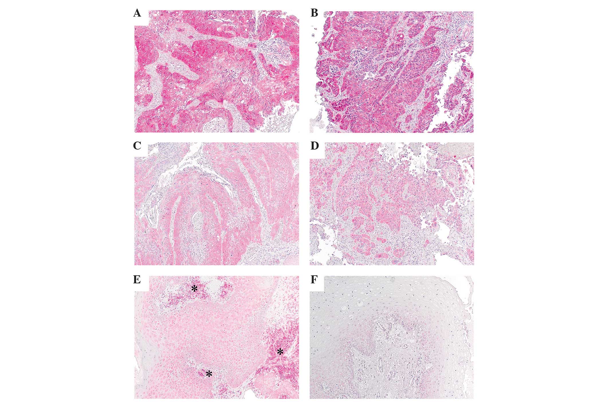
Strong Sec62 staining intensity was observed in
12/35 cases (34%) and was primarily identified in OSCC (10/24
tumors). Furthermore, in 8 (23%) and 11 HNSCC cases (31%), moderate
and weak staining was observed, respectively, whereas only 4 cases
(11%) were Sec62-negative in the analyzed representative tumor
tissue samples (Table II; Fig. 1). The detailed results of Sec62
immunostaining with regard to IRS and tumor site are presented in
Table III and Fig. 2. There was no association between
tumor site and Sec62 expression level (χ2, P=0.458).
Normal squamous epithelium demonstrated weak Sec62 staining in
regenerating basal cells (stratum germinativum), while no
immunoreactivity was observed in the more superficial cell layers
(Fig. 1). As described previously
(31), plasma cells within
inflammatory infiltrates demonstrated strong Sec62 staining in all
samples, irrespective of the Sec62 signals of adjacent tumor cells,
due to their large amount of rough ER, thereby serving as internal
positive controls (Fig. 1).
| Table III.Immunohistochemical expression of
Sec62 in the cytoplasm on the basis of the IRS according to HNSCC
site. |
Table III.
Immunohistochemical expression of
Sec62 in the cytoplasm on the basis of the IRS according to HNSCC
site.
|
| HNSCC localization
site |
|---|
|
|
|
|---|
| Sec62 expression
level | Oropharynx | Hypopharynx | Nasopharynx | Larynx |
|---|
| Strong (IRS
9–12) | 10 | 2 | – | – |
| Moderate (IRS
6–8) | 4 | 3 | – | 1 |
| Weak (IRS 3–4) | 7 | 1 | 1 | 2 |
| Negative (IRS
0–2) | 3 | – | 1 | – |
Correlation of protein expression with
clinicopathological parameters
Analysis of clinicopathological parameters in the
CRT-treated patient cohort revealed no statistically significant
differences in OS with regard to gender, tumor localization,
Karnofsky score, tumor grading and lymph node metastases (Table I).
A detailed analysis of potential correlations
between the analyzed protein expression scores and the
clinicopathological parameters of the patients was performed. With
regard to tumor localization, only p16 overexpression was
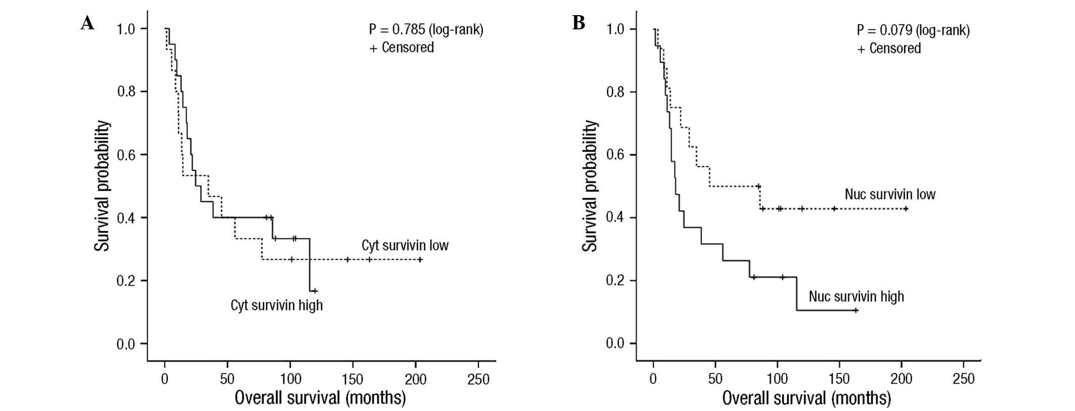
restricted to OSCCs (5/24 tumors; χ2, P=0.593; Table II). Regarding survival, the
expression levels of EGFR, cytoplasmic survivin or p16 were not
significantly associated with OS (P=0.585, P=0.785 and P=0.557,
respectively) or PFS (P=0.847, P=0.943 and P=0.560, respectively).
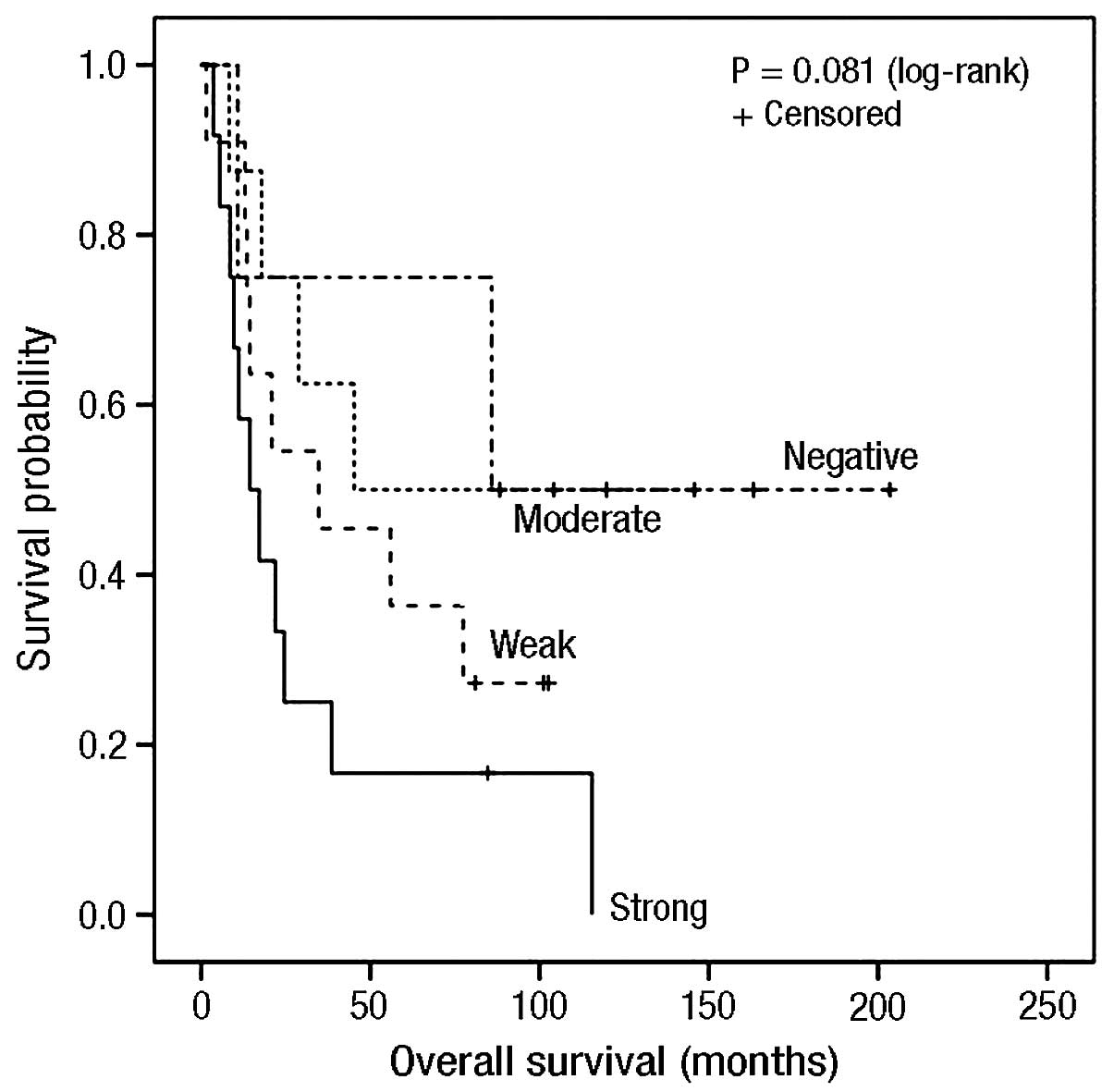
However, strong nuclear expression of survivin showed a weak trend
for poorer OS (P=0.079; Fig. 3), but
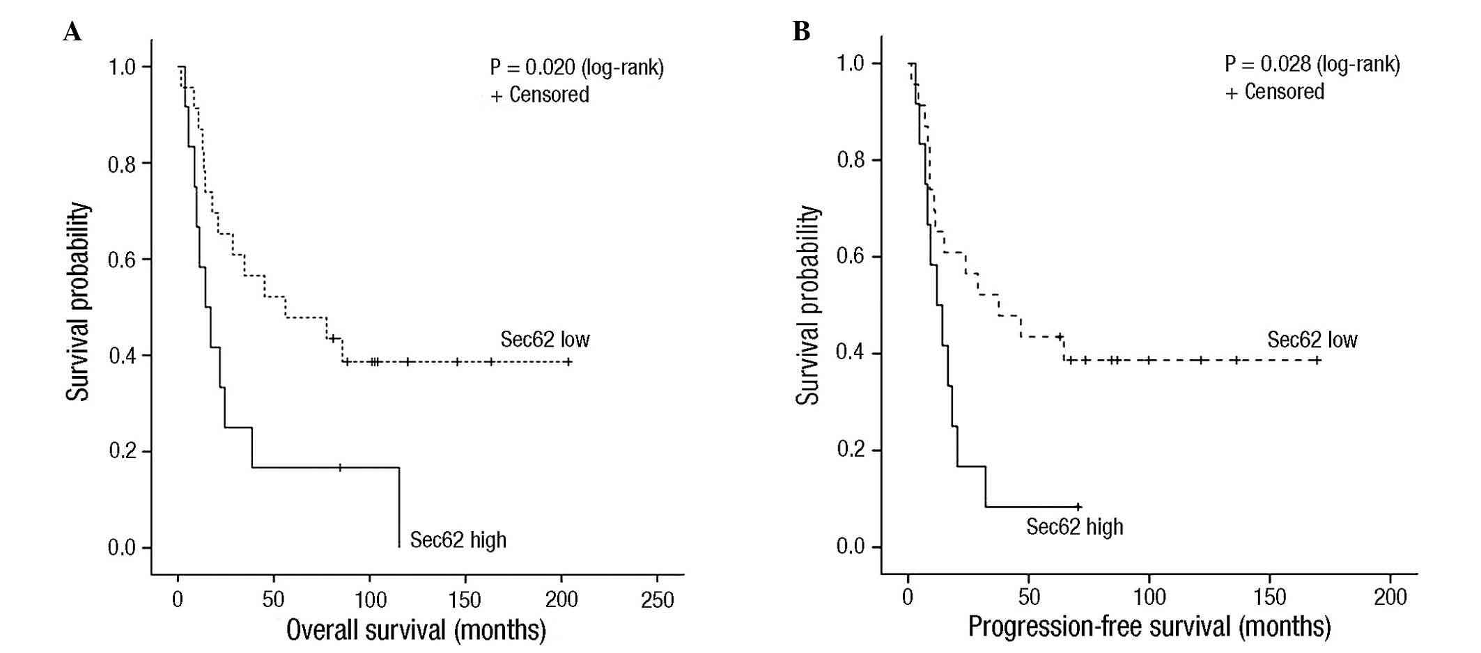
not PFS (P=0.103). Notably, high Sec62 IRS was significantly
associated with poorer OS and PFS rates in the present patient
cohort (P=0.020 and P=0.028, respectively; Table IV; Fig.
4).
| Table IV.Summary of immunohistochemical
expression analysis and influence on survival time in head and neck
squamous cell carcinoma patients. |
Table IV.
Summary of immunohistochemical
expression analysis and influence on survival time in head and neck
squamous cell carcinoma patients.
|
| Patients | P-value |
|---|
|
|
|
|
|---|
| Expression
level | n | % | Overall
survival | Progression-free
survival |
|---|
| Sec62 |
|
| 0.020 | 0.028 |
|
High | 12 | 34 |
|
|
|
Low | 23 | 66 |
|
|
| Survivin nuc |
|
| 0.079 | 0.103 |
|
High | 19 | 54 |
|
|
|
Low | 16 | 46 |
|
|
| Survivin cyt |
|
| 0.785 | 0.943 |
|
High | 20 | 57 |
|
|
|
Low | 15 | 43 |
|
|
| Survivin
nuc+cyt |
|
| 0.338 | 0.270 |
|
High | 28 | 80 |
|
|
|
Low | 7 | 20 |
|
|
| p16 |
|
|
|
|
|
Oropharynx, high/low | 5/19 | 21 | 0.248 | 0.245 |
| All
cases, high/low | 5/30 | 14 | 0.557 | 0.560 |
| EGFR |
|
| 0.585 | 0.847 |
|
High | 17 | 49 |
|
|
|
Low | 18 | 51 |
|
|
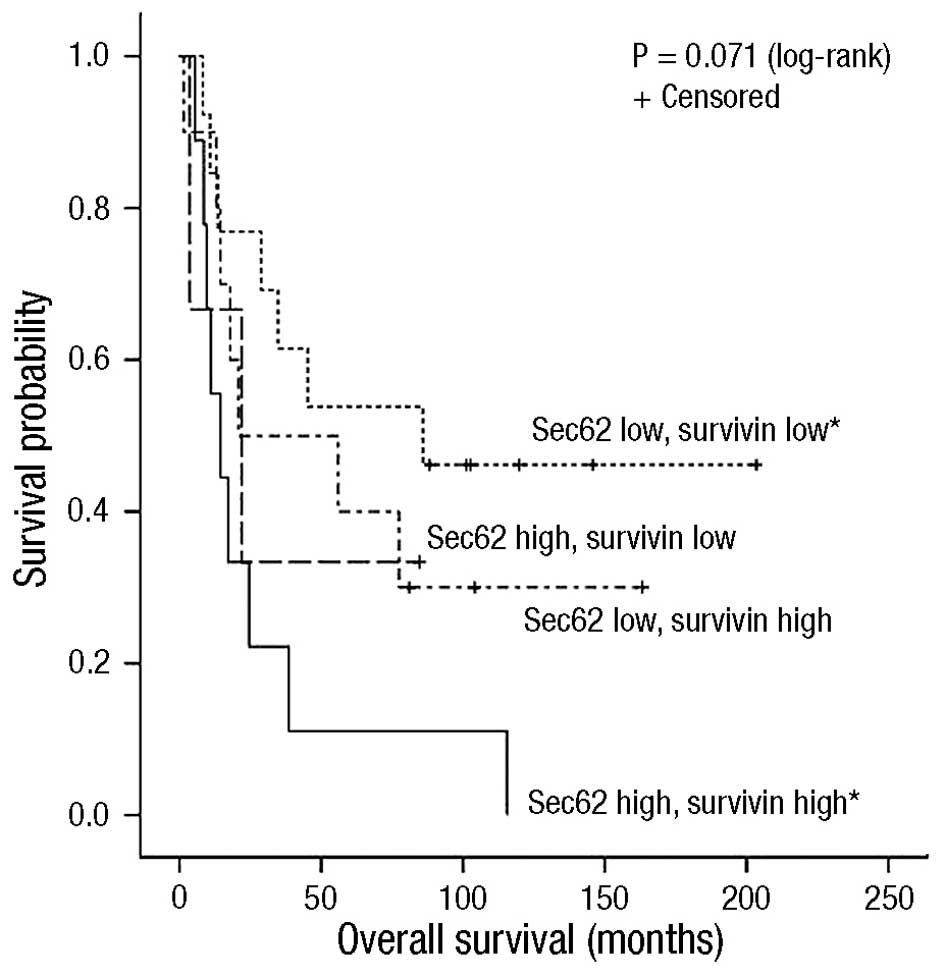
Subsequently, whether the expression levels of a
combination of these markers were associated with a greater
difference in PFS and OS compared with the expression of each
single protein was analyzed. Due to the relatively small number of
clinical samples, only combinations of two different proteins were
analyzed, and not combinations of three or more. Initially, the
combination of high Sec62 and high nuclear survivin expression
appeared to indicate a poorer prognosis for HNSCC patients (PFS,
P=0.071; OS, P=0.071; Fig. 5);
however, statistical significance was achieved only when
considering patients with low Sec62 and low nuclear survivin
expression vs. high Sec62 and high nuclear survivin expression
(PFS, P=0.006; OS, P=0.009). No combinatorial effect was observed
for Sec62 and EGFR (PFS, P=0.091; OS, P=0.097), Sec62 and p16 (PFS,
P=0.133; OS, P=0.095), EGFR and p16 (PFS, P=0.928; OS, P=0.841) or
EGFR and survivin (PFS, P=0.297; OS, P=0.431).
Discussion
The purpose of the present study was to characterize
the role of Sec62 as a novel potential biomarker for HNSCC, as well
as expression of the already extensively discussed biomarkers EGFR,
p16 and survivin in a cohort of patients with locally advanced
HNSCC treated with CRT.
EGFR is expressed in normal epithelium and has been
observed to be upregulated in a number of types of carcinoma
(19). High expression levels of EGFR
are also a common feature of HNSCC across various anatomical sites
to differing degrees (21,35). However, previous studies focusing on
the prognostic impact of EGFR expression levels in HNSCC reported
varying results (11,12). A number of reasons may account for the
discrepant results in terms of the prognostic impact of EGFR
expression in HNSCC. These include differences in the anatomical
sub-sites of HNSCC, varying sample compositions (biopsies and whole
tumor sections) and sample sizes, differing EGFR expression
measurement methods, different EGFR immunohistochemical staining
protocols and varying scoring methods (36,37). In
the present study, no significant correlation between EGFR
expression and OS time was observed, which contrasted with the
results of the majority of studies that have analyzed and compared
EGFR expression in HNSCC (11,12).
However, there are results that support the present observations,
particularly studies that identified no prognostic significance of
EGFR expression when comparing the treatment of HNSCC with
radiotherapy or CRT (38–40).
Previous studies have revealed that patients
exhibiting HPV-positive HNSCC demonstrate an improved prognosis
compared with patients exhibiting virus-negative tumors (3,28,41–43).
Previous work by a number of groups has demonstrated that
significantly more tumors originating in the oropharynx express
p16, a surrogate marker for high-risk HPV infection, compared with
non-oropharyngeal sites (3,41,43).
Furthermore, previous studies have revealed the prognostic impact
of p16 positivity for tumor collectives treated with various
treatment modalities, including surgery, radiotherapy and CRT
(3,4,41,43), or OSCC treated by various modalities
(that were not used to distinguish patients) (28,42,44). This
favorable prognosis is hypothesized to be due to a combination of
tumor biology, favorable patient characteristics (younger patients
with limited comorbidity and positive performance status, and
reduced likelihood of tobacco and alcohol abuse in these patients)
and an apparent reduced risk of experiencing secondary primary
tumors (28,45).
In the present study, p16 expression was restricted
to the oropharyngeal sub-site; however, a prognostic influence on
survival was not observed. One of the limitations of the present
study was the small number of p16-positive tumors (n=5), which may
explain the non-significant difference in survival time. The
slightly reduced rate as compared with other studies may
additionally be the result of varying socioeconomic profiles and
risk factors of the patients. Furthermore, previous studies have
suggested that the biological behavior of HPV-positive tumors may
be altered by tobacco use and may render positive tumors less
responsive to therapy (44,45). As smoking status was inconsistently
recorded in the current patient cohort, this influence could not be
analyzed. However, the importance of determining the HPV status in
HNSCC, particularly in oropharyngeal cancer, has been extensively
demonstrated in the relevant literature and should be considered in
additional studies (3–5,28,41–45).
As previously mentioned, survivin is overexpressed
in the majority of human tumors; however, is undetectable or
present at very low levels in the majority of normal adult tissues
(23). Survivin expression has been
associated with poor prognosis, disease progression and drug
resistance in various types of malignancy, including HNSCC, and
particularly in those exhibiting high nuclear survivin expression
(14,17,46).
However, a number of reports have revealed survivin expression to
be associated with favorable prognosis, including in gastric
(47) and breast cancer (48), as well as in HNSCC, regardless of the
various nuclear and cytoplasmic staining patterns of survivin
(15,16). In concordance with the results of
several previous studies (13–15,17,49),
the present study identified that survivin exists in two
subcellular pools, cytoplasmic and nuclear. Studies considering
these differences reported a consistent association between high
levels of nuclear survivin expression and poor prognosis (17,49). Due
potentially to the limited number of patients investigated, the
present study could demonstrate only a weak trend between nuclear
survivin expression and a reduced OS time, whereas cytoplasmic
survivin expression had no prognostic influence.
Multiple reports have described 3q amplification,
particularly within the region from 3q22 to 3qter, as one of the
most common genomic alterations in HNSCC (8,10), and
this amplification has been demonstrated to be associated with more
aggressive tumor behaviors and to be predictive of a poorer
prognosis (9,10). In NSCLC, the clinical relevance of
Sec62, another tumor entity characterized by 3q amplification, has
already been demonstrated. A significantly increased Sec62 content
was observed in tumors exhibiting lymph node metastases compared
with non-metastasized tumors, as well as in poorly differentiated
(grade 3) compared with moderately differentiated (grade 2) tumors
(31). Furthermore, SEC62
overexpression was associated with significantly reduced OS time in
NSCLC patients who underwent curative surgery, particularly in
patients exhibiting squamous cell lung carcinoma (32).
In addition to its correlation with a cancerous
phenotype and adverse clinical parameters, SEC62
overexpression has been demonstrated to serve a significant
functional role in molecular carcinogenesis in vitro;
SEC62 silencing by small interfering RNA transfection
resulted in a markedly decreased migration capability, as well as
increased sensitivity towards ER stress in various cell lines
(29,31). Furthermore, artificial SEC62
overexpression in HEK293 cells induced a potential to migrate in
this otherwise scarcely migrating cell line (32). Thus, based on current knowledge
regarding Sec62 biology in cancer, tumor cells may benefit from an
increased Sec62 level with regard to an increased migration
potential, as well as an enhanced ability to cope with ER stress
(50,51). However, the exact molecular mechanisms
by which Sec62 is able to influence the processes of cell migration
and ER stress tolerance remain to be elucidated. As Sec62 is known
to have a role in protein translocation into the ER, and thereby in
the biosynthesis of numerous secretory and membrane proteins
(52–54), it may be speculated that the
inhibition of cell migration associated with decreased Sec62
protein content may be due to a reduced biogenesis of specific
membrane or secretory proteins that are involved in cell migration
and ER stress-associated mechanisms. Furthermore, the
tumor-promoting effects of Sec62 in cancer cells appear to be
mediated, at least in part, by Ca2+-dependent signaling
mechanisms (32). A previous study
examining various putative oncogenes within 3q26 by systematic
loss- and gain-of-function experiments was able to identify
SEC62/TLOC1 as a major cancer driver within this
frequently amplified region (55).
Notably, in the present study, patients with tumors
exhibiting an elevated level of Sec62 and nuclear survivin
demonstrated the poorest prognosis. The interaction of survivin
with other molecules, such as X-linked inhibitor of apoptosis
protein, is known to inhibit the catalytic activity of caspase-9,
thereby preventing apoptosis (46).
Caspase-9 activation represents a major component of ER
stress-induced apoptosis (56),
providing an association with Sec62, which is thought to have a
significant role in the regulation of Ca2+ leakage from
the ER, via indirectly mediating the interaction of the
Sec61-channel in the ER membrane with calmodulin on the cytoplasmic
side and binding immunoglobulin protein on the ER-luminal side
(32). Thus, survivin and Sec62 may
synergistically act as potent apoptosis inhibitors, conferring a
proliferation advantage to cancer cells. Based on the results of
the present study, the simultaneous therapeutic targeting of these
two cancer drivers in HNSCC may be suggested to provide a
potentially promising therapeutic approach.
The conclusions of the present study indicate that
Sec62 is a promising novel candidate for a prognostic marker in
HNSCC patients, and additional studies should include larger
populations, as well as a comparison of various treatment
strategies.
Acknowledgements
The present study was supported by the Homburger
Research Promotion Program HOMFOR 2010 grant (Saarland University
Medical Center, Homburg, Germany).
References
|
1
|
Jemal A, Bray F, Center MM, Ferlay J, Ward
E and Forman D: Global cancer statistics. CA Cancer J Clin.
61:69–90. 2011. View Article : Google Scholar : PubMed/NCBI
|
|
2
|
Hashibe M, Brennan P, Chuang SC, Boccia S,
Castellsague X, Chen C, Curado MP, Dal Maso L, Daudt AW, Fabianova
E, et al: Interaction between tobacco and alcohol use and the risk
of head and neck cancer: Pooled analysis in the International Head
and Neck Cancer Epidemiology Consortium. Cancer Epidemiol
Biomarkers Prev. 18:541–550. 2009. View Article : Google Scholar : PubMed/NCBI
|
|
3
|
Fakhry C, Westra WH, Li S, Cmelak A, Ridge
JA, Pinto H, Forastiere A and Gillison ML: Improved survival of
patients with human papillomavirus-positive head and neck squamous
cell carcinoma in a prospective clinical trial. J Natl Cancer Inst.
100:261–269. 2008. View Article : Google Scholar : PubMed/NCBI
|
|
4
|
Ang KK, Harris J, Wheeler R, Weber R,
Rosenthal DI, Nguyen-Tân PF, Westra WH, Chung CH, Jordan RC, Lu C,
et al: Human papillomavirus and survival of patients with
oropharyngeal cancer. N Engl J Med. 363:24–35. 2010. View Article : Google Scholar : PubMed/NCBI
|
|
5
|
Chung CH and Gillison ML: Human
papillomavirus in head and neck cancer: Its role in pathogenesis
and clinical implications. Clin Cancer Res. 15:6758–6762. 2009.
View Article : Google Scholar : PubMed/NCBI
|
|
6
|
Nevins JR: The Rb/E2F pathway and cancer.
Hum Mol Genet. 10:699–703. 2001. View Article : Google Scholar : PubMed/NCBI
|
|
7
|
Burri RJ and Lee NY: Concurrent
chemotherapy and radiotherapy for head and neck cancer. Expert Rev
Anticancer Ther. 9:293–302. 2009. View Article : Google Scholar : PubMed/NCBI
|
|
8
|
Bauer VL, Braselmann H, Henke M, Mattern
D, Walch A, Unger K, Baudis M, Lassmann S, Huber R, Wienberg J, et
al: Chromosomal changes characterize head and neck cancer with poor
prognosis. J Mol Med (Berl). 86:1353–1365. 2008. View Article : Google Scholar : PubMed/NCBI
|
|
9
|
Wreesmann VB, Shi W, Thaler HT, Poluri A,
Kraus DH, Pfister D, Shaha AR, Shah JP, Rao PH and Singh B:
Identification of novel prognosticators of outcome in squamous cell
carcinoma of the head and neck. J Clin Oncol. 22:3965–3972. 2004.
View Article : Google Scholar : PubMed/NCBI
|
|
10
|
Bockmühl U, Schlüns K, Küchler I, Petersen
S and Petersen I: Genetic imbalances with impact on survival in
head and neck cancer patients. Am J Pathol. 157:369–375. 2000.
View Article : Google Scholar : PubMed/NCBI
|
|
11
|
Zhu X, Zhang F, Zhang W, He J, Zhao Y and
Chen X: Prognostic role of epidermal growth factor receptor in head
and neck cancer: A meta-analysis. J Surg Oncol. 108:387–397. 2013.
View Article : Google Scholar : PubMed/NCBI
|
|
12
|
Keren S, Shoude Z, Lu Z and Beibei Y: Role
of EGFR as a prognostic factor for survival in head and neck
cancer: A meta-analysis. Tumour Biol. 35:2285–2295. 2014.
View Article : Google Scholar : PubMed/NCBI
|
|
13
|
Li F, Yang J, Ramnath N, Javle MM and Tan
D: Nuclear or cytoplasmic expression of survivin: What is the
significance? Int J Cancer. 114:509–512. 2005. View Article : Google Scholar : PubMed/NCBI
|
|
14
|
Lo Muzio L, Pannone G, Leonardi R,
Staibano S, Mignogna MD, De Rosa G, Kudo Y, Takata T and Altieri
DC: Survivin, a potential early predictor of tumor progression in
the oral mucosa. J Dent Res. 82:923–928. 2003. View Article : Google Scholar : PubMed/NCBI
|
|
15
|
Freier K, Pungs S, Sticht C,
Flechtenmacher C, Lichter P, Joos S and Hofele C: High survivin
expression is associated with favorable outcome in advanced primary
oral squamous cell carcinoma after radiation therapy. Int J Cancer.
120:942–946. 2007. View Article : Google Scholar : PubMed/NCBI
|
|
16
|
Farnebo L, Tiefenböck K, Ansell A, Thunell
LK, Garvin S and Roberg K: Strong expression of survivin is
associated with positive response to radiotherapy and improved
overall survival in head and neck squamous cell carcinoma patients.
Int J Cancer. 133:1994–2003. 2013. View Article : Google Scholar : PubMed/NCBI
|
|
17
|
Preuss SF, Weinell A, Molitor M, Semrau R,
Stenner M, Drebber U, Wedemeyer I, Hoffmann TK, Guntinas-Lichius O
and Klussmann JP: Survivin and epidermal growth factor receptor
expression in surgically treated oropharyngeal squamous cell
carcinoma. Head Neck. 30:1318–1324. 2008. View Article : Google Scholar : PubMed/NCBI
|
|
18
|
Herbst RS: Review of epidermal growth
factor receptor biology. Int J Radiat Oncol Biol Phys. 59(Suppl 2):
S21–S26. 2004. View Article : Google Scholar
|
|
19
|
Nicholson RI, Gee JM and Harper ME: EGFR
and cancer prognosis. Eur J Cancer. 37(Suppl 4): S9–S15. 2001.
View Article : Google Scholar : PubMed/NCBI
|
|
20
|
Grandis Rubin J, Tweardy DJ and Melhem MF:
Asynchronous modulation of transforming growth factor alpha and
epidermal growth factor receptor protein expression in progression
of premalignant lesions to head and neck squamous cell carcinoma.
Clin Cancer Res. 4:13–20. 1998.PubMed/NCBI
|
|
21
|
Dassonville O, Formento JL, Francoual M,
Ramaioli A, Santini J, Schneider M, Demard F and Milano G:
Expression of epidermal growth factor receptor and survival in
upper aerodigestive tract cancer. J Clin Oncol. 11:1873–1878.
1993.PubMed/NCBI
|
|
22
|
Li F, Ambrosini G, Chu EY, Plescia J,
Tognin S, Marchisio PC and Altieri DC: Control of apoptosis and
mitotic spindle checkpoint by survivin. Nature. 396:580–584. 1998.
View Article : Google Scholar : PubMed/NCBI
|
|
23
|
Ambrosini G, Adida C and Altieri DC: A
novel anti-apoptosis gene, survivin, expressed in cancer and
lymphoma. Nat Med. 3:917–921. 1997. View Article : Google Scholar : PubMed/NCBI
|
|
24
|
Jung V, Kindich R, Kamradt J, Jung M,
Müller M, Schulz WA, Engers R, Unteregger G, Stöckle M, Zimmermann
R and Wullich B: Genomic and expression analysis of the 3q25-q26
amplification unit reveals TLOC1/SEC62 as a probable target gene in
prostate cancer. Mol Cancer Res. 4:169–176. 2006. View Article : Google Scholar : PubMed/NCBI
|
|
25
|
Dehan E, Ben-Dor A, Liao W, Lipson D,
Frimer H, Rienstein S, Simansky D, Krupsky M, Yaron P, Friedman E,
et al: Chromosomal aberrations and gene expression profiles in
non-small cell lung cancer. Lung Cancer. 56:175–184. 2007.
View Article : Google Scholar : PubMed/NCBI
|
|
26
|
Chen J, Guo L, Peiffer DA, Zhou L, Chan
OT, Bibikova M, Wickham-Garcia E, Lu SH, Zhan Q, Wang-Rodriguez J,
et al: Genomic profiling of 766 cancer-related genes in archived
esophageal normal and carcinoma tissues. Int J Cancer.
122:2249–2254. 2008. View Article : Google Scholar : PubMed/NCBI
|
|
27
|
Thomas LK, Bermejo JL, Vinokurova S,
Jensen K, Bierkens M, Steenbergen R, Bergmann M, von Knebel
Doeberitz M and Reuschenbach M: Chromosomal gains and losses in
human papillomavirus-associated neoplasia of the lower genital
tract - a systematic review and meta-analysis. Eur J Cancer.
50:85–98. 2014. View Article : Google Scholar : PubMed/NCBI
|
|
28
|
Klussmann JP, Mooren JJ, Lehnen M,
Claessen SM, Stenner M, Huebbers CU, Weissenborn SJ, Wedemeyer I,
Preuss SF, Straetmans JM, et al: Genetic signatures of HPV-related
and unrelated oropharyngeal carcinoma and their prognostic
implications. Clin Cancer Res. 15:1779–1786. 2009. View Article : Google Scholar : PubMed/NCBI
|
|
29
|
Greiner M, Kreutzer B, Lang S, Jung V,
Cavalié A, Unteregger G, Zimmermann R and Wullich B: Sec62 protein
level is crucial for the ER stress tolerance of prostate cancer.
Prostate. 71:1074–1083. 2011. View Article : Google Scholar : PubMed/NCBI
|
|
30
|
Greiner M, Kreutzer B, Jung V, Grobholz R,
Hasenfus A, Stöhr RF, Tornillo L, Dudek J, Stöckle M, Unteregger G,
et al: Silencing of the SEC62 gene inhibits migratory and invasive
potential of various tumor cells. Int J Cancer. 128:2284–2295.
2011. View Article : Google Scholar : PubMed/NCBI
|
|
31
|
Linxweiler M, Linxweiler J, Barth M,
Benedix J, Jung V, Kim YJ, Bohle RM, Zimmermann R and Greiner M:
Sec62 bridges the gap from 3q amplification to molecular cell
biology in non-small cell lung cancer. Am J Pathol. 180:473–483.
2012. View Article : Google Scholar : PubMed/NCBI
|
|
32
|
Linxweiler M, Schorr S, Schäuble N, Jung
M, Linxweiler J, Langer F, Schäfers HJ, Cavalié A, Zimmermann R and
Greiner M: Targeting cell migration and the endoplasmic reticulum
stress response with calmodulin antagonists: A clinically tested
small molecule phenocopy of SEC62 gene silencing in human tumor
cells. BMC Cancer. 13:574–588. 2013. View Article : Google Scholar : PubMed/NCBI
|
|
33
|
Sobin LH and Wittekind C: International
Union Against Cancer system (UICC). TNM Classification of Malignant
Tumours (6th). (Hoboken, New Jersey). John Wiley & Sons.
2002.
|
|
34
|
Remmele W and Stegner HE: Recommendation
for uniform definition of an immunoreactive score (IRS) for
immunohistochemical estrogen receptor detection (ER-ICA) in breast
cancer tissue. Pathologe. 8:138–140. 1987.(In German). PubMed/NCBI
|
|
35
|
Bentzen SM, Atasoy BM, Daley FM, Dische S,
Richman PI, Saunders MI, Trott KR and Wilson GD: Epidermal growth
factor receptor expression in pretreatment biopsies from head and
neck squamous cell carcinoma as a predictive factor for a benefit
from accelerated radiation therapy in a randomized controlled
trial. J Clin Oncol. 23:5560–5567. 2005. View Article : Google Scholar : PubMed/NCBI
|
|
36
|
Atkins D, Reiffen KA, Tegtmeier CL,
Winther H, Bonato MS and Störkel S: Immunohistochemical detection
of EGFR in paraffin-embedded tumor tissues: Variation in staining
intensity due to choice of fixative and storage time of tissue
sections. J Histochem Cytochem. 52:893–901. 2004. View Article : Google Scholar : PubMed/NCBI
|
|
37
|
Fischer C, Zlobec I, Stöckli E, Probst S,
Storck C, Tornillo L, Lugli A, Wolfensberger M and Terracciano L:
Is immunohistochemical epidermal growth factor receptor expression
overestimated as a prognostic factor in head-neck squamous cell
carcinoma? A retrospective analysis based on a tissue microarray of
365 carcinomas. Hum Pathol. 39:1527–1534. 2008. View Article : Google Scholar : PubMed/NCBI
|
|
38
|
Numico G, Russi EG, Colantonio I, Lantermo
RA, Silvestris N, Vitiello R, Comino A, Abrate M, Zavattero C,
Melano A and Merlano M: EGFR status and prognosis of patients with
locally advanced head and neck cancer treated with
chemoradiotherapy. Anticancer Res. 30:671–676. 2010.PubMed/NCBI
|
|
39
|
Eriksen JG, Steiniche T, Askaa J, Alsner J
and Overgaard J: The prognostic value of epidermal growth factor
receptor is related to tumor differentiation and the overall
treatment time of radiotherapy in squamous cell carcinomas of the
head and neck. Int J Radiat Oncol Biol Phys. 58:561–566. 2004.
View Article : Google Scholar : PubMed/NCBI
|
|
40
|
Aebersold DM, Froehlich SC, Jonczy M, Beer
KT, Laissue J, Greiner RH and Djonov V: Expression of transforming
growth factor-alpha, epidermal growth factor receptor and
platelet-derived growth factors A and B in oropharyngeal cancers
treated by curative radiation therapy. Radiother Oncol. 63:275–283.
2002. View Article : Google Scholar : PubMed/NCBI
|
|
41
|
Lassen P, Eriksen JG, Hamilton-Dutoit S,
Tramm T, Alsner J and Overgaard J: Effect of HPV-associated
p16INK4A expression on response to radiotherapy and survival in
squamous cell carcinoma of the head and neck. J Clin Oncol.
27:1992–1998. 2009. View Article : Google Scholar : PubMed/NCBI
|
|
42
|
Fischer CA, Zlobec I, Green E, Probst S,
Storck C, Lugli A, Tornillo L, Wolfensberger M and Terracciano LM:
Is the improved prognosis of p16 positive oropharyngeal squamous
cell carcinoma dependent of the treatment modality? Int J Cancer.
126:1256–1262. 2010.PubMed/NCBI
|
|
43
|
Deng Z, Hasegawa M, Aoki K, Matayoshi S,
Kiyuna A, Yamashita Y, Uehara T, Agena S, Maeda H, Xie M and Suzuki
M: A comprehensive evaluation of human papillomavirus positive
status and p16INK4a overexpression as a prognostic biomarker in
head and neck squamous cell carcinoma. Int J Oncol. 45:67–76.
2014.PubMed/NCBI
|
|
44
|
Hafkamp HC, Manni JJ, Haesevoets A, Voogd
AC, Schepers M, Bot FJ, Hopman AH, Ramaekers FC and Speel EJ:
Marked differences in survival rate between smokers and nonsmokers
with HPV 16-associated tonsillar carcinomas. Int J Cancer.
122:2656–2664. 2008. View Article : Google Scholar : PubMed/NCBI
|
|
45
|
Gillison ML, Zhang Q, Jordan R, Xiao W,
Westra WH, Trotti A, Spencer S, Harris J, Chung CH and Ang KK:
Tobacco smoking and increased risk of death and progression for
patients with p16-positive and p16-negative oropharyngeal cancer. J
Clin Oncol. 30:2102–2111. 2012. View Article : Google Scholar : PubMed/NCBI
|
|
46
|
Mita AC, Mita MM, Nawrocki ST and Giles
FJ: Survivin: Key regulator of mitosis and apoptosis and novel
target for cancer therapeutics. Clin Cancer Res. 14:5000–5005.
2008. View Article : Google Scholar : PubMed/NCBI
|
|
47
|
Okada E, Murai Y, Matsui K, Isizawa S,
Cheng C, Masuda M and Takano Y: Survivin expression in tumor cell
nuclei is predictive of a favorable prognosis in gastric cancer
patients. Cancer Lett. 163:109–116. 2001. View Article : Google Scholar : PubMed/NCBI
|
|
48
|
Kennedy SM, O'Driscoll L, Purcell R,
Fitz-Simons N, McDermott EW, Hill AD, O'Higgins NJ, Parkinson M,
Linehan R and Clynes M: Prognostic importance of survivin in breast
cancer. Br J Cancer. 88:1077–1083. 2003. View Article : Google Scholar : PubMed/NCBI
|
|
49
|
Qi G, Kudo Y, Ando T, Tsunematsu T,
Shimizu N, Siriwardena SB, Yoshida M, Keikhaee MR, Ogawa I and
Takata T: Nuclear Survivin expression is correlated with malignant
behaviors of head and neck cancer together with Aurora-B. Oral
Oncol. 46:263–270. 2010. View Article : Google Scholar : PubMed/NCBI
|
|
50
|
Koumenis C: ER stress, hypoxia tolerance
and tumor progression. Curr Mol Med. 6:55–69. 2006. View Article : Google Scholar : PubMed/NCBI
|
|
51
|
Bi M, Naczki C, Koritzimsky M, Fels D,
Blais J, Hu N, Harding H, Novoa I, Varia M, Raleigh J, et al: ER
stress-regulated translation increases tolerance to extreme hypoxia
and promotes tumor growth. EMBO J. 24:3470–3481. 2005. View Article : Google Scholar : PubMed/NCBI
|
|
52
|
Lang S, Benedix J, Fedeles SV, Schorr S,
Schirra C, Schäuble N, Jalal C, Greiner M, Hassdenteufel S, Tatzelt
J, et al: Different effects of Sec61α, Sec62 and Sec63 depletion on
transport of polypeptides into the endoplasmic reticulum of
mammalian cells. J Cell Sci. 125:1958–1969. 2012. View Article : Google Scholar : PubMed/NCBI
|
|
53
|
Lakkaraju AK, Thankappan R, Mary C,
Garrison JL, Taunton J and Strub K: Efficient secretion of small
proteins in mammalian cells relies on Sec62-dependent
posttranslational translocation. Mol Biol Cell. 23:2712–2722. 2012.
View Article : Google Scholar : PubMed/NCBI
|
|
54
|
Reithinger JH, Kim JE and Kim H: Sec62
protein mediates membrane insertion and orientation of moderately
hydrophobic signal anchor proteins in the endoplasmic reticulum
(ER). J Biol Chem. 288:18058–18067. 2013. View Article : Google Scholar : PubMed/NCBI
|
|
55
|
Hagerstrand D, Tong A, Schumacher SE, Ilic
N, Shen RR, Cheung HW, Vazquez F, Shrestha Y, Kim SY, Giacomelli
AO, et al: Systematic interrogation of 3q26 identifies TLOC1 and
SKIL as cancer drivers. Cancer Discov. 3:1044–1057. 2013.
View Article : Google Scholar : PubMed/NCBI
|
|
56
|
Jimbo A, Fujita E, Kouroku Y, Ohnishi J,
Inohara N, Kuida K, Sakamaki K, Yonehara S and Momoi T: ER stress
induces caspase-8 activation, stimulating cytochrome c
release and caspase-9 activation. Exp Cell Res. 283:156–166. 2003.
View Article : Google Scholar : PubMed/NCBI
|