Spandidos Publications Logo
  • About
    • About Spandidos
    • Aims and Scopes
    • Abstracting and Indexing
    • Editorial Policies
    • Reprints and Permissions
    • Job Opportunities
    • Terms and Conditions
    • Contact
  • Journals
    • All Journals
    • Oncology Letters
      • Oncology Letters
      • Information for Authors
      • Editorial Policies
      • Editorial Board
      • Aims and Scope
      • Abstracting and Indexing
      • Bibliographic Information
      • Archive
    • International Journal of Oncology
      • International Journal of Oncology
      • Information for Authors
      • Editorial Policies
      • Editorial Board
      • Aims and Scope
      • Abstracting and Indexing
      • Bibliographic Information
      • Archive
    • Molecular and Clinical Oncology
      • Molecular and Clinical Oncology
      • Information for Authors
      • Editorial Policies
      • Editorial Board
      • Aims and Scope
      • Abstracting and Indexing
      • Bibliographic Information
      • Archive
    • Experimental and Therapeutic Medicine
      • Experimental and Therapeutic Medicine
      • Information for Authors
      • Editorial Policies
      • Editorial Board
      • Aims and Scope
      • Abstracting and Indexing
      • Bibliographic Information
      • Archive
    • International Journal of Molecular Medicine
      • International Journal of Molecular Medicine
      • Information for Authors
      • Editorial Policies
      • Editorial Board
      • Aims and Scope
      • Abstracting and Indexing
      • Bibliographic Information
      • Archive
    • Biomedical Reports
      • Biomedical Reports
      • Information for Authors
      • Editorial Policies
      • Editorial Board
      • Aims and Scope
      • Abstracting and Indexing
      • Bibliographic Information
      • Archive
    • Oncology Reports
      • Oncology Reports
      • Information for Authors
      • Editorial Policies
      • Editorial Board
      • Aims and Scope
      • Abstracting and Indexing
      • Bibliographic Information
      • Archive
    • Molecular Medicine Reports
      • Molecular Medicine Reports
      • Information for Authors
      • Editorial Policies
      • Editorial Board
      • Aims and Scope
      • Abstracting and Indexing
      • Bibliographic Information
      • Archive
    • World Academy of Sciences Journal
      • World Academy of Sciences Journal
      • Information for Authors
      • Editorial Policies
      • Editorial Board
      • Aims and Scope
      • Abstracting and Indexing
      • Bibliographic Information
      • Archive
    • International Journal of Functional Nutrition
      • International Journal of Functional Nutrition
      • Information for Authors
      • Editorial Policies
      • Editorial Board
      • Aims and Scope
      • Abstracting and Indexing
      • Bibliographic Information
      • Archive
    • International Journal of Epigenetics
      • International Journal of Epigenetics
      • Information for Authors
      • Editorial Policies
      • Editorial Board
      • Aims and Scope
      • Abstracting and Indexing
      • Bibliographic Information
      • Archive
    • Medicine International
      • Medicine International
      • Information for Authors
      • Editorial Policies
      • Editorial Board
      • Aims and Scope
      • Abstracting and Indexing
      • Bibliographic Information
      • Archive
  • Articles
  • Information
    • Information for Authors
    • Information for Reviewers
    • Information for Librarians
    • Information for Advertisers
    • Conferences
  • Language Editing
Spandidos Publications Logo
  • About
    • About Spandidos
    • Aims and Scopes
    • Abstracting and Indexing
    • Editorial Policies
    • Reprints and Permissions
    • Job Opportunities
    • Terms and Conditions
    • Contact
  • Journals
    • All Journals
    • Biomedical Reports
      • Information for Authors
      • Editorial Policies
      • Editorial Board
      • Aims and Scope
      • Abstracting and Indexing
      • Bibliographic Information
      • Archive
    • Experimental and Therapeutic Medicine
      • Information for Authors
      • Editorial Policies
      • Editorial Board
      • Aims and Scope
      • Abstracting and Indexing
      • Bibliographic Information
      • Archive
    • International Journal of Epigenetics
      • Information for Authors
      • Editorial Policies
      • Editorial Board
      • Aims and Scope
      • Abstracting and Indexing
      • Bibliographic Information
      • Archive
    • International Journal of Functional Nutrition
      • Information for Authors
      • Editorial Policies
      • Editorial Board
      • Aims and Scope
      • Abstracting and Indexing
      • Bibliographic Information
      • Archive
    • International Journal of Molecular Medicine
      • Information for Authors
      • Editorial Policies
      • Editorial Board
      • Aims and Scope
      • Abstracting and Indexing
      • Bibliographic Information
      • Archive
    • International Journal of Oncology
      • Information for Authors
      • Editorial Policies
      • Editorial Board
      • Aims and Scope
      • Abstracting and Indexing
      • Bibliographic Information
      • Archive
    • Medicine International
      • Information for Authors
      • Editorial Policies
      • Editorial Board
      • Aims and Scope
      • Abstracting and Indexing
      • Bibliographic Information
      • Archive
    • Molecular and Clinical Oncology
      • Information for Authors
      • Editorial Policies
      • Editorial Board
      • Aims and Scope
      • Abstracting and Indexing
      • Bibliographic Information
      • Archive
    • Molecular Medicine Reports
      • Information for Authors
      • Editorial Policies
      • Editorial Board
      • Aims and Scope
      • Abstracting and Indexing
      • Bibliographic Information
      • Archive
    • Oncology Letters
      • Information for Authors
      • Editorial Policies
      • Editorial Board
      • Aims and Scope
      • Abstracting and Indexing
      • Bibliographic Information
      • Archive
    • Oncology Reports
      • Information for Authors
      • Editorial Policies
      • Editorial Board
      • Aims and Scope
      • Abstracting and Indexing
      • Bibliographic Information
      • Archive
    • World Academy of Sciences Journal
      • Information for Authors
      • Editorial Policies
      • Editorial Board
      • Aims and Scope
      • Abstracting and Indexing
      • Bibliographic Information
      • Archive
  • Articles
  • Information
    • For Authors
    • For Reviewers
    • For Librarians
    • For Advertisers
    • Conferences
  • Language Editing
Login Register Submit
  • This site uses cookies
  • You can change your cookie settings at any time by following the instructions in our Cookie Policy. To find out more, you may read our Privacy Policy.

    I agree
Search articles by DOI, keyword, author or affiliation
Search
Advanced Search
presentation
Oncology Letters
Join Editorial Board Propose a Special Issue
Print ISSN: 1792-1074 Online ISSN: 1792-1082
Journal Cover
April-2016 Volume 11 Issue 4

Full Size Image

Sign up for eToc alerts
Recommend to Library

Journals

International Journal of Molecular Medicine

International Journal of Molecular Medicine

International Journal of Molecular Medicine is an international journal devoted to molecular mechanisms of human disease.

International Journal of Oncology

International Journal of Oncology

International Journal of Oncology is an international journal devoted to oncology research and cancer treatment.

Molecular Medicine Reports

Molecular Medicine Reports

Covers molecular medicine topics such as pharmacology, pathology, genetics, neuroscience, infectious diseases, molecular cardiology, and molecular surgery.

Oncology Reports

Oncology Reports

Oncology Reports is an international journal devoted to fundamental and applied research in Oncology.

Experimental and Therapeutic Medicine

Experimental and Therapeutic Medicine

Experimental and Therapeutic Medicine is an international journal devoted to laboratory and clinical medicine.

Oncology Letters

Oncology Letters

Oncology Letters is an international journal devoted to Experimental and Clinical Oncology.

Biomedical Reports

Biomedical Reports

Explores a wide range of biological and medical fields, including pharmacology, genetics, microbiology, neuroscience, and molecular cardiology.

Molecular and Clinical Oncology

Molecular and Clinical Oncology

International journal addressing all aspects of oncology research, from tumorigenesis and oncogenes to chemotherapy and metastasis.

World Academy of Sciences Journal

World Academy of Sciences Journal

Multidisciplinary open-access journal spanning biochemistry, genetics, neuroscience, environmental health, and synthetic biology.

International Journal of Functional Nutrition

International Journal of Functional Nutrition

Open-access journal combining biochemistry, pharmacology, immunology, and genetics to advance health through functional nutrition.

International Journal of Epigenetics

International Journal of Epigenetics

Publishes open-access research on using epigenetics to advance understanding and treatment of human disease.

Medicine International

Medicine International

An International Open Access Journal Devoted to General Medicine.

Journal Cover
April-2016 Volume 11 Issue 4

Full Size Image

Sign up for eToc alerts
Recommend to Library

  • Article
  • Citations
    • Cite This Article
    • Download Citation
    • Create Citation Alert
    • Remove Citation Alert
    • Cited By
  • Similar Articles
    • Related Articles (in Spandidos Publications)
    • Similar Articles (Google Scholar)
    • Similar Articles (PubMed)
  • Download PDF
  • Download XML
  • View XML
Case Report

An uncommon recurrence of an immature teratoma: A case report

  • Authors:
    • Xuelu Li
    • Dandan Zhu
    • Li Lv
    • Jian Yu
  • View Affiliations / Copyright

    Affiliations: Department of Breast Surgery, The Second Hospital of Dalian Medical University, Dalian, Liaoning 116023, P.R. China, Department of Critical Care Medicine, The Second Hospital of Dalian Medical University, Dalian, Liaoning 116023, P.R. China, Department of Pathology, The Second Hospital of Dalian Medical University, Dalian, Liaoning 116023, P.R. China
  • Pages: 2453-2456
    |
    Published online on: February 19, 2016
       https://doi.org/10.3892/ol.2016.4254
  • Expand metrics +
Metrics: Total Views: 0 (Spandidos Publications: | PMC Statistics: )
Metrics: Total PDF Downloads: 0 (Spandidos Publications: | PMC Statistics: )
Cited By (CrossRef): 0 citations Loading Articles...

This article is mentioned in:


Abstract

A 24-year-old female who was previously diagnosed with an immature, right ovarian teratoma was referred to The Second Hospital of Dalian Medical University (Dalian, China) due to abdominal pain. The patient had previously undergone a laparotomy and right ovarian oophorectomy. Despite this, at 10 years post‑surgery, the patient developed extensive abdominal metastases, in conjunction with elevated serum cancer antigen (CA)-125 and CA19-9 levels. The patient had a high risk of recurrence, and the liver and spleen metastases were subsequently diagnosed as mature teratomas. In order to prevent the possibility of disease recurrence, frequent post‑surgical follow-up, consisting of imaging analysis and the testing of serum markers, is required. Regarding the published literature, there have been no other patients presenting with a giant teratoma with associated metastasis. Due to the lack of regular follow-up appointments in China, the tumor in the present study was not recognized until too late. Therefore, to increase the successful detection of recurrent teratomas, a longer follow‑up period is necessary for patients who present with a high risk of recurrence.

Introduction

Teratomas may be categorized as either mature or immature, and originate from the three pluripotent germ cell layers: The ectoderm (skin and neural tissue), the endoderm (gastrointestinal and bronchial epithelium, and thyroid tissue), and the mesoderm (fat, bone, muscle and cartilage) (1). Immature teratomas are rare tumors that develop in the ovaries, accounting for 0.25–3% of tumors that develop in the ovaries, with individuals at a greater risk during their first two decades of life (1). By contrast, mature teratomas occur most commonly in women aged between 20–40 years (2). Teratomas have high recurrence and metastasis rates, and immature tumor tissues may be converted into mature tissues following post-surgical recurrence. The conversion of immature teratoma is characterized by slow growth, so symptoms are not typical. Clinical doctors often neglect the diagnosis of teratomas. If the size of the tumor is >6 cm, it is common for surgeons to utilize a surgical treatment. Recurrence typically develops subsequent to surgical excision of the tumor, often within the first year of primary therapy (3). The present study describes the case of a patient who underwent excision of an immature teratoma, which later developed into mature teratomas found as extensive abdominal metastases to the liver, spleen and pelvic cavity, identified after 10 years.

Case report

A 24-year-old female was referred to the Department of General Surgery, The Second Hospital of Dalian Medical University (Dalian, China), on 24 April 2013, due to intermittent upper abdominal pain. Upon physical examination of the right abdomen, a hard, large mass that moved under palpation was identified. Abdominal tenderness, rebound pain and muscular tension were impalpable, with no other positive signs. When examining the medical history of the patient in detail, an immature right ovarian teratoma that had been diagnosed 10 years previously was noted, for which the patient had undergone a laparotomy and right ovarian oophorectomy. The post-surgical pathological analysis had identified an immature teratoma, with an official diagnosis of grade 2 immature teratoma [Norris grading system (4)]. Adjuvant therapy had been administered, consisting of 3 cycles of bleomycin (20 U/m2), etoposide (1,675 mg/m2) and cisplatin (20 mg/m2), following surgery. At the last chemotherapy session, no residual tumor had been observed. The patient had subsequently been lost to follow-up.

Based on the medical history of the patient, recurrence of the teratoma was now suspected. Notably, laboratory tests exhibited elevated levels of the serum markers cancer antigen (CA)-125 and CA19-9 (Table I). In order to assess the giant tumor in the abdomen, the patient underwent a sonographic examination and computed tomography (CT) 
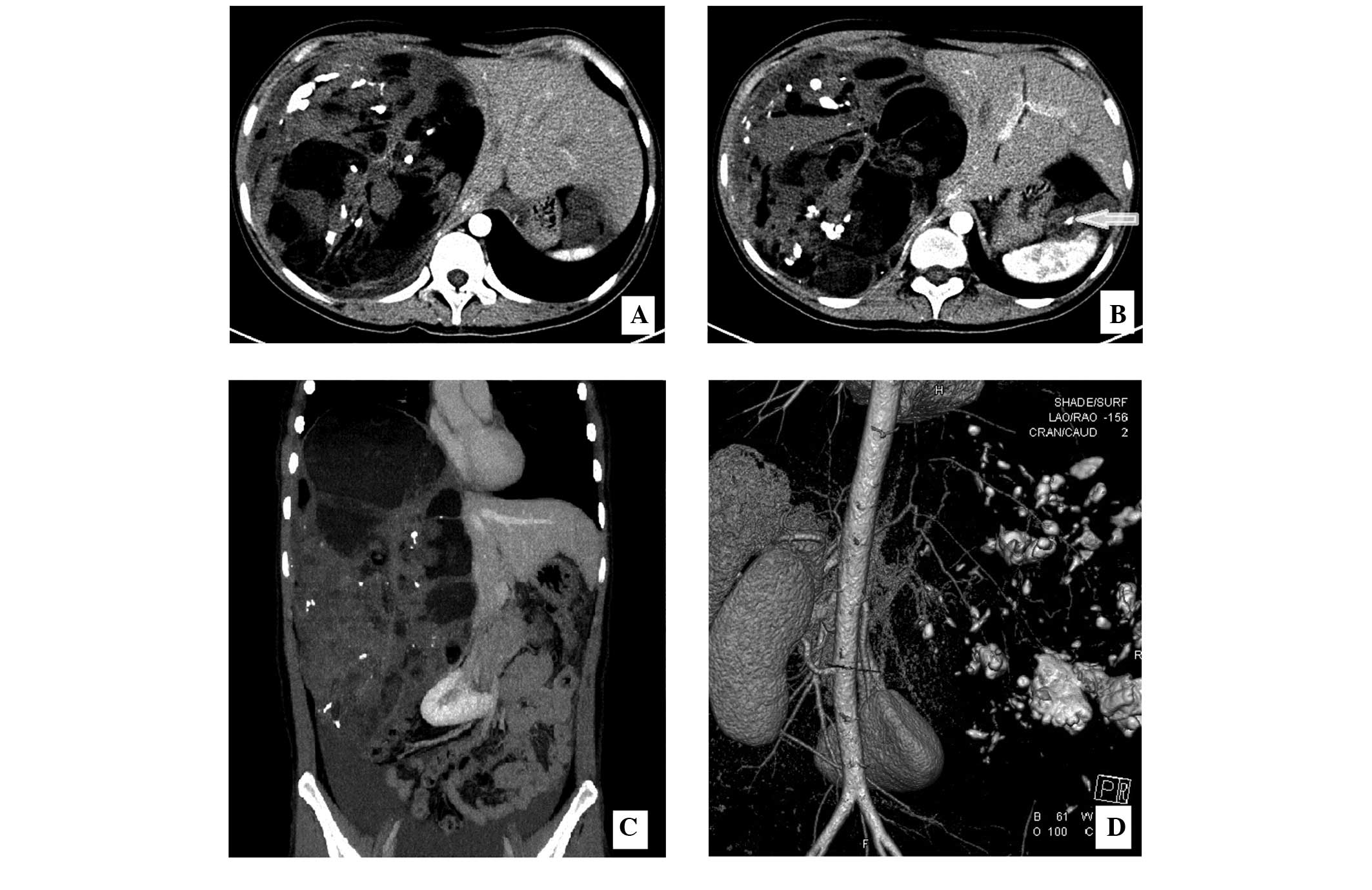
scan. The contrast-enhanced total abdominal CT scan revealed a complex cystic mass, measuring 18.6×15.7 cm2, on the right lobe of the liver (Fig. 1A). Additionally, a further tumor that exhibited the same properties as the giant mass was detected on the spleen (Fig. 1B).

Figure 1.

(A) Abdominal computed tomography enhancement scanning. A mass (18.6×15.7 cm2) was identified on the right liver lobe, exhibiting calcification and cystic changes. (B) A mass was also identified on the surface of the spleen, exhibiting calcification (white arrow). (C) An abdominal coronal section demonstrated that the compressed right ventricle, right kidney and pancreas had shifted to the left, and ascites was observed in the pelvic cavity. (D) Vascular remodeling.

Table I.

Dynamic change of tumor markers.

Table I.

Dynamic change of tumor markers.

Level post-surgery, kU/l

Tumor markerLevel prior to surgery, kU/l7 days30 days180 daysNormal range, kU/l
CA19–9>7000.001927.51250.5023.000.00–35.00
CA12–5440.63   201.70   40.00  5.200.00–35.00

[i] CA, cancer antigen.

With consideration of the tumor size, the compression symptoms (the right ventricle, right kidney and pancreas had shifted to the left; Fig. 1C and D) and the elevated serum markers, a diagnosis of an immature teratoma was the most probable. The patient subsequently underwent a laparotomy. Possible invasion of the tumor into the liver could not be excluded, therefore, a cardiopulmonary blood bypass was prepared. Following entry into the peritoneal cavity, the mass was removed from the right lobe of the liver, the first porta hepatis was blocked and the right hepatic vein was resected where the tumor had infiltrated. Subsequently, complete removal of 
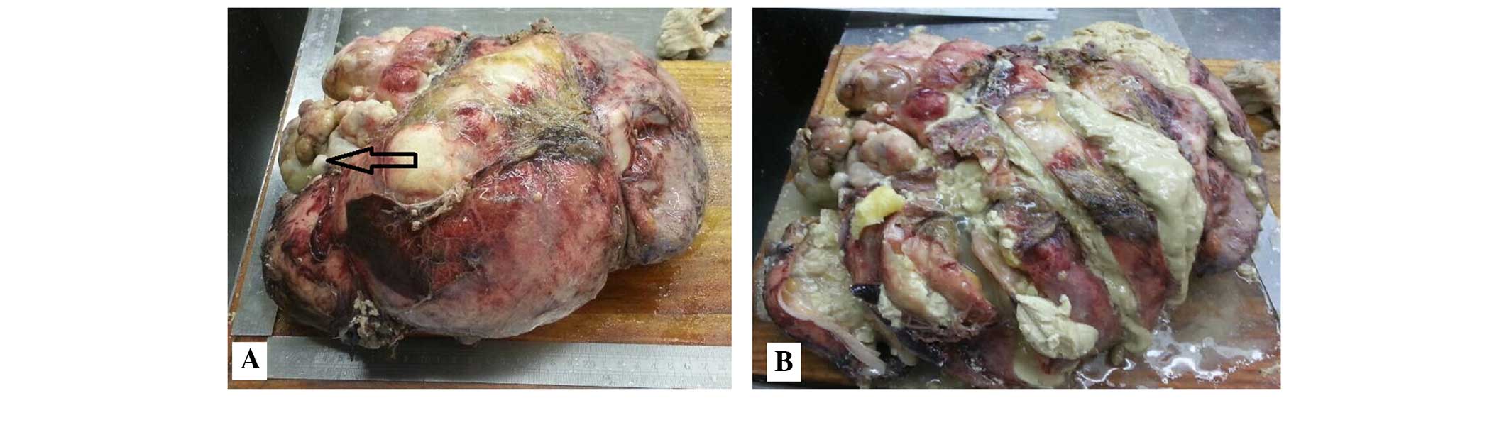
the mass from the spleen surface was successful. The giant tumor was rich in sebaceous materials, with hair shaft tissues and teeth identified inside the mass (Fig. 2A and 
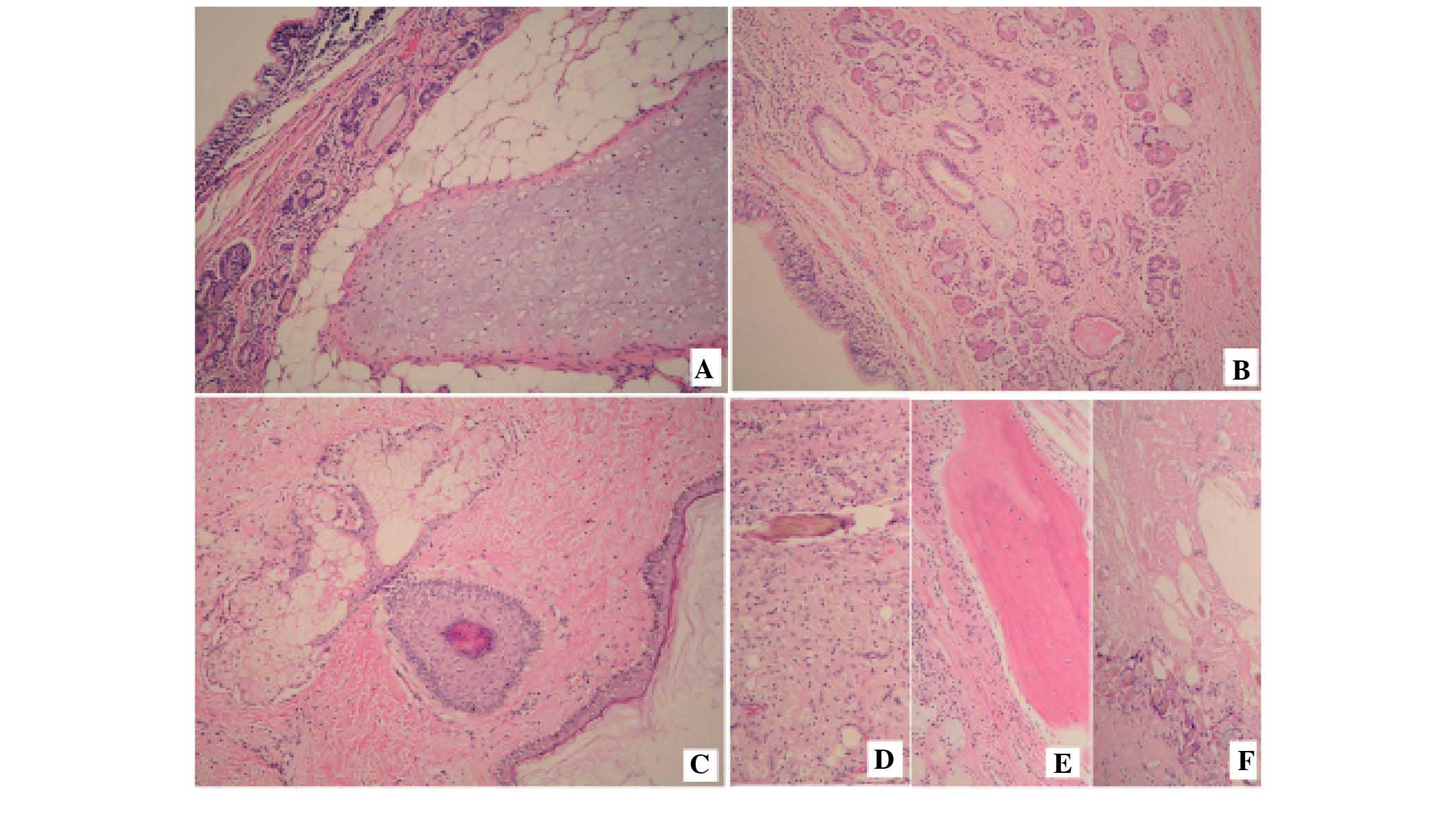
B). The pathology report confirmed that all tumors on the surface of the liver and the spleen, and in the pelvic cavity were mature teratomas (Fig. 3). Following tumor resection, there was no evidence of recurrence or metastasis during the 1 and 6 month follow-up evaluations and ongoing follow-up examinations every 3–6 months have been planned.

Figure 2.

Gross specimen removed from the liver. (A) The tumor (29×24 cm2) contained hair shaft tissues and teeth (black arrow). (B) The inside of the mass contained abundant, gelatinous and sebaceous materials, hair shaft tissues and teeth.

Figure 3.

Hematoxylin and eosin staining features. (A) Mature components of the bronchial epithelial cells, the bronchial glands and the bronchial cartilage. (B) Typical bronchial epithelium. Photomicrographs showing the skin tissue with (C) sebaceous glands, and (D) hair, (E) lamellar bone and (F) calcification. Magnification, x100.

Discussion

Immature teratomas of the ovary are rare tumors, accounting for <3% of all teratomas located in the ovaries. The disease typically occurs during the first two decades of life, and the tumors are composed of tissue derived two or more germ cell layers (ectoderm, mesoderm and/or endoderm), containing immature and embryonal structures (1). The current case presented with the classic symptoms and histological appearance associated with immature teratomas. Excision of such tumors is often followed by local recurrence within the first year of primary therapy initiation (3). The present patient was lost to follow-up subsequent to initial chemotherapy, but developed rare mature teratomas and extensive abdominal metastases after 10 years.

Mature teratomas are typically benign, however, malignant transformation may occur, albeit rarely. The peak incidence of such tumors is reported in women between the ages of 20–40 years. The disease is characterized by slow growth, with a growth rate of 1.8 mm per year, and when the size of the tumor is <6 cm, it is common for surgeons to utilize a non-surgical treatment (1). Growing teratoma syndrome is an uncommon condition present in men and women, with appropriately treated germ cell tumors characterized by the persistence or development of enlarging masses following adjuvant chemotherapy, normal tumor marker values and the presence of mature teratomas in the specimen. If a diagnosis of growing teratoma syndrome is confirmed, the surgeon must determine the operability of the tumor, whilst also weighing up the risks and benefits of surgery (5). This is necessary to allow for the optimal management of the disease and to prevent unnecessary chemotherapy. In the present case, the serum makers, CA-125 and CA19-9, exhibited particularly high levels when compared with levels observed in growing teratoma syndrome. Excision was performed on tumors that were <2 cm in size. Following the surgery, pathological sections from different locations in the tumor showed no immature elements. No further treatment was administered to the patient. To the best of our knowledge, this metastasis of a mature teratoma to the liver, the spleen and the pelvic cavity is particularly rare.

When forming a differential diagnosis for teratomas, the measurement of the serum levels of CA-125 and CA19-9 is a common method used for early detection. Ustunyurt et al (6) reported that serum CA19-9 is the most reliable tumor marker of mature ovarian cystic teratomas, and that the levels of serum CA19-9 are often associated with the size of the teratoma; however, this does not appear to have high specificity or sensitivity. Bast et al (7) reported that serum CA-125 is a reliable marker, and that in combination with elevated CA19-9, it may be a differential character of malignant tumors. Notably, in the present case, the variation in serum CA-125 and CA19-9 levels was greater compared with the typical change (Table I).

Harada et al (8) hypothesized that a young age (<30 years), a large cyst size (>8 cm) and the bilateral occurrence of mature teratomas are predictive factors for recurrence. In the present case, the patient was at a high risk of recurrence. In order to prevent residual disease, frequent post-surgical imaging analysis and the testing of serum markers is required. In the literature, to the best of our knowledge, no giant teratoma patients have been described who have presented with the same uncommon metastasis as the patient in the current case. Due to the lack of follow-up appointments in China, the tumor was recognized too late. To increase the likelihood of detecting recurrent teratomas, longer follow-up periods are required for patients who are at a high risk of recurrence.

References

1 

Saba L, Guerriero S, Sulcis R, Virgilio B, Melis G and Mallarini G: Mature and immature ovarian teratomas: CT, US and MR imaging characteristics. Eur J Radiol. 72:454–463. 2009. View Article : Google Scholar : PubMed/NCBI

2 

Chang CF and Lin CK: A case of recurrent, bilateral ovarian mature teratoma in a young woman. BMC Womens Health. 14:572014. View Article : Google Scholar : PubMed/NCBI

3 

Barwad A, Dey P and Shivalingam J: Metastatic of mature component in a treated case of immature teratoma diagnosed on fine-needle aspiration cytology of the liver. Diagn Cytopathol. 39:711–713. 2011. View Article : Google Scholar : PubMed/NCBI

4 

Norris HJ, Zirkin HJ and Benson WL: Immature (malignant) teratoma of the ovary: A clinical and pathologic study of 58 cases. Cancer. 37:2359–2372. 1976. View Article : Google Scholar : PubMed/NCBI

5 

Byrd K, Stany MP, Herbold NC, Leath CA III and Hamilton CA: Growing teratoma syndrome: Brief communication and algorithm for management. Aust NZ J Obstet Gynaecol. 53:318–321. 2013. View Article : Google Scholar

6 

Ustunyurt E, Gungor T, Iskender C, Ustunyurt BO, Bilge U and Mollamahmutoglu L: Tumor markers in mature cystic teratomas of the ovary. Arch Gynecol Obstet. 279:145–147. 2009. View Article : Google Scholar : PubMed/NCBI

7 

Bast RC Jr, Badgwell D, Lu Z, Marquez R, Rosen D, Liu J, Baggerly KA, Atkinson EN, Skates S, Zhang Z, et al: New tumor markers: CA125 and beyond. Int J Gynecol Cancer. 15(Suppl 3): 274–281. 2005. View Article : Google Scholar : PubMed/NCBI

8 

Harada M, Osuga Y, Fujimoto A, Fujimoto A, Fujii T, Yano T and Kozuma S: Predictive factors for recurrence of ovarian mature cystic teratomas after surgical excision. Eur J Obstet Gynecol Reprod Biol. 171:325–328. 2013. View Article : Google Scholar : PubMed/NCBI

Related Articles

  • Abstract
  • View
  • Download
  • Twitter
Copy and paste a formatted citation
Spandidos Publications style
Li X, Zhu D, Lv L and Yu J: An uncommon recurrence of an immature teratoma: A case report. Oncol Lett 11: 2453-2456, 2016.
APA
Li, X., Zhu, D., Lv, L., & Yu, J. (2016). An uncommon recurrence of an immature teratoma: A case report. Oncology Letters, 11, 2453-2456. https://doi.org/10.3892/ol.2016.4254
MLA
Li, X., Zhu, D., Lv, L., Yu, J."An uncommon recurrence of an immature teratoma: A case report". Oncology Letters 11.4 (2016): 2453-2456.
Chicago
Li, X., Zhu, D., Lv, L., Yu, J."An uncommon recurrence of an immature teratoma: A case report". Oncology Letters 11, no. 4 (2016): 2453-2456. https://doi.org/10.3892/ol.2016.4254
Copy and paste a formatted citation
x
Spandidos Publications style
Li X, Zhu D, Lv L and Yu J: An uncommon recurrence of an immature teratoma: A case report. Oncol Lett 11: 2453-2456, 2016.
APA
Li, X., Zhu, D., Lv, L., & Yu, J. (2016). An uncommon recurrence of an immature teratoma: A case report. Oncology Letters, 11, 2453-2456. https://doi.org/10.3892/ol.2016.4254
MLA
Li, X., Zhu, D., Lv, L., Yu, J."An uncommon recurrence of an immature teratoma: A case report". Oncology Letters 11.4 (2016): 2453-2456.
Chicago
Li, X., Zhu, D., Lv, L., Yu, J."An uncommon recurrence of an immature teratoma: A case report". Oncology Letters 11, no. 4 (2016): 2453-2456. https://doi.org/10.3892/ol.2016.4254
Follow us
  • Twitter
  • LinkedIn
  • Facebook
About
  • Spandidos Publications
  • Careers
  • Cookie Policy
  • Privacy Policy
How can we help?
  • Help
  • Live Chat
  • Contact
  • Email to our Support Team