Introduction
Pancreatic cancer is an aggressive and highly fatal
malignancy affecting a large number of individuals worldwide, and
has a relative 5-year survival rate of <5% (1,2). It is the
fourth leading cause of cancer-associated mortality in the United
States, with ~46,420 new cases and 39,590 fatalities occurring as a
result of the disease during 2014 (3). Surgical resection is currently the only
treatment available that may cure pancreatic cancer (4); however, the majority of patients are
diagnosed late in the natural course of the disease, with ~80%
presenting with metastasis (5,6).
The drug 2′,2′-difluorodeoxycytidine, also known as
gemcitabine, is the first-line chemotherapeutic agent for
pancreatic cancer treatment that is approved by the Food and Drug
Administration (7,8). It exerts antitumor activity depending on
several inhibitory actions of DNA synthesis, functioning to impair
DNA repair and induce apoptosis (9).
Gemcitabine has been demonstrated to effectively inhibit pancreatic
cancer cells that are insensitive to other drugs, including
fluorouracil, doxorubicin and cisplatin (10). Although gemcitabine exhibits effective
inhibition on pancreatic cancer cell growth in vitro, its
clinical efficacy remains low (11).
Thus, novel therapies are urgently required for the treatment of
this extremely aggressive malignancy.
Triptolide is a diterpenoid triepoxide derived from
the herb Tripterygium wilfordii, which has been utilized as
a natural agent in China for centuries (12). Triptolide has been successfully
applied as an immunosuppressant for clinical treatment of
inflammation, autoimmune diseases and organ transplantations
(13–15). In addition to immunosuppressive
activity, triptolide has been demonstrated to possess potent
antitumor activity and induce apoptosis in a variety of tumor types
in vivo and in vitro (16–18).
Furthermore, triptolide has been reported to synergistically
increase the antitumor activities of conventional chemotherapies
(19,20). Synergistic antitumor interactions
between triptolide and other chemotherapeutic drugs, including
cisplatin, artesunate and hydroxycamptothecin, have been previously
observed in pancreatic cancer cells (21–23). In
addition, triptolide is currently undergoing clinical trials for
its antitumor and proapoptotic activities on primary cultures of
human prostatic epithelial cells (24).
The present study hypothesized that triptolide
synergistically enhances the antitumor activity of gemcitabine by
inducing apoptosis in pancreatic cancer cells. The results
demonstrate that triptolide inhibits pancreatic cell growth, and
cooperatively augments gemcitabine-induced apoptosis by increasing
gemcitabine-induced S phase arrest and DNA double-strand breaks
(DSBs). This suggests that triptolide may possess therapeutic
potential for the treatment of pancreatic cancer, particularly when
administered in combination with gemcitabine.
Materials and methods
Chemicals
Triptolide was purchased from the National
Institutes for Food and Drug Control (Beijing, China), and
gemcitabine (GEM; Gemzar®) was purchased from Lilly
(Fegersheim, France). All other chemicals were of analytical grade
and obtained commercially.
Cell culture
The human pancreatic cancer BxPC-3 and PANC-1 cell
lines were obtained from the Shanghai Institute of Biochemistry and
Cell Biology, Chinese Academy of Sciences. (Shanghai, China). The
cell lines were maintained in Dulbecco's modified Eagle's medium
(DMEM; Gibco; Thermo Fisher Scientific, Inc., Waltham, MA, USA)
supplemented with 10% fetal bovine serum (HyClone; GE Healthcare
Life Sciences, Logan, UT, USA), penicillin/streptomycin (100 U/ml
each; Shijiazhuang No. 4 Pharmaceutical Co., Ltd., Shijiazhuang,
China), sodium bicarbonate (2 g/l; Sinopharm Chemical Reagent Co.,
Ltd., Shanghai, China) and HEPES (2.4 g/l; Amresco, LLC, Solon, OH,
USA), and were incubated at 37°C with 5% CO2.
In vitro cytotoxicity assays
In vitro cytotoxicities of triptolide and
gemcitabine, alone or in combination, in the pancreatic cancer cell
lines were determined by
3-(4,5-dimethylthiazol-2-yl)-2,5-diphenyltetrazolium bromide (MTT)
assay (25). Control groups were
treated without gemcitabine and triptolide, and cultured for up 120
h. Briefly, BxPC-3 or PANC-1 cells were plated in 96-well plates
(Corning Inc., Corning, NY, USA; 3×10³ cells/well) and
were treated with triptolide or gemcitabine at the indicated
concentrations, alone (triptolide: 0, 6.25, 12.5, 25 and 50 nM;
gemcitabine: 0, 5, 10, 20 and 40 nM or sequentially combined
[gemcitabine treatment (0, 5, 10, 20 and 40 nM) for 48 h followed
by triptolide treatment (0, 6.25, 12.5 and 25 nM) for 72 h], for up
to 120 h. MTT (Amresco, LLC) was subsequently added to a final
concentration of 1 mM. Following 4 h of incubation, the formazan
crystals were dissolved by the addition of 100 µl 10% sodium
dodecyl sulfate (SDS; Amresco, LLC) in 10 mM hydrogen chloride
(Sinopharm Chemical Reagent Co., Ltd.). Optical densities were
measured with a visible microplate reader (SpectraMax®
M5; Molecular Devices, LLC, Sunnyvale, CA, USA) at 570 nm. The
extent and direction of antitumor interactions between the two
agents were determined by calculating the combination index (CI)
using the CalcuSyn software version 2.1 (Biosoft, Cambridge, UK)
(26). The CI indices were
categorized as follows: CI <1, synergistic effect; CI=1,
additive effect; and CI>1, antagonistic effect.
Cell cycle analysis
The effects of triptolide and gemcitabine treatment,
alone or in combination, on cell cycle distribution in the
pancreatic cancer cell lines were analyzed using propidium iodide
staining (Sigma-Aldrich, St. Louis, MO, USA) and flow cytometry
(Cytomics FC 500; Beckman Coulter, Inc., Brea, CA, USA). A total of
5×10⁵ BxPC-3 and PANC-1 cells were seeded in each well,
and were subsequently treated with 25 nM triptolide and 40 nM
gemcitabine alone for 72 h. For the combined treatment, the cells
were treated with gemcitabine for 24 h, followed by triptolide for
up to 72 h. Cell medium and serum were the same as previously
described in cell culture, and 70% alcohol (Sinopharm
Chemical Reagent Co., Ltd.) was used as a fixative. Cells were
treated with trypsin (Amresco, LCC) for 2 min, washed with
phophate-buffered saline (PBS; 8 g sodium chloride, 0.2 g potassium
chloride, 1.44 g disodium hydrogen phosphate and 0.24 g potassium
dihydrogen phosphate, dissolved in 1 liter saline; Sinopharm
Chemical Reagent Co., Ltd.) and DNA was stained with 50 µg/ml
propidium iodide for 30 min in the dark. Cell cycle analysis was
performed using the ModFit LT™ 3.0 DNA analysis software (Verity
Software House, Inc., Topsham, ME, USA).
Assessment of baseline and
drug-induced apoptosis
The effects of triptolide and gemcitabine treatment,
alone or in combination, on apoptosis in the BxPC-3 or PANC-1 cell
lines were analyzed using flow cytometry, as previously described
(27). Briefly, the treated cells
were harvested and apoptosis was determined using an Apoptosis
Annexin V-FITC kit (Nanjing KeyGen Biotechnology Co. Ltd., Nanjing,
China) and flow cytometric analysis. The results are presented as
the percentage of Annexin V+ cells (mean ± standard
error).
Western blot analysis
BxPC-3 or PANC-1 cells treated with triptolide and
gemcitabine, alone or in combination, were washed three times with
PBS (5 min each time; Sinopharm Chemical Reagent Co., Ltd.) and
lysed in a lysis buffer [20 mM Tris (pH, 7.5; Amresco, LLC), 150 mM
sodium chloride (Sinopharm Chemical Reagent Co., Ltd.), 1% Triton
X-100 (Sigma-Aldrich), 1 mM ethylenediaminetetraacetic acid
(Sinopharm Chemical Reagent Co., Ltd.), 1 mM sodium orthovanadate,
10 mM NaF, 1 mM phenylmethylsulfonyl fluoride (Amresco, LLC) and
protease inhibitor cocktail (dilution, 1:100; Sigma-Aldrich)] for
20 min on ice. The protein lysates were clarified by centrifugation
at 12,000 × g for 20 min at 4°C and quantified using a Pierce BCA
Protein Assay kit (product no. 23227; Thermo Fisher Scientific
Inc.). Protein was subsequently subjected to SDS-polyacrylamide gel
electrophoresis and transferred onto an Immobilon®-P
PVDF Membrane (EMD Millipore, Billerica, MA, USA), with at least 50
µg protein added to each lane. The membrane was immunoblotted with
monoclonal rabbit anti-B-cell lymphoma 2 (Bcl-2; #2870), monoclonal
rabbit anti-caspase 3 (#9665), monoclonal rabbit anti-phospho-H2AX
(γH2AX; #9718), monoclonal rabbit anti-glyceraldehyde 3-phosphate
dehydrogenase (#2118) (dilutions, 1:1,000; Cell Signaling
Technology, Inc., Danvers, MA, USA), polyclonal rabbit anti-myeloid
cell leukemia 1 (Mcl-1; #sc-819) or polyclonal rabbit
anti-checkpoint kinase 1 (CHK1; #sc-7898) (dilutions, 1:100; Santa
Cruz Biotechnology Inc., Dallas, TX, USA) antibodies.
Immunoreactive proteins were visualized using the
Odyssey® CLx Imaging system (Li-Cor, Lincoln, NE,
USA).
Analysis of mitochondrial membrane
potential (MMP)
MMP was assessed by retention of Rhodamine 123
within the pancreatic cancer cells (28). BxPC-3 and PANC-1 cells were treated
with triptolide or gemcitabine, either alone or in combination,
washed three times with PBS (5 min each time) and incubated with
serum-free DMEM containing Rhodamine 123 (1 µM; Beyotime
Biotechnology, Nantong, China) at 37°C for 30 min in the dark. The
cells were subsequently washed three times with PBS (5 min each
time), and the fluorescence was measured using a fluorescence
spectrophotometer (SpectraMax® M5; Molecular Devices,
LLC) with an excitation wavelength of 507 nm and an emission
wavelength of 529 nm.
Statistical analysis
The differences between the two experimental groups
were analyzed by Student's t-test, with the statistical tests
performed using GraphPad Prism version 5.0 (GraphPad Software,
Inc., La Jolla, CA, USA). P<0.05 was considered to indicate a
statistically significant difference.
Results
Triptolide induces growth arrest and
synergistically enhances the cytotoxicity of gemcitabine in
pancreatic cancer cells
To investigate the antiproliferative activity of
triptolide in pancreatic cancer cells, PANC-1 or BxPC-3 cells were
treated with various concentrations of triptolide (0, 6.25, 12.5,
25 and 50 nM) for up to 96 h, and cell viability was analyzed by
MTT assay. Treatment effects were determined as the percentage
viability compared with untreated cells. Treatment with triptolide
resulted in growth inhibition of the PANC-1 and BxPC-3 cell lines
in a time- and dose-dependent manner compared with untreated
control cells (Fig. 1A and B).
Triptolide was observed to inhibit cell growth even at the lowest
dose of 6.25 nM. Compared with control cells (0% cell inhibition),
cell viability was significantly reduced by 99.2±0.3 and 85.4±0.7%
when treated with 50 nM triptolide for 96 h in the BxPC-3 and
PANC-1 cells, respectively. Triptolide has been demonstrated to
possess antitumor activity and to synergistically enhance the
anti-proliferative abilities of conventional chemotherapeutic drugs
in pancreatic cancer cells (21–23).
Therefore, it may be conceivable that triptolide is able to exert
similar effects on gemcitabine in pancreatic cancer cells. To
establish the effects of triptolide on gemcitabine cytotoxicity,
the present study tested three drug administration schedules.
Notably, pretreatment of the BxPC-3 and PANC-1 cell lines with
gemcitabine for 48 h followed by triptolide treatment for 72 h was
observed to be the optimal procedure for drug administration (data
not shown). As presented in Fig. 1C and
D, triptolide significantly increased gemcitabine-induced
growth inhibition (2.7- to 6.5-fold in the BxPC-3 cells and 1.7- to
12.8-fold in the PANC-1 cells; P<0.05). The integrated effects
of triptolide and gemcitabine were markedly synergistic, as
determined by calculating a CI value of <1 for each of the drug
combinations (Fig. 1E and F).
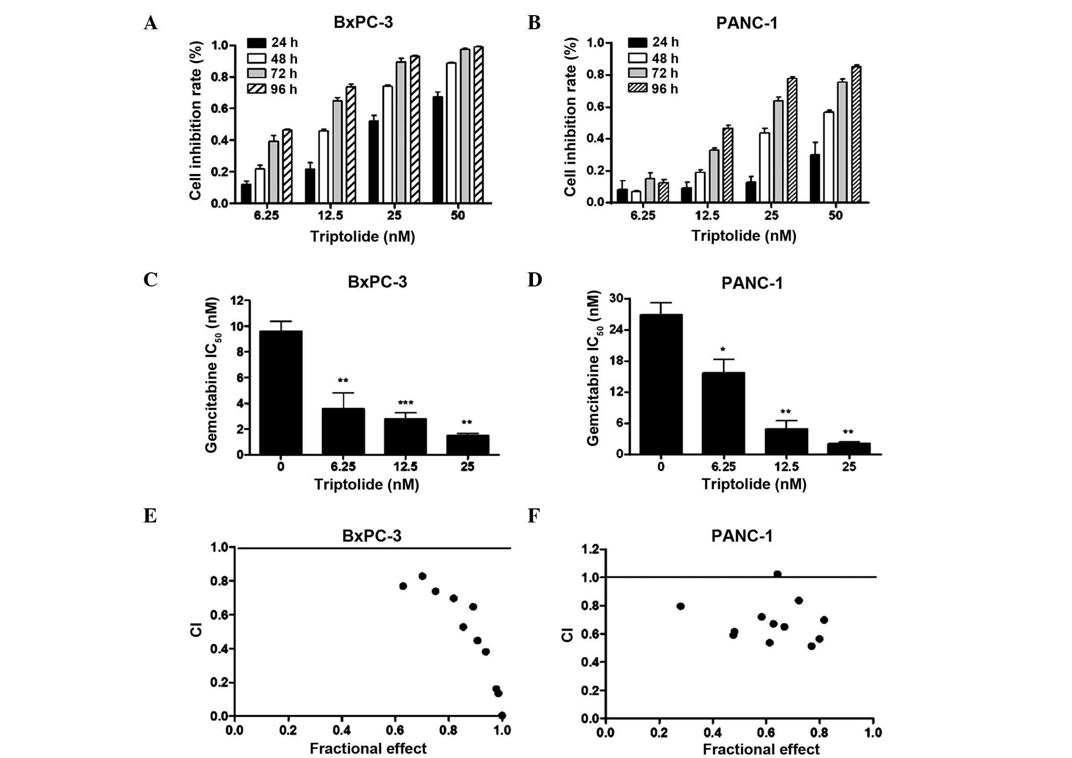 | Figure 1.Triptolide treatment results in growth
arrest, and synergistically enhances the cytotoxicity of
gemcitabine in pancreatic cancer cells. (A and B) BxPC-3 or PANC-1
cells were treated with vehicle control or triptolide at the
indicated doses for up to 96 h. Cell viability was determined by
3-(4,5-dimethylthiazol-2-yl)-2,5-diphenyltetrazolium bromide assay.
The data are presented as the mean inhibition rate ± standard error
from at least three independent experiments. (C and D) Gemcitabine
IC50 of BxPC-3 or PANC-1 cells was determined in the
absence or presence of sequential triptolide treatment (gemcitabine
followed by triptolide) (presented as the mean ± standard error).
(E and F) BxPC-3 or PANC-1 cells were treated with gemcitabine (4
varying concentrations: 5, 10, 20 and 40 nM) or triptolide (3
varying concentrations: 6.25, 12.5 and 25 nM) alone or combined
sequentially (12 combined groups). CI values were calculated with
CalcuSyn software (Biosoft, Cambridge, UK). *P<0.05, **P<0.01
and ***P<0.001 vs. vehicle control. IC50, half
maximal inhibitory concentration; CI, combination index. |
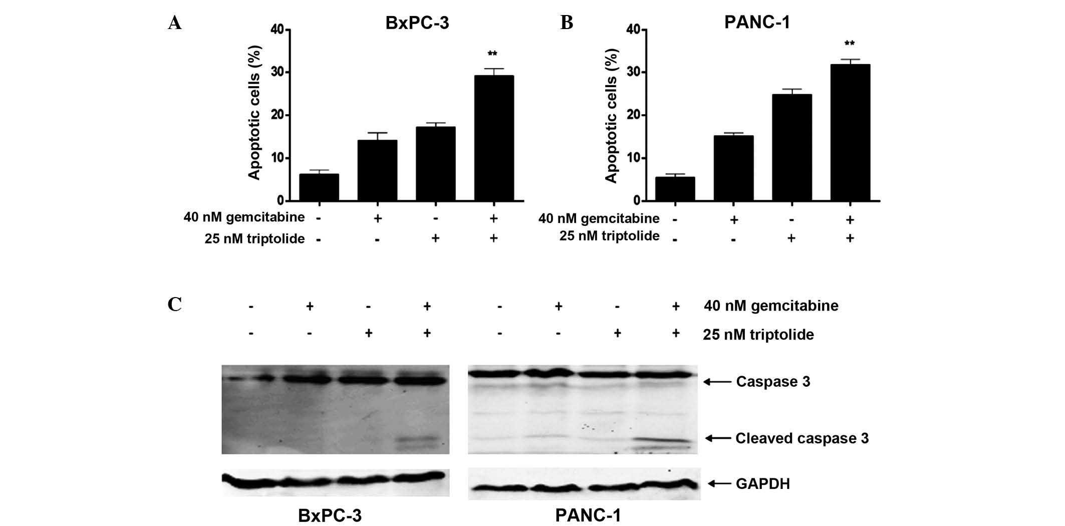
Triptolide augments
gemcitabine-induced apoptosis in pancreatic cancer cells,
accompanied by activation of caspase 3
The present study investigated whether the
synergistic antitumor effects of combined gemcitabine and
triptolide treatment were a result of apoptosis induction. Flow
cytometry was used to determine the apoptosis induced by triptolide
combined with gemcitabine in pancreatic cancer cells, and the
activation of caspase 3 was analyzed by immunoblotting. Following
treatment with the two agents, triptolide significantly augmented
the gemcitabine-induced apoptosis in the pancreatic cancer cell
lines, particularly in the BxPC-3 cells (Fig. 2A and B). Although triptolide alone was
observed to exert a modest apoptotic effect in the BxPC-3 cells at
a concentration of 25 nM (17.3±0.9%), when combined with 40 nM of
gemcitabine, it significantly enhanced gemcitabine-induced
apoptosis (29.3±1.6%; (P<0.001). Similar results were
additionally observed in the PANC-1 cells, although to a lesser
extent (P<0.01). In addition, combined treatment cooperatively
activated the cleavage of caspase 3 from 35 kDa to 17/19 kDa when
compared to treatment with each drug individually (Fig. 2C).
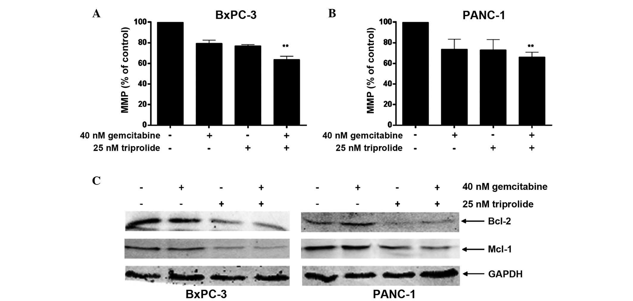
Triptolide and gemcitabine
cooperatively induce the loss of MMP in pancreatic cancer
cells
Mitochondria serve a crucial function in the
regulation of apoptosis, with apoptosis often associated with a
decrease in MMP (29). To establish
the effects of gemcitabine, triptolide and the drugs in combination
on mitochondrial function, the present study determined the MMP
induced by each treatment using the Rhodamine 123 staining method.
The expression levels of Bcl-2 family proteins were analyzed by
immunoblot analysis. Treatment with gemcitabine or triptolide alone
resulted in a reduction in MMP in each cell line (P<0.05;
Fig. 3A and B). Consistent with the
results of apoptosis analysis (Fig. 2A
and B), the combined treatment of the cell lines with
triptolide and gemcitabine resulted in an additional reduction in
MMP when compared with gemcitabine or triptolide treatment alone
(Fig. 3A and B). As presented in
Fig. 3C, combined treatment
cooperatively downregulated the expression of Mcl-1 and Bcl-2.
These results indicate that gemcitabine and triptolide
cooperatively regulate the expression of Bcl-2 family proteins,
resulting in MMP reduction and apoptosis in pancreatic cancer
cells.
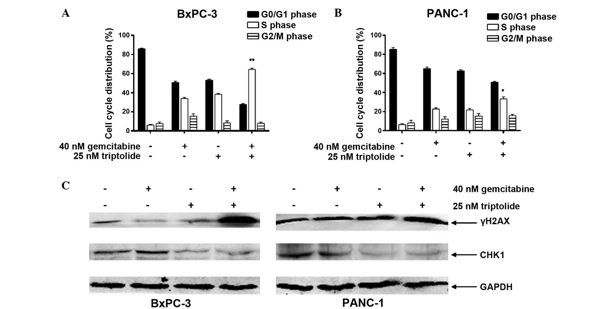
Triptolide synergistically enhances
gemcitabine-induced cell cycle arrest and DNA DSBs by suppressing
CHK1 expression in pancreatic cancer cells
Cell cycle arrest may additionally contribute to the
synergistic antitumor activity of gemcitabine and triptolide in
pancreatic cancer cell lines. BxPC-3 and PANC-1 cells treated with
gemcitabine or triptolide alone demonstrated S phase arrest.
Notably, the combined treatment of the cell lines with the two
agents resulted in cooperative induction of S phase arrest, with a
larger number of cells remaining in S phase (P<0.05; Fig. 4A and B). As a deoxycytidine analogue,
the anticancer activity of gemcitabine is exerted to inhibit DNA
synthesis and induce DNA damage (9).
The cooperative induction of S phase arrest suggests that
triptolide may increase gemcitabine-induced DNA damage and promote
apoptosis in pancreatic cancer cells. As presented in Fig. 4C, triptolide significantly augmented
DNA DSBs induced by gemcitabine represented by the induction of
γH2AX. Notably, triptolide inhibited the expression of CHK1, a
vital protein in DNA DSB repair and cell cycle checkpoint pathways
(30), which was induced following
gemcitabine treatment (Fig. 4C).
These results indicate that triptolide cooperates with gemcitabine
in pancreatic cancer cells to induce DNA DSBs by repressing the
expression of CHK1.
Discussion
As an immunosuppressant, triptolide has been
successfully applied for the clinical treatment of inflammation,
autoimmune diseases and organ transplantation (13–15,31).
Previous research has demonstrated that the drug additionally
possesses potent antitumor activity, and is able to synergistically
enhance the antitumor activities of conventional chemotherapeutic
drugs (19,20). In the present study, the cooperative
antitumor activities of triptolide and gemcitabine were
investigated in vitro in pancreatic cancer cells. The
results demonstrated that triptolide treatment initiated growth
inhibition in BxPC-3 and PANC-1 cell lines and synergistically
enhanced gemcitabine-induced apoptosis. This was accompanied by the
cooperative induction of DNA DSBs when the two agents were
combined, and suppression of CHK1 when treated with triptolide
alone. These results suggest that a shared mechanism may underlie
the synergistic antitumor interactions between triptolide and DNA
damaging agents in cancer cells. Triptolide may overcome the
chemoresistance to gemcitabine which the first-line chemotherapy
drugs for pancreatic cancer and improve the clinical therapy.
Antiproliferation assays demonstrated that
triptolide treatment inhibited pancreatic cell growth in a time-
and dose-dependent manner. When applied in combination with
gemcitabine, triptolide synergistically increased the cytotoxicity
of gemcitabine in the BxPC-3 and PANC-1 cell lines. It may be
assumed that this synergistic cytotoxicity occurs as a result of
cell death as the cooperative induction of apoptosis by the two
agents was detected. Triptolide has previously been demonstrated to
induce apoptosis in cancer cells by regulating members of the Bcl-2
family, resulting in MMP loss and activation of caspase-3 (12,32).
Notably, combined treatment with triptolide and gemcitabine
resulted in cooperative MMP loss in the present study, as well as
downregulation of Mcl-1 and Bcl-2. These results strongly suggest
that gemcitabine and triptolide cooperatively regulate the
expression of Bcl-2 family proteins, which leads to a reduction in
MMP and induction of apoptosis.
Triptolide has been demonstrated to increase S phase
arrest of cancer cells and sensitize cancer cells to conventional
anti-cancer drugs (12,33). Similarly, in the present study,
triptolide and gemcitabine collectively induced S phase arrest,
indicating that triptolide may increase the DNA damage induced by
gemcitabine. Triptolide and gemcitabine cooperatively induced DNA
DSBs, represented by an increase in γH2AX in the PANC-1 and BxPC-3
cell lines, which may serve as an explanation for the collective
induction of S phase arrest. The results of the present study
indicate that triptolide increases gemcitabine-induced DNA DSBs,
possibly resulting in S phase arrest, regulation of the expression
of Bcl-2 family proteins, and subsequent MMP loss and
apoptosis.
Triptolide has been demonstrated to repress DNA
repair genes (including ataxia telangiectasia mutated, ataxia
telangiectasia and Rad3 related, breast cancer 1 and DNA-dependent
protein kinase), subsequently increasing radiation sensitivity in
human malignant melanoma cells (34).
It is possible that triptolide may additionally downregulate DNA
repair genes in pancreatic cancer cells and enhance
gemcitabine-induced DNA DSBs. Triptolide significantly suppressed
CHK1, which is induced by gemcitabine and has a significant role in
the repair of DNA DSBs and cell cycle checkpoint pathways (30).
In conclusion, the current study demonstrates that
triptolide promotes growth arrest of pancreatic cancer cells, and
enhances gemcitabine-induced DNA DSBs and apoptosis. Such results
indicate that triptolide possesses antitumor potential against
pancreatic cancer cells, particularly when administered in
combination with gemcitabine. These in vitro findings
require follow-up in vivo studies for validation, and the
data from the present study suggests that the co-treatment of
pancreatic cancer with gemcitabine and triptolide may provide
additional clinical benefits.
Acknowledgements
The present study was supported by the National
Scientific and Technological Major Project for ‘Significant New
Drugs Development’, Beijing, China (grant no. 2011ZXJ09302) and
China's 12th Five Year Plan, Beijing, China (grant no.
2012ZX10001003).
References
|
1
|
Shaib YH, Davila JA and El-Serag B: The
epidemiology of pancreatic cancer in the United States: Changes
below the surface. Aliment Pharmacol Ther. 24:87–94. 2006.
View Article : Google Scholar : PubMed/NCBI
|
|
2
|
Rosenberg L: Pancreatic cancer: A review
of emerging therapies. Drugs. 59:1071–1089. 2000. View Article : Google Scholar : PubMed/NCBI
|
|
3
|
Siegel R, Ma J, Zou Z and Jemal A: Cancer
statistics, 2014. CA Cancer J Clin. 64:9–29. 2014. View Article : Google Scholar : PubMed/NCBI
|
|
4
|
Freelove R and Walling AD: Pancreatic
cancer: Diagnosis and management. Am Fam Physician. 73:485–492.
2006.PubMed/NCBI
|
|
5
|
Bilimoria KY, Bentrem DJ, Ko CY, Stewart
AK, Winchester DP and Talamonti MS: National failure to operate on
early stage pancreatic cancer. Ann Surg. 246:173–180. 2007.
View Article : Google Scholar : PubMed/NCBI
|
|
6
|
Zuckerman DS and Ryan DP: Adjuvant therapy
for pancreatic cancer: A review. Cancer. 112:243–249. 2008.
View Article : Google Scholar : PubMed/NCBI
|
|
7
|
Rothenberg ML, Moore MJ, Cripps MC,
Andersen JS, Portenoy RK, Burris HA III, Green MR, Tarassoff PG,
Brown TD, Casper ES, et al: A phase II trial of gemcitabine in
patients with 5-FU-refractory pancreas cancer. Ann Oncol.
7:347–353. 1996. View Article : Google Scholar : PubMed/NCBI
|
|
8
|
Burris HA III, Moore MJ, Andersen J, et
al: Improvements in survival and clinical benefit with gemcitabine
as first-line therapy for patients with advanced pancreas cancer: A
randomized trial. J Clin Oncol. 15:2403–2413. 1997.PubMed/NCBI
|
|
9
|
Mini E, Nobili S, Caciagli B, Landini I
and Mazzei T: Cellular pharmacology of gemcitabine. Ann Oncol.
17(Suppl 5): v7–v12. 2006. View Article : Google Scholar : PubMed/NCBI
|
|
10
|
Schultz RM, Merriman RL, Toth JE,
Zimmermann JE, Hertel LW, Andis SL, Dudley DE, Rutherford PG,
Tanzer LR and Grindey GB: Evaluation of new anticancer agents
against the MIA PaCa-2 and PANC-1 human pancreatic carcinoma
xenografts. Oncol Res. 5:223–228. 1993.PubMed/NCBI
|
|
11
|
Nakano Y, Tanno S, Koizumi K, Nishikawa T,
Nakamura K, Minoguchi M, Izawa T, Mizukami Y, Okumura T and Kohgo
Y: Gemcitabine chemoresistance and molecular markers associated
with gemcitabine transport and metabolism in human pancreatic
cancer cells. Br J Cancer. 96:457–463. 2007. View Article : Google Scholar : PubMed/NCBI
|
|
12
|
Xu B, Guo X, Mathew S, Armesilla AL,
Cassidy J, Darling JL and Wang W: Triptolide simultaneously induces
reactive oxygen species, inhibits NF-kappaB activity and sensitizes
5-fluorouracil in colorectal cancer cell lines. Cancer Lett.
291:200–208. 2010. View Article : Google Scholar : PubMed/NCBI
|
|
13
|
Qiu D, Zhao G, Aoki Y, Shi L, Uyei A,
Nazarian S, Ng JC and Kao PN: Immunosuppressant PG490 (triptolide)
inhibits T-cell interleukin-2 expression at the level of
purine-box/nuclear factor of activated T-cells and NF-kappaB
transcriptional activation. J Biol Chem. 274:13443–13450. 1999.
View Article : Google Scholar : PubMed/NCBI
|
|
14
|
Liu Q: Triptolide and its expanding
multiple pharmacological functions. Int Immunopharmacol.
11:377–383. 2011. View Article : Google Scholar : PubMed/NCBI
|
|
15
|
Brinker AM, Ma J, Lipsky PE and Raskin I:
Medicinal chemistry and pharmacology of genus Tripterygium
(Celastraceae). Phytochemistry. 68:732–766. 2007. View Article : Google Scholar : PubMed/NCBI
|
|
16
|
Huang W, He T, Chai C, Yang Y, Zheng Y,
Zhou P, Qiao X, Zhang B, Liu Z, Wang J, et al: Triptolide inhibits
the proliferation of prostate cancer cells and down-regulates
SUMO-specific protease 1 expression. PLoS One. 7:e376932012.
View Article : Google Scholar : PubMed/NCBI
|
|
17
|
Phillips PA, Dudeja V, McCarroll JA,
Borja-Cacho D, Dawra RK, Grizzle WE, Vickers SM and Saluja AK:
Triptolide induces pancreatic cancer cell death via inhibition of
heat shock protein 70. Cancer Res. 67:9407–9416. 2007. View Article : Google Scholar : PubMed/NCBI
|
|
18
|
Shamon LA, Pezzuto JM, Graves JM, Mehta
RR, Wangcharoentrakul S, Sangsuwan R, Chaichana S, Tuchinda P,
Cleason P and Reutrakul V: Evaluation of the mutagenic, cytotoxic
and antitumor potential of triptolide, a highly oxygenated
diterpene isolated from Tripterygium wilfordii. Cancer Lett.
112:113–117. 1997. View Article : Google Scholar : PubMed/NCBI
|
|
19
|
Li CJ, Chu CY, Huang LH, Wang MH, Sheu LF,
Yeh JI and Hsu HY: Synergistic anticancer activity of triptolide
combined with cisplatin enhances apoptosis in gastric cancer in
vitro and in vivo. Cancer Lett. 319:203–213. 2012.
View Article : Google Scholar : PubMed/NCBI
|
|
20
|
Chen YW, Lin GJ, Chuang YP, Chia WT, Hueng
DY, Lin CK, Nieh S and Sytwu HK: Triptolide circumvents
drug-resistant effect and enhances 5-fluorouracil antitumor effect
on KB cells. Anticancer Drugs. 21:502–513. 2010. View Article : Google Scholar : PubMed/NCBI
|
|
21
|
Zhu W, Li J, Wu S, Li S, Le L, Su X, Qiu
P, Hu H and Yan G: Triptolide cooperates with Cisplatin to induce
apoptosis in gemcitabine-resistant pancreatic cancer. Pancreas.
41:1029–1038. 2012. View Article : Google Scholar : PubMed/NCBI
|
|
22
|
Liu Y and Cui YF: Synergism of
cytotoxicity effects of triptolide and artesunate combination
treatment in pancreatic cancer cell lines. Asian Pac J Cancer Prev.
14:5243–5248. 2013. View Article : Google Scholar : PubMed/NCBI
|
|
23
|
Yang SW, Wang W, Xie XY, Zhu WP and Li FQ:
In vitro synergistic cytotoxic effect of triptolide combined
with hydroxycamptothecin on pancreatic cancer cells. Am J Chin Med.
39:121–134. 2011. View Article : Google Scholar : PubMed/NCBI
|
|
24
|
Kiviharju TM, Lecane PS, Sellers RG and
Peehl DM: Antiproliferative and proapoptotic activities of
triptolide (PG490), a natural product entering clinical trials, on
primary cultures of human prostatic epithelial cells. Clin Cancer
Res. 8:2666–2674. 2002.PubMed/NCBI
|
|
25
|
Qiao Z, Ren S, Li W, Wang X, He M, Guo Y,
Sun L, He Y, Ge Y and Yu Q: Chidamide, a novel histone deacetylase
inhibitor, synergistically enhances gemcitabine cytotoxicity in
pancreatic cancer cells. Biochem Biophys Res Commun. 434:95–101.
2013. View Article : Google Scholar : PubMed/NCBI
|
|
26
|
Donadelli M, Costanzo C, Beghelli S,
Scupoli MT, Dandrea M, Bonora A, Piacentini P, Budillon A, Caraglia
M, Scarpa A and Palmieri M: Synergistic inhibition of pancreatic
adenocarcinoma cell growth by trichostatin A and gemcitabine.
Biochim Biophys Acta. 1773:1095–1106. 2007. View Article : Google Scholar : PubMed/NCBI
|
|
27
|
Xie C, Edwards H, Xu X, Zhou H, Buck SA,
Stout ML, Yu Q, Rubnitz JE, Matherly LH, Taub JW and Ge Y:
Mechanisms of synergistic antileukemic interactions between
valproic acid and cytarabine in pediatric acute myeloid leukemia.
Clin Cancer Res. 16:5499–5510. 2010. View Article : Google Scholar : PubMed/NCBI
|
|
28
|
Xiang M, Qian ZY, Zhou CH, Liu J and Li
WN: Crocetin inhibits leukocyte adherence to vascular endothelial
cells induced by AGEs. J Ethnopharmacol. 107:25–31. 2006.
View Article : Google Scholar : PubMed/NCBI
|
|
29
|
Kinnally KW and Antonsson B: A tale of two
mitochondrial channels, MAC and PTP, in apoptosis. Apoptosis.
12:857–868. 2007. View Article : Google Scholar : PubMed/NCBI
|
|
30
|
Dai Y and Grant S: New insights into
checkpoint kinase 1 in the DNA damage response signaling network.
Clin Cancer Res. 16:376–383. 2010. View Article : Google Scholar : PubMed/NCBI
|
|
31
|
Kupchan SM, Court WA, Dailey RG
Jr..Gilmore CJ and Bryan RF: Triptolide and tripdiolide, novel
antileukemic diterpenoid triepoxides from Tripterygium
wilfordii. J Am Chem Soc. 94:7194–7195. 1972. View Article : Google Scholar : PubMed/NCBI
|
|
32
|
Hu YP, Tan ZJ, Wu XS, Liu TY, Jiang L, Bao
RF, Shu YJ, Li ML, Weng H, Ding Q, Tao F and Liu YB: Triptolide
induces s phase arrest and apoptosis in gallbladder cancer cells.
Molecules. 2014.19(2): 2612–2628. View Article : Google Scholar : PubMed/NCBI
|
|
33
|
Ho JN, Byun SS, Lee S, Oh JJ, Hong SK, Lee
SE and Yeon JS: Synergistic antitumor effect of triptolide and
cisplatin in cisplatin resistant human bladder cancer cells. The
Journal of urology. 2015.193(3): 1016–1022, 14. View Article : Google Scholar : PubMed/NCBI
|
|
34
|
Chueh FS, Chen YL, Hsu SC, Yang JS, Hsueh
SC, Ji BC, Lu HF and Chung JG: Triptolide induced DNA damage in
A375.S2 human malignant melanoma cells is mediated via reduction of
DNA repair genes. Oncol Rep. 29:613–618. 2013.PubMed/NCBI
|