Spandidos Publications Logo
  • About
    • About Spandidos
    • Aims and Scopes
    • Abstracting and Indexing
    • Editorial Policies
    • Reprints and Permissions
    • Job Opportunities
    • Terms and Conditions
    • Contact
  • Journals
    • All Journals
    • Oncology Letters
      • Oncology Letters
      • Information for Authors
      • Editorial Policies
      • Editorial Board
      • Aims and Scope
      • Abstracting and Indexing
      • Bibliographic Information
      • Archive
    • International Journal of Oncology
      • International Journal of Oncology
      • Information for Authors
      • Editorial Policies
      • Editorial Board
      • Aims and Scope
      • Abstracting and Indexing
      • Bibliographic Information
      • Archive
    • Molecular and Clinical Oncology
      • Molecular and Clinical Oncology
      • Information for Authors
      • Editorial Policies
      • Editorial Board
      • Aims and Scope
      • Abstracting and Indexing
      • Bibliographic Information
      • Archive
    • Experimental and Therapeutic Medicine
      • Experimental and Therapeutic Medicine
      • Information for Authors
      • Editorial Policies
      • Editorial Board
      • Aims and Scope
      • Abstracting and Indexing
      • Bibliographic Information
      • Archive
    • International Journal of Molecular Medicine
      • International Journal of Molecular Medicine
      • Information for Authors
      • Editorial Policies
      • Editorial Board
      • Aims and Scope
      • Abstracting and Indexing
      • Bibliographic Information
      • Archive
    • Biomedical Reports
      • Biomedical Reports
      • Information for Authors
      • Editorial Policies
      • Editorial Board
      • Aims and Scope
      • Abstracting and Indexing
      • Bibliographic Information
      • Archive
    • Oncology Reports
      • Oncology Reports
      • Information for Authors
      • Editorial Policies
      • Editorial Board
      • Aims and Scope
      • Abstracting and Indexing
      • Bibliographic Information
      • Archive
    • Molecular Medicine Reports
      • Molecular Medicine Reports
      • Information for Authors
      • Editorial Policies
      • Editorial Board
      • Aims and Scope
      • Abstracting and Indexing
      • Bibliographic Information
      • Archive
    • World Academy of Sciences Journal
      • World Academy of Sciences Journal
      • Information for Authors
      • Editorial Policies
      • Editorial Board
      • Aims and Scope
      • Abstracting and Indexing
      • Bibliographic Information
      • Archive
    • International Journal of Functional Nutrition
      • International Journal of Functional Nutrition
      • Information for Authors
      • Editorial Policies
      • Editorial Board
      • Aims and Scope
      • Abstracting and Indexing
      • Bibliographic Information
      • Archive
    • International Journal of Epigenetics
      • International Journal of Epigenetics
      • Information for Authors
      • Editorial Policies
      • Editorial Board
      • Aims and Scope
      • Abstracting and Indexing
      • Bibliographic Information
      • Archive
    • Medicine International
      • Medicine International
      • Information for Authors
      • Editorial Policies
      • Editorial Board
      • Aims and Scope
      • Abstracting and Indexing
      • Bibliographic Information
      • Archive
  • Articles
  • Information
    • Information for Authors
    • Information for Reviewers
    • Information for Librarians
    • Information for Advertisers
    • Conferences
  • Language Editing
Spandidos Publications Logo
  • About
    • About Spandidos
    • Aims and Scopes
    • Abstracting and Indexing
    • Editorial Policies
    • Reprints and Permissions
    • Job Opportunities
    • Terms and Conditions
    • Contact
  • Journals
    • All Journals
    • Biomedical Reports
      • Information for Authors
      • Editorial Policies
      • Editorial Board
      • Aims and Scope
      • Abstracting and Indexing
      • Bibliographic Information
      • Archive
    • Experimental and Therapeutic Medicine
      • Information for Authors
      • Editorial Policies
      • Editorial Board
      • Aims and Scope
      • Abstracting and Indexing
      • Bibliographic Information
      • Archive
    • International Journal of Epigenetics
      • Information for Authors
      • Editorial Policies
      • Editorial Board
      • Aims and Scope
      • Abstracting and Indexing
      • Bibliographic Information
      • Archive
    • International Journal of Functional Nutrition
      • Information for Authors
      • Editorial Policies
      • Editorial Board
      • Aims and Scope
      • Abstracting and Indexing
      • Bibliographic Information
      • Archive
    • International Journal of Molecular Medicine
      • Information for Authors
      • Editorial Policies
      • Editorial Board
      • Aims and Scope
      • Abstracting and Indexing
      • Bibliographic Information
      • Archive
    • International Journal of Oncology
      • Information for Authors
      • Editorial Policies
      • Editorial Board
      • Aims and Scope
      • Abstracting and Indexing
      • Bibliographic Information
      • Archive
    • Medicine International
      • Information for Authors
      • Editorial Policies
      • Editorial Board
      • Aims and Scope
      • Abstracting and Indexing
      • Bibliographic Information
      • Archive
    • Molecular and Clinical Oncology
      • Information for Authors
      • Editorial Policies
      • Editorial Board
      • Aims and Scope
      • Abstracting and Indexing
      • Bibliographic Information
      • Archive
    • Molecular Medicine Reports
      • Information for Authors
      • Editorial Policies
      • Editorial Board
      • Aims and Scope
      • Abstracting and Indexing
      • Bibliographic Information
      • Archive
    • Oncology Letters
      • Information for Authors
      • Editorial Policies
      • Editorial Board
      • Aims and Scope
      • Abstracting and Indexing
      • Bibliographic Information
      • Archive
    • Oncology Reports
      • Information for Authors
      • Editorial Policies
      • Editorial Board
      • Aims and Scope
      • Abstracting and Indexing
      • Bibliographic Information
      • Archive
    • World Academy of Sciences Journal
      • Information for Authors
      • Editorial Policies
      • Editorial Board
      • Aims and Scope
      • Abstracting and Indexing
      • Bibliographic Information
      • Archive
  • Articles
  • Information
    • For Authors
    • For Reviewers
    • For Librarians
    • For Advertisers
    • Conferences
  • Language Editing
Login Register Submit
  • This site uses cookies
  • You can change your cookie settings at any time by following the instructions in our Cookie Policy. To find out more, you may read our Privacy Policy.

    I agree
Search articles by DOI, keyword, author or affiliation
Search
Advanced Search
presentation
Oncology Letters
Join Editorial Board Propose a Special Issue
Print ISSN: 1792-1074 Online ISSN: 1792-1082
Journal Cover
June-2016 Volume 11 Issue 6

Full Size Image

Sign up for eToc alerts
Recommend to Library

Journals

International Journal of Molecular Medicine

International Journal of Molecular Medicine

International Journal of Molecular Medicine is an international journal devoted to molecular mechanisms of human disease.

International Journal of Oncology

International Journal of Oncology

International Journal of Oncology is an international journal devoted to oncology research and cancer treatment.

Molecular Medicine Reports

Molecular Medicine Reports

Covers molecular medicine topics such as pharmacology, pathology, genetics, neuroscience, infectious diseases, molecular cardiology, and molecular surgery.

Oncology Reports

Oncology Reports

Oncology Reports is an international journal devoted to fundamental and applied research in Oncology.

Experimental and Therapeutic Medicine

Experimental and Therapeutic Medicine

Experimental and Therapeutic Medicine is an international journal devoted to laboratory and clinical medicine.

Oncology Letters

Oncology Letters

Oncology Letters is an international journal devoted to Experimental and Clinical Oncology.

Biomedical Reports

Biomedical Reports

Explores a wide range of biological and medical fields, including pharmacology, genetics, microbiology, neuroscience, and molecular cardiology.

Molecular and Clinical Oncology

Molecular and Clinical Oncology

International journal addressing all aspects of oncology research, from tumorigenesis and oncogenes to chemotherapy and metastasis.

World Academy of Sciences Journal

World Academy of Sciences Journal

Multidisciplinary open-access journal spanning biochemistry, genetics, neuroscience, environmental health, and synthetic biology.

International Journal of Functional Nutrition

International Journal of Functional Nutrition

Open-access journal combining biochemistry, pharmacology, immunology, and genetics to advance health through functional nutrition.

International Journal of Epigenetics

International Journal of Epigenetics

Publishes open-access research on using epigenetics to advance understanding and treatment of human disease.

Medicine International

Medicine International

An International Open Access Journal Devoted to General Medicine.

Journal Cover
June-2016 Volume 11 Issue 6

Full Size Image

Sign up for eToc alerts
Recommend to Library

  • Article
  • Citations
    • Cite This Article
    • Download Citation
    • Create Citation Alert
    • Remove Citation Alert
    • Cited By
  • Similar Articles
    • Related Articles (in Spandidos Publications)
    • Similar Articles (Google Scholar)
    • Similar Articles (PubMed)
  • Download PDF
  • Download XML
  • View XML
Case Report Open Access

Sphenoid sinus organized hematoma with cranial neuropathies masquerading as a malignancy: A case report

  • Authors:
    • Yu‑Hsuan Lin
    • Po‑Chin Wang
    • Yaoh‑Shiang Lin
  • View Affiliations / Copyright

    Affiliations: Department of Otolaryngology ‑ Head and Neck Surgery, Kaohsiung Veterans General Hospital, Kaohsiung 813, Taiwan, R.O.C., Department of Radiology, Kaohsiung Veterans General Hospital, Kaohsiung 813, Taiwan, R.O.C.
    Copyright: © Lin et al. This is an open access article distributed under the terms of Creative Commons Attribution License.
  • Pages: 3571-3574
    |
    Published online on: April 20, 2016
       https://doi.org/10.3892/ol.2016.4475
  • Expand metrics +
Metrics: Total Views: 0 (Spandidos Publications: | PMC Statistics: )
Metrics: Total PDF Downloads: 0 (Spandidos Publications: | PMC Statistics: )
Cited By (CrossRef): 0 citations Loading Articles...

This article is mentioned in:


Abstract

Sinonasal organized hematoma (SNOH) is rarely encountered in clinical practice. The disease demonstrates a high tendency for occurrence in East Asian individuals, and in the majority of cases, is located in the maxillary sinus. The current report presents the case of an 81‑year‑old female who developed a space‑occupying lesion, which masqueraded as a skull base malignancy, following surgery for the treatment of isolated sphenoid sinus aspergilloma. Subsequent endoscopic endonasal surgery confirmed the diagnosis of an OH of the sphenoid sinus. The patient recovered from all neurological deficits within two months, with the exception of the loss of visual perception. Although SNOH presents a diagnostic challenge, when physicians possess knowledge of its typical imaging features, this facilitates the achievement of a correct diagnosis and the prescription of optimal treatment.

Introduction

Sinonasal organized hematoma (SNOH) was initially described in 1917 by Tadokoro (1) as a benign lesion characterized by an expansile nature and locally aggressive behavior, capable of mimicking a potentially dangerous neoplastic process (2–5). The diagnosis of SNOH is challenging, even given the integration of multiple informative symptoms (2). Computed tomography (CT) and magnetic resonance imaging (MRI) are useful for the assessment of potential cases of SNOH (2,5), and usually reveal typical bony changes and a distinct speckled pattern that exhibits various signal intensities, respectively. The hallmarks of SNOH are typically a dark peripheral rim on T2-weighted imaging (T2WI), and a nodular and patchy enhancement in post-contrast T1-weighted imaging (T1WI) (2,5). The successful treatment of SNOH relies on complete surgical excision (3,5). However, one notable and unexplained anomaly is that the disease demonstrates a high tendency for occurrence in the maxillary sinus (3,5–7). Only one case involving the sphenoid sinus, an intricately structured space in close vicinity to vital structures, has been reported to date (4). To the best of our knowledge, the current report presents the first case of an OH of the sphenoid sinus with multiple cranial nerve involvement, which was identified following surgery for the treatment of isolated sphenoid sinus aspergillosis (ISSA). The disease was successfully treated using endoscopic endonasal surgery, yielding a positive outcome for the patient.

Case report

An 81-year-old woman presented as an inpatient to the Kaohsiung Veterans General Hospital (Kaohsiung, Taiwan) in October 2013, with a 2-year history of an excruciating headache, which was described as deep-seated and throbbing. The patient had subsequently developed progressive deterioration of visual acuity in the right eye and drooping of the right eyelid, although the total duration of these symptoms was uncertain. The medical history of the patient noted the use of endoscopic sinus surgery for the treatment of ISSA 3 years previously, and a long-standing history of chronic kidney disease. Upon examination, besides optic atrophy, the patient's right eye exhibited complete ptosis and mydriasis with a sluggish pupillary reaction. The eye was positioned looking down and out, and the orbit could not be adducted. These symptoms were compatible with a diagnosis of compressive occulomotor nerve 
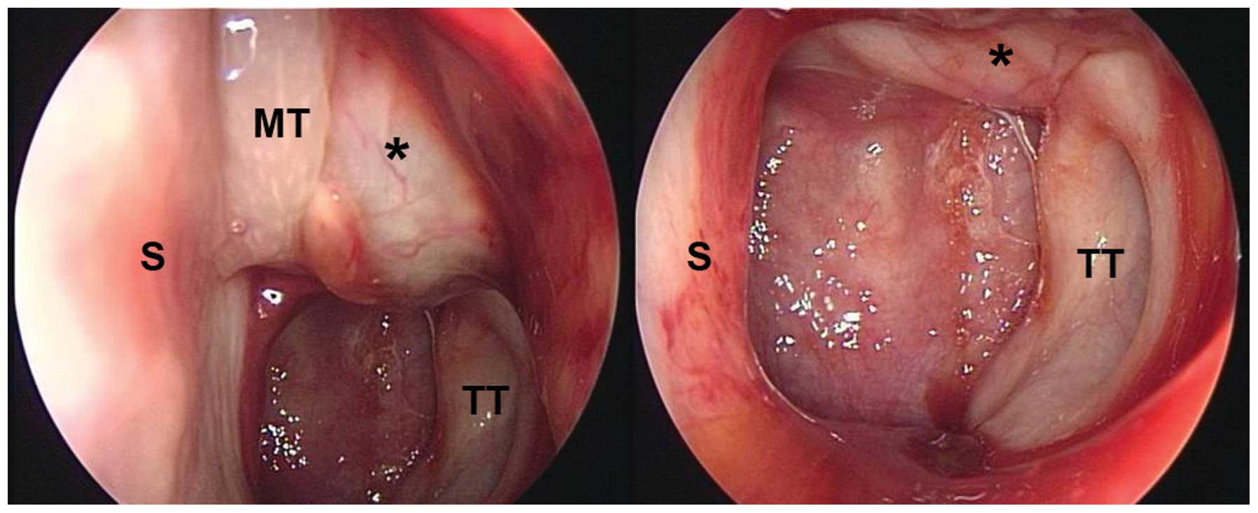
palsy. The nasal endoscopy revealed a friable mass with superficial telangiectasia straddling the torus tubarius and extending forward 
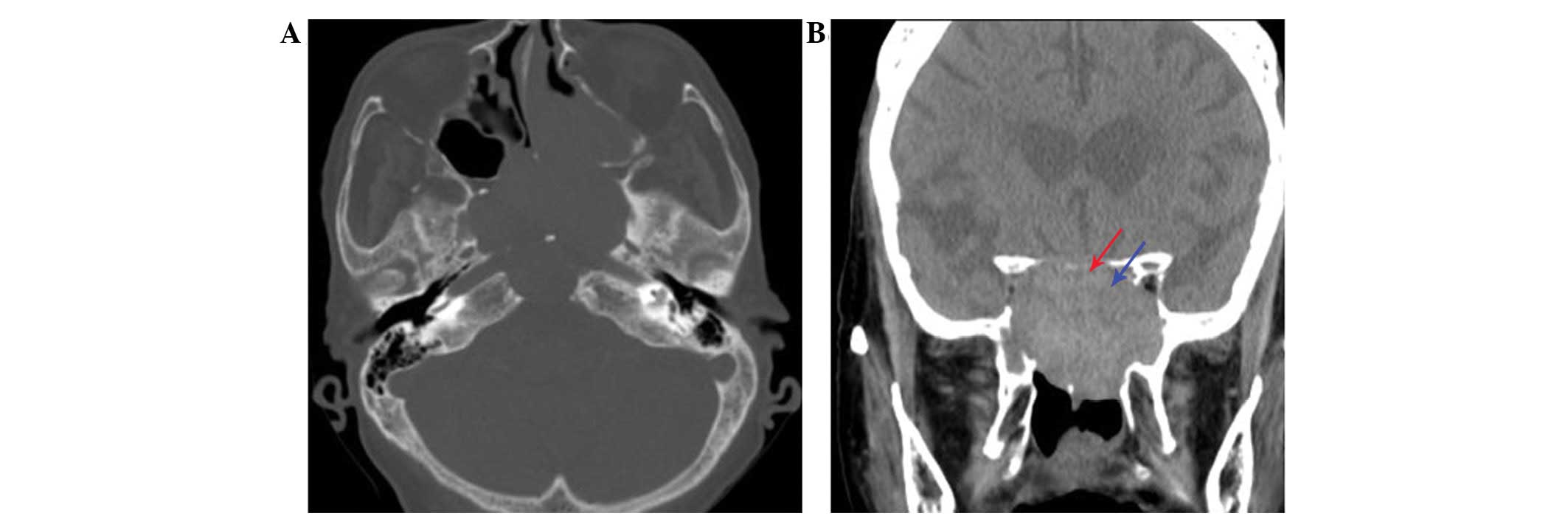
(Fig. 1). Non-contrast CT revealed an aggressively enlarging lesion in the midline skull base, with destructive bony structures (Fig. 2). Distinctive internal heterogeneous hyperintensity and a hypointense peripheral rim on T2WI, and a patchy enhancement pattern on post-contrast T1W1 were observed (Fig. 3). Routine laboratory data revealed normal coagulation and pituitary function profiles, including human growth hormone (0.23 ng/ml; normal range, 0.0–16 ng/ml), luteinizing hormone (31.35 mIU/ml; normal range, 11.3–38.7 mIU/ml), follicle stimulating hormone (74.09 mIU/ml; normal range, 31.8–134 mIU/ml) and free thyroxine (1.58 ng/dl; normal range, 0.8–1.9 ng/dl). Only high sensitivity-thyroid-stimulating hormone levels were abnormal (0.245 uIU/ml; normal range, 0.4–4.0 uIU/ml).

Figure 1.

Sinoscopic examination at patient presentation revealing a friable lesion (*) between the MT and the lateral wall of the nasal cavity, and straddling the TT. S, septum; MT, middle turbinate; TT, torus tubarius.

Figure 2.

CT images. (A) Bony algorithm CT image revealing an expansile neoplasm of the sphenoid sinus, extending forward to the left ethmoid sinus, with erosion of adjacent skeletal structures. (B) Coronal non-contrast CT image demonstrating partial fat tissue effacement of the right superior orbital fissure and total obliteration of the right optic canal, compatible with clinical optic and oculomotor compressive neuropathies. Red arrow, left optic canal. Blue arrow, left superior orbital fissure. CT, computed tomography.

Figure 3.

MRI images. (A) Simultaneous axial T1WI revealing heterogeneously mixed signals within the lesion, and typical patchy and (B) nodular enhancement following administration of gadolinium. (C) A dark peripheral rim with distinct internal mixture of varying signals on T2WI. (D) Contrast-enhanced sagittal T1WI revealing the extensive nature of the sinonasal organized hematoma, with the expansile tumor pushing the pituitary gland (red arrow) upwards, and concomitant caudal involvement of the clivus (blue arrow). T1WI, T1-weighted images; T2WI, T2-weighted images.

In order to optimize surgical planning, the patient underwent a biopsy, which yielded a negative result. The patient subsequently underwent endoscopic endonasal surgery to resect the lesion; a posterior ethmoidectomy, sphenoidotomy and septoplasty were required in order to gain complete surgical access. A well-capsulated, russet-colored and blood clot-containing tumor was removed in a piece-by-piece manner, leaving the periosteal layer of dura mater that protected the intracranial structures intact. The thinned bony components located above the right orbital apex and right optic canal were gently flaked off. The surgical extent was explored inferiorly to the upper clivus. A sample of the lesion sent for pathological analysis revealed an OH without any evidence of fungal hyphae (Fig. 4). The immediate post-operative course was uncomplicated, with resolution of the patient's headache. At the 2-month follow-up, the subject had recovered fully, with the exception of the lost vision.

Figure 4.

Pathological photomicrograph revealing a lesion consisting of numerous red blood cells mixed with neovascularization and granulation tissue. Hematoxylin and eosin stain; magnification, ×40.

Written informed consent was obtained from the patient for publication of the present case report and any accompanying images.

Discussion

Sphenoid disease is primarily a result of the extension of disease anteriorly from the ethmoid complex or posteriorly from the adjacent anatomical sites, including the sella turcica and the surrounding skull base structures (8). Isolated sphenoid sinus disease is rarely encountered and the associated symptoms are generally non-specific (8). Sphenoid disease frequently leads to nerve involvement, and the effects of masses causing compression, destruction or invasion lead to varying levels of impact on the extent and degree of neurological deficits (8). Etiologically, non-neoplastic lesions constitute the majority of cases, while fungal processes are the third most common cause (8). ISSA is a type of fungal sinus infection that is distinct from other categories of fungal rhinosinusitis, and is characterized by higher levels of occurrence in older women (9). A sphenoid sinusotomy is regarded to be the primary therapy (8,9). To the best of our knowledge, the occurrence of an OH of the sphenoid sinus following ISSA surgery has not been previously reported (2–6,8,9).

SNOH is rarely encountered in clinical practice (2–6). When it does occur, it is usually locally invasive and demonstrates a high tendency to occur in the maxillary sinus (3,5,6,10). The incidence of SNOH appears to be most frequent in the population of East Asia (3,5,6,10). The exact underlying etiological mechanisms remain to be elucidated (4,5,6). Ozaki et al (11) advanced what is now the most widely-accepted hypothesis, the negative spiral theory (3,6). The negative spiral theory assumes that an OH results from persistent negative intraluminal pressure following an initial episodic hemorrhage into a semi-closed cavity, leading to repeated rupturing of fragile mucosal vessels. Subsequent formation of a superficial fibrotic capsule prevents further reabsorption of the hematoma. Under the succession of biological healing processes, an OH emerges (3,6). The progressive expansion of the OH results in augmentation of the sinus antrum, and therefore results in demineralization of the adjacent skeletal structures (3,5).

Macroscopically, SNOH is described as a slow-growing, well-circumscribed and friable brownish mass. The histopathological findings reveal a mixture of neovascularization, fibrosis, hemorrhaging and hemosiderin deposition (5). A number of studies have attempted to identify an association between SNOH and sinonasal angiomatous polyps, a rare subtype of inflammatory sinonasal polyp, using radiology and histopathology (10,12). However, this association remains to be fully elucidated. Returning to the present case, the causative etiology may be attributed to an obstructed sinus cavity with equivalent volume to the maxillary sinus, and repetitive post-operative hemorrhaging due to an inflammatory vascular injury. To the best of our knowledge, such a case was previously unknown, and has not been reported in the relevant literature (5). It may be postulated that the incidence of post-operative SNOH is underestimated. Long-term follow-up for patients undergoing endoscopic sinus surgery is necessary to provide an answer to this hypothesis.

CT possesses an advantage over MRI in terms of its ability to analyze the integrity of bone in detail (5). The density in unenhanced CT scans of OH is frequently hyperattenuated compared with masticator muscles. The hallmarks of OH are mucoperiosteal thickening, occasional calcification, convex bowing of natural skeletal architecture, cortical thinning or direct extension to adjacent structures sparing frank osseous destruction (5,6). However, CT alone does not provide enough information to allow for the differentiation of OH from locally aggressive neoplasms. MRI exhibits marked superiority over CT in terms of its ability to determine the margin and true extent of tumor expansion. In addition, MRI possesses an advantage with regard to distinguishing adjacent secondary inflammation and nasal secretions from a tumor mass, and has the ability to display the corresponding pathological components within the lesion (2,5). Typically, SNOH exhibits a mosaic of varying signal intensities in T1WI and T2WI, and heterogeneous enhancement in a patchy pattern following contrast administration (2,5,10). For SNOH, the most conclusive diagnostic finding is a hypointense zone surrounding the lesion on T2WI, indicating the pathological feature of a fibrous capsule (2,5,10). Therefore, obtaining knowledge of the distinctive characteristic observations acquired using CT and MRI (2,5,10) offers clinicians valuable anatomical and diagnostic informative clues that may allow for the achievement of a correct diagnosis of SNOH.

To the best of our knowledge, the present study reports the first case of an OH of the sphenoid sinus in a patient who previously underwent ISSA surgery. The case highlights the requirement for physicians to include SNOH in the differential diagnosis for expansile sphenoid sinus disease, regardless of its low prevalence. Advances in techniques mean that the performance of endoscopic endonasal skull base surgery is clinically feasible and reliable for the treatment of patients exhibiting SNOH (7), particularly for cases in which patients exhibit compressive neuropathy, and timely intervention is required in order to avoid permanent sequelae (13). The present case additionally suggests that a preexisting inflammatory process and performance of previous surgery may have a role in multifaceted SNOH pathogenesis. However, increased experience with regard to the diagnosis and treatment of sphenoid SNOH is required in order to fully elucidate the underlying mechanisms of the disease.

In conclusion, although the pathogenesis and epidemiology of SNOH are not fully understood, the application of CT and MRI, combined with the involvement of a diagnosing physician who possesses familiarity with the classical imaging findings, allows for an accurate pre-operative diagnosis of SNOH. Care should be exercised when interpreting the abnormal imaging results acquired from a sinus that has been operated on, in order to ensure that SNOH is not misidentified as recurrence or residual disease. Once a diagnosis has been reached, following consultation of the CT and MRI scans, the current understanding is that a complete surgical excision is the optimal treatment for SNOH, yielding positive patient outcomes and rarely resulting in recurrence later in life.

Glossary

Abbreviations

Abbreviations:

SNOH

sinonasal organized hematoma

OH

organized hematoma

CT

computed tomography

MRI

magnetic resonance imaging

T1WI

T1-weighted imaging

T2WI

T2-weighted imaging

ISSA

isolated sphenoid sinus aspergilloma

References

1 

Tadokoro K: Jogakudo ketsuryu ni tsuite. Dainichijibi. 23:359–360. 1917.(In Japanese).

2 

Wu AW, Ting JY, Borgie RC, Busaba NY, Sadow PM, Juliano AF, Gray ST and Holbrook EH: Diagnostic characteristics of sinonasal organizing hematomas: Avoiding misdiagnosis. Int Forum Allergy Rhinol. 3:598–602. 2013. View Article : Google Scholar : PubMed/NCBI

3 

Omura G, Watanabe K, Fujishiro Y, Ebihara Y, Nakao K and Asakage T: Organized hematoma in the paranasal sinus and nasal cavity - imaging diagnosis and pathological findings. Auris Nasus Larynx. 37:173–177. 2010. View Article : Google Scholar : PubMed/NCBI

4 

Nakagawa T, Kawai Y, Sakamoto T and Ito J: Organised haematoma of the sphenoid sinus mimicking a pituitary tumour. J Laryngol Otol. 124:83–85. 2010. View Article : Google Scholar : PubMed/NCBI

5 

Kim EY, Kim HJ, Chung SK, Dhong HJ, Kim HY, Yim YJ, Kim ST, Jeon P and Ko YH: Sinonasal organized hematoma: CT and MR imaging findings. AJNR Am J Neuroradiol. 29:1204–1208. 2008. View Article : Google Scholar : PubMed/NCBI

6 

Lee PK, Wu JK and Ludemann JP: Hemorrhagic pseudotumour of the maxillary sinus. J Otolaryngol. 33:206–208. 2004. View Article : Google Scholar : PubMed/NCBI

7 

Lee LA, Huang CC and Lee TJ: Prolonged visual disturbance secondary to isolated sphenoid sinus disease. Laryngoscope. 114:986–990. 2004. View Article : Google Scholar : PubMed/NCBI

8 

Ng YH and Sethi DS: Isolated sphenoid sinus disease: Differential diagnosis and management. Curr Opin Otolaryngol Head Neck Surg. 19:16–20. 2011. View Article : Google Scholar : PubMed/NCBI

9 

Chakrabarti A, Denning DW, Ferguson BJ, Ponikau J, Buzina W, Kita H, Marple B, Panda N, Vlaminck S, Kauffmann-Lacroix C, et al: Fungal rhinosinusitis: A categorization and definitional schema addressing current controversies. Laryngoscope. 119:1809–1818. 2009. View Article : Google Scholar : PubMed/NCBI

10 

Wang YZ, Yang BT, Wang ZC, Song L and Xian JF: MR evaluation of sinonasal angiomatous polyp. AJNR Am J Neuroradiol. 33:767–772. 2012. View Article : Google Scholar : PubMed/NCBI

11 

Ozaki M, Sakai S and Ikeda H: Hemangioma of the nasal cavity and sinuses - a report of twenty five cases. Otolaryngol Head Neck Surg (Tokyo). 49:53–58. 1977.

12 

Yfantis HG, Drachenberg CB, Gray W and Papadimitriou JC: Angiectatic nasal polyps that clinically simulate a malignant process: report of 2 cases and review of the literature. Arch Pathol Lab Med. 124:406–410. 2000.PubMed/NCBI

13 

Castelnuovo P, Dallan I, Battaglia P and Bignami M: Endoscopic endonasal skull base surgery: Past, present and future. Eur Arch Otorhinolaryngol. 267:649–663. 2010. View Article : Google Scholar : PubMed/NCBI

Related Articles

  • Abstract
  • View
  • Download
  • Twitter
Copy and paste a formatted citation
Spandidos Publications style
Lin YH, Wang PC and Lin YS: Sphenoid sinus organized hematoma with cranial neuropathies masquerading as a malignancy: A case report. Oncol Lett 11: 3571-3574, 2016.
APA
Lin, Y., Wang, P., & Lin, Y. (2016). Sphenoid sinus organized hematoma with cranial neuropathies masquerading as a malignancy: A case report. Oncology Letters, 11, 3571-3574. https://doi.org/10.3892/ol.2016.4475
MLA
Lin, Y., Wang, P., Lin, Y."Sphenoid sinus organized hematoma with cranial neuropathies masquerading as a malignancy: A case report". Oncology Letters 11.6 (2016): 3571-3574.
Chicago
Lin, Y., Wang, P., Lin, Y."Sphenoid sinus organized hematoma with cranial neuropathies masquerading as a malignancy: A case report". Oncology Letters 11, no. 6 (2016): 3571-3574. https://doi.org/10.3892/ol.2016.4475
Copy and paste a formatted citation
x
Spandidos Publications style
Lin YH, Wang PC and Lin YS: Sphenoid sinus organized hematoma with cranial neuropathies masquerading as a malignancy: A case report. Oncol Lett 11: 3571-3574, 2016.
APA
Lin, Y., Wang, P., & Lin, Y. (2016). Sphenoid sinus organized hematoma with cranial neuropathies masquerading as a malignancy: A case report. Oncology Letters, 11, 3571-3574. https://doi.org/10.3892/ol.2016.4475
MLA
Lin, Y., Wang, P., Lin, Y."Sphenoid sinus organized hematoma with cranial neuropathies masquerading as a malignancy: A case report". Oncology Letters 11.6 (2016): 3571-3574.
Chicago
Lin, Y., Wang, P., Lin, Y."Sphenoid sinus organized hematoma with cranial neuropathies masquerading as a malignancy: A case report". Oncology Letters 11, no. 6 (2016): 3571-3574. https://doi.org/10.3892/ol.2016.4475
Follow us
  • Twitter
  • LinkedIn
  • Facebook
About
  • Spandidos Publications
  • Careers
  • Cookie Policy
  • Privacy Policy
How can we help?
  • Help
  • Live Chat
  • Contact
  • Email to our Support Team