Introduction
Breast cancer has become the most common cancer in
women in China, accounting for 12.2% of all newly diagnosed breast
cancers and 9.6% of all mortalities from breast cancer worldwide
(1). Early detection of breast cancer
is the key to successful treatment and patient survival.
Mammographies, magnetic resonance imaging and ultrasound are being
used for the screening of breast cancer, but these techniques have
limitations such as low sensitivity and specificity (2). Finding effective biomarkers is vital for
the screening of breast cancer.
MicroRNAs (miRNAs/miRs) are a group of small
non-coding RNAs (3), which can bind
to the 3′ untranslated region (UTR), the 5′ UTR (4) or the coding region (5) of target mRNA. miRNAs play important
roles in the negative regulation of gene expression in a
post-transcriptional manner, and are involved in a number of signal
transduction pathways, including cell proliferation,
differentiation, apoptosis, immune response and angiogenesis
(6). Previous studies have revealed
the abnormal expression of miRNAs in the development and
progression of breast cancer (7), and
miRNAs are closely linked with tumor-associated signal transduction
pathways (8). In our previous study,
10 miRNAs that were significantly differentially expressed were
found by the second generation of high-throughput sequencing
technology, Illumina Hiseq2500 (9).
In the present study, 4 miRNAs, miR-382-3p, −598-3p, −1246 and
−184, were further investigated. The purpose of this study was to
investigate the serum levels of these miRNAs in breast cancer
patients and to assess their feasibility as biomarkers for breast
cancer screening.
Materials and methods
Subjects
A total of 140 serum samples were obtained in
Taizhou Central Hospital (Taizhou, Zhejiang, China) between January
2013 and September 2014, including 100 serum samples from breast
cancer patients and 40 samples from 40 age-matched healthy women.
The clinical characteristics of the breast cancer patients are
listed in Table I. The present study
was approved by the Ethics Committee of Taizhou Central Hospital,
Taizhou, China.
 | Table I.Clinical features of the breast cancer
patients. |
Table I.
Clinical features of the breast cancer
patients.
| Patient
characteristics | Value |
|---|
| Mean age (range),
years | 50.17 (25–80) |
| Stages, n |
|
| I | 15 |
| II | 46 |
| III | 24 |
| IV | 6 |
|
Unknown | 9 |
| Lymph nodes, n |
|
|
Positive | 37 |
|
Negative | 57 |
|
Unknown | 6 |
| ER, n |
|
|
Positive | 54 |
|
Negative | 30 |
|
Unknown | 16 |
| PR, n |
|
|
Positive | 46 |
|
Negative | 38 |
|
Unknown | 16 |
| HER2, n |
|
|
Positive | 56 |
|
Negative | 26 |
|
Unknown | 18 |
Reagents and instruments
The QIAzol Lysis Reagent, miRNeasy Serum/Plasma kit,
miScript II Reverse Transcription (RT) kit, miScript Primer assays
and miScript SYBR Green Polymerase Chain Reaction (PCR) kit were
purchased from Qiagen GmbH (Hilden, Germany). Ethanol and
chloroform were purchased from Adamas-beta (Shanghai, China).
Primers were purchased from Sangon Biotech (Shanghai, China) and
the StepOnePlus PCR thermal cycler was purchased form Applied
Biosystems (Foster City, CA, USA).
RNA extraction and purification
Total RNA was isolated from 200 µl of plasma using
the miRNeasy Serum/Plasma kit according to the manufacturer's
protocols. RNA was dissolved in RNase-free water. Following the use
of QIAzol Lysis Reagent, exogenous cel-miR-39 (Sangon Biotech) was
added as an internal standard.
RT-quantitative (q)PCR
RT-qPCR was performed using the ABI StepOne system
(Qiagen GmbH). All experiments were performed as specified in the
manufacturer's protocols. RT reactions were performed with a
reaction mixture that included 4 µl 5X miScript HiSpec Buffer, 2 µl
10X miScript Nucleics Mix, 10 µl RNase-free water, 2 µl miScript
Reverse Transcriptase Mix and 2 µl RNA. The reaction occurred at
37°C for 60 min and then 95°C for 5 min. The obtained cDNA was
stored at −80°C.
Amplifications were performed using the miScript
SYBR Green PCR kit (Qiagen GmbH) with a reaction mixture that
included 12.5 µl 2X QuantiTect SYBR Green PCR Master Mix, 2.5 µl
10X miScript Universal Primer, 2.5 µl primers, 5 µl RNase-free
water and 2.5 µl cDNA. The PCR conditions were as follows: Initial
denaturation at 95°C for 15 min; then 94°C for 15 sec, 55°C for 30
sec and 70°C for 30 sec for 40 cycles. The melting curve was
obtained from 65°C to 95°C. A quantitation cycle (Cq) value >35
was regarded as impossible for amplification. qPCR primers are
shown in Table II. Each sample was
analyzed in triplicate. The relative expression of the miRNA
(10) was calculated using the
2−ΔΔCq method as follows: ΔΔCq = (CqmiRNA -
CqmiR-39)cancer group - (CqmiRNA -
CqmiR-39)control group.
| Table II.Polymerase chain reaction primers. |
Table II.
Polymerase chain reaction primers.
| Products | Primers (5′-3′) |
|---|
| miR-382–3p-F |
CTGCAATCATTCACGGACAAC |
| miR-598–3p-F |
AGCTACGTCATCGTTGTCATC |
| miR-1246-F |
GCCGAATGGATTTTTGGAGC |
| miR-184-F |
ATGGACGGAGAACTGATAAGG |
| Reverse primer |
GTGTCGTCGAGTCGGCAATTC |
Statistical analysis
Statistical analyses were performed using SPSS 17.0.
software (SPSS Inc., Chicago, IL, USA). An independent sample
t-test, Mann-Whitney U test and Kruskal-Wallis t-test were
performed accordingly. P<0.05 was considered to indicate a
statistically significant difference.
Results
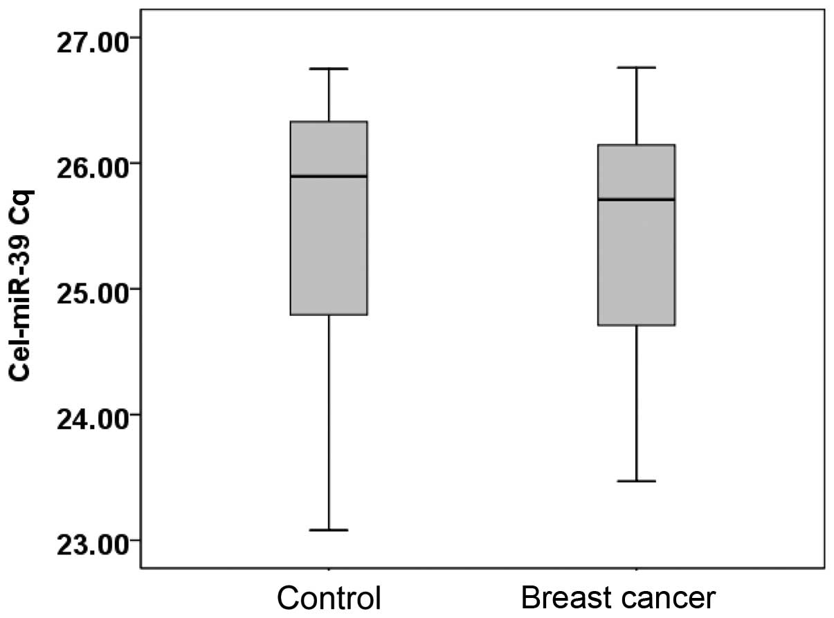
Serum levels of cel-miR-39
The serum levels of cel-miR-39 in 20 randomly
selected breast cancer patients and 20 healthy individuals were
examined. The median Cq value for cel-miR-39 in the healthy
individuals was 25.90 (range, 23.08–26.61), while in patients with
breast cancer, the value was 25.71 (range, 23.47–26.76) (Fig. 1).
Cel-miR-39 expression was calculated using the
2−Cq equation (11). The
Mann-Whitney U test showed that there was no statistically
significant difference between the healthy controls and breast
cancer patients (P=0.543). Cel-miR-39 expression is stable, and can
be used as an exogenous reference in qPCR.
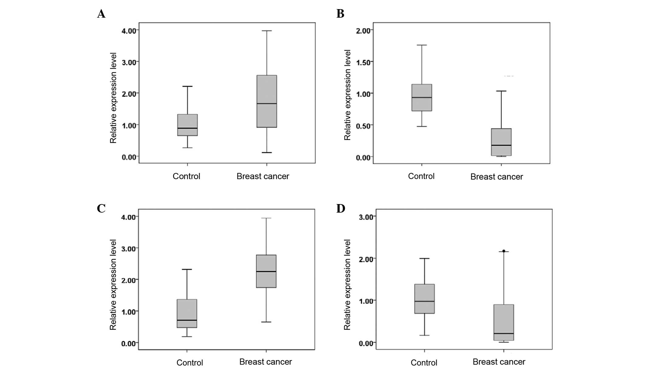
Serum levels of miR-382-3p, −598-3p,
−1246 and −184
The serum miR-382-3p, −598-3p, −1246 and −184
expression levels in 100 cases of breast cancer and 40 healthy
individuals are shown in Fig. 2.
Compared with the serum of the healthy controls, the serum of the
breast cancer patients exhibited upregulated levels of miR-382-3p
and −1246, while the levels of miR-598-3p and −184 were
downregulated (P<0.05).
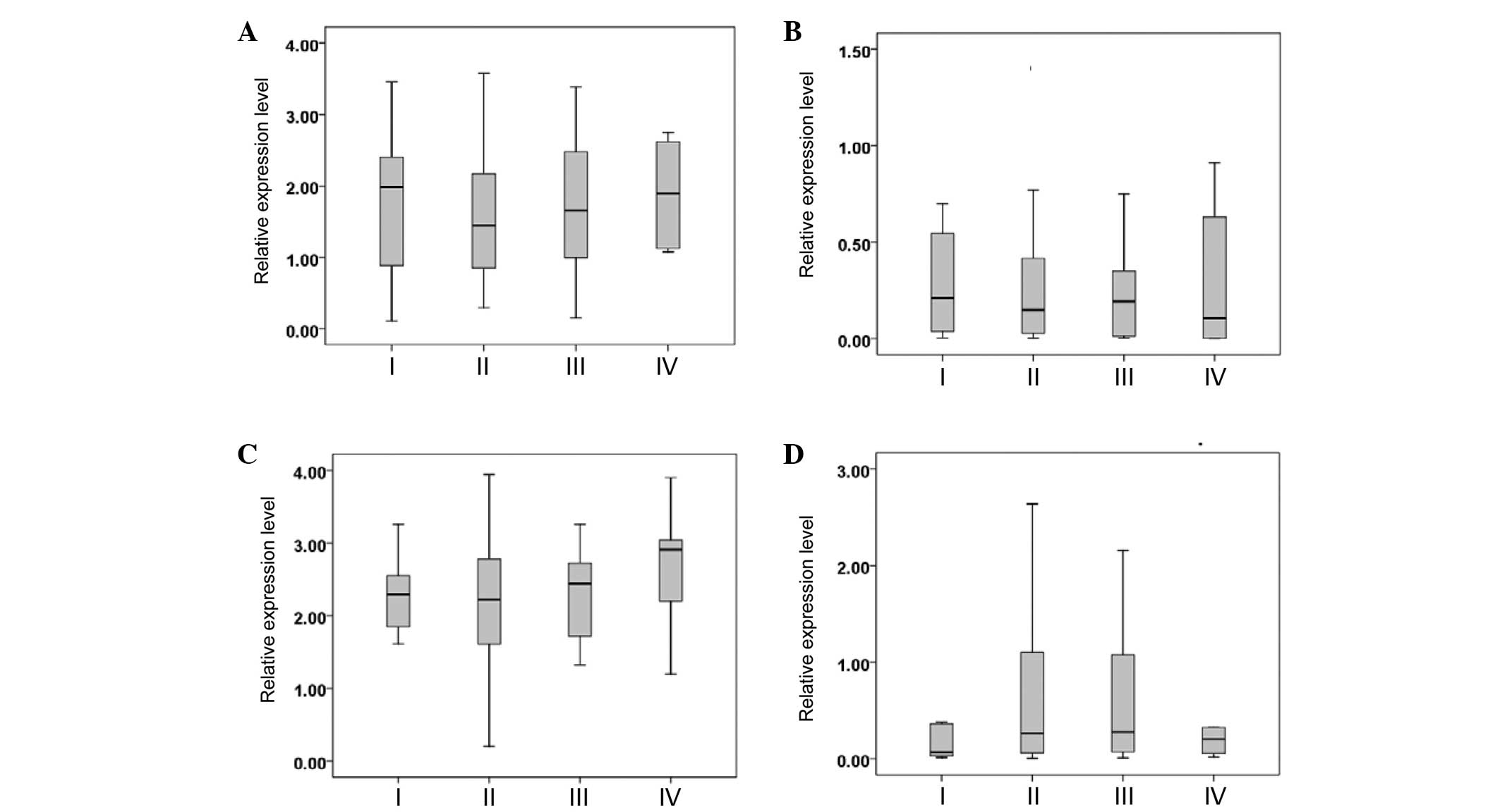
Serum miRNAs in breast cancer patients
with different tumor-node-metastasis (TNM) stages
The breast cancer patients were divided into TNM
stages I, II, III and IV according to the National Comprehensive
Cancer Network (NCCN) Clinical Practice Guidelines in Oncology:
Breast Cancer (version 3.2014) (12).
The serum miR-382-3p, −598-3p, −1246 and −184 levels were not
significantly different between the patients with different TNM
stages (P>0.05) (Fig. 3).
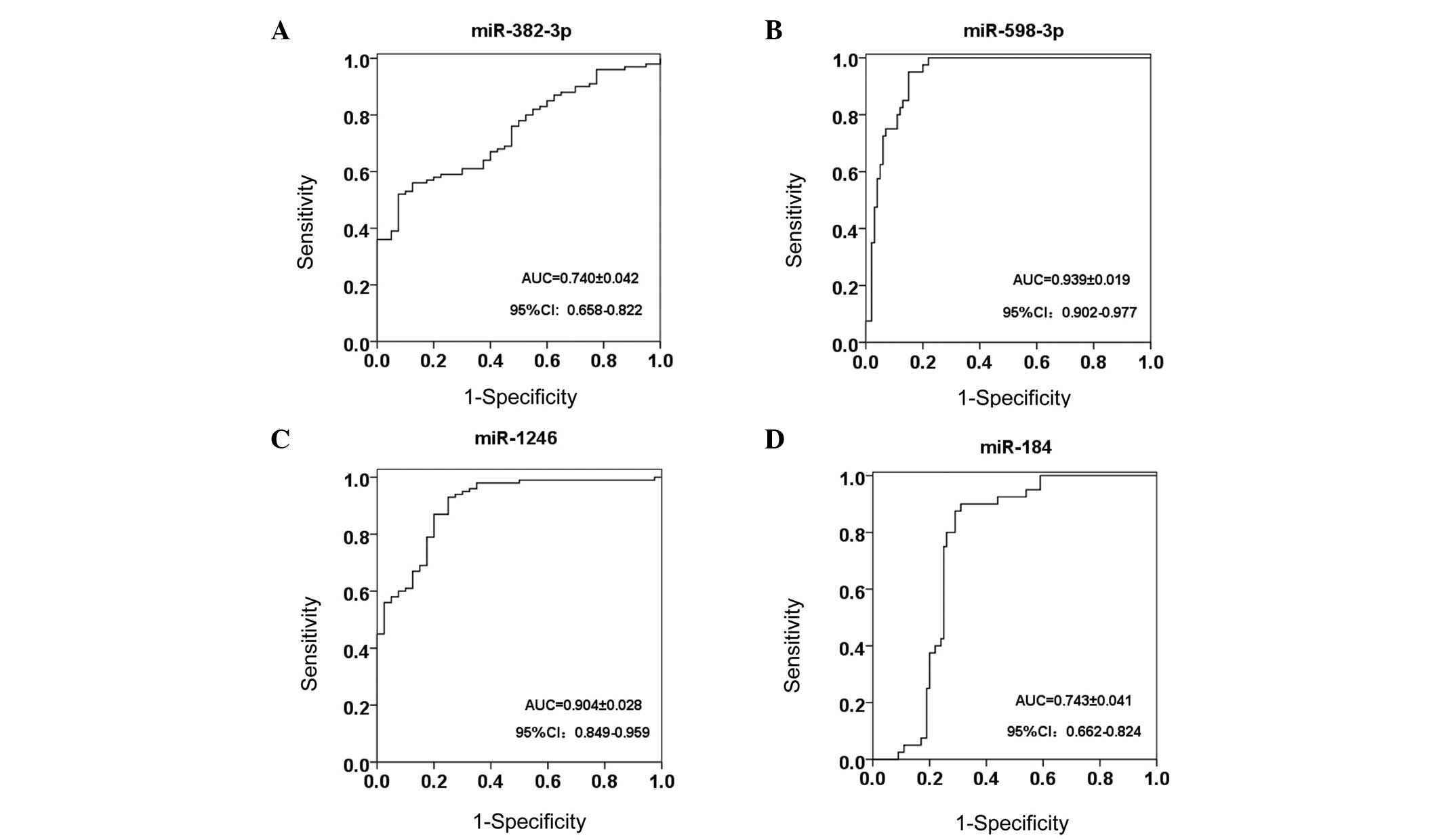
Receiver operating characteristic
(ROC) curve analysis
The area under the ROC curve for miR-382-3p,
−598-3p, −1246 and −184 was 0.740, 0.939, 0.904 and 0.743,
respectively (Fig. 4). Notably, serum
miR-598-3p showed the highest sensitivity and specificity of 95.0
and 85.0%, respectively, while miR-1246 exhibited a lower
specificity of 75.0%, and miR-382-3p showed the lowest sensitivity
at 52.0% (Table III).
| Table III.Diagnostic performance of serum
miR-382–3p, −598–3p, −1246 and −184. |
Table III.
Diagnostic performance of serum
miR-382–3p, −598–3p, −1246 and −184.
| miRNA | AUC | 95% CI | Sensitivity, % | Specificity, % | Cut-off value |
|---|
| miR-382–3p | 0.740 | 0.658–0.822 | 52.0 | 92.5 | 1.611 |
| miR-598–3p | 0.939 | 0.902–0.977 | 95.0 | 85.0 | 0.549 |
| miR-1246 | 0.904 | 0.849–0.959 | 93.0 | 75.0 | 1.318 |
| miR-184 | 0.743 | 0.663–0.824 | 87.5 | 71.0 | 0.484 |
Discussion
The early detection of breast cancer remains a
challenge. Mammography X-ray photography is widely used for breast
cancer screening and detection, but this technology has
limitations, such as irradiation and a high false-positive rate.
miRNAs are expressed in the serum and a variety of other bodily
fluids (13). The molecules are
stable enough for long-term preservation (14) and can tolerate repeated freezing and
thawing, which are promising characteristics for tumor markers. A
number of studies have shown that serum miRNAs are promising
biomarkers for cancer detection (15). Sun et al showed that serum
miRNA-155 can be used as a diagnostic marker for breast cancer
(16). It has also been demonstrated
that serum miRNA-10b expression is significantly higher in breast
cancer patients with bone metastases, and that it can therefore be
used as a tumor marker for breast cancer bone metastasis (17). However, previous studies on serum
miRNAs have not provided a standardized internal standard. A number
of studies have used miR-16 as a standardized internal control for
serum miRNA experiments (18,19), however, the expression of serum miR-16
is not stable in a number of tumors (20). Furthermore, in miR-16 derived from red
blood cells, the occurrence of hemolysis would result in 20–30-fold
increase in blood miR-16, which would affect the results (21). In the present study, exogenous RNA
cel-miR-39 was added during serum RNA extraction. The results
showed that cel-miR-39 expression was stable in the serum of the
breast cancer patients and the healthy controls. Therefore,
cel-miR-39 can be used as a standard for serum miRNA detection.
The upregulation of miR-598 was previously found in
patients with esophageal cancer (22). miR-184 was observed to be highly
expressed in prostate cancer, while it was significantly
downregulated in renal cell carcinoma patients, and was not
correlated with patient age and gender or clinical stage of renal
cell carcinoma (23). The study by
Mar-Aguilar et al confirmed the upregulation of miR-382 in
the serum of patients with breast cancer; a sensitivity of 94.40%
and a specificity of 90.00% was found for the diagnosis of breast
cancer (24). A study by Li et
al showed that miR-382-5p expression was significantly
downregulated in patients with ductal carcinoma in situ
(25), while miR-1246 was
demonstrated to exhibit high expression in colorectal cancer
(26), esophageal squamous cell
carcinoma (27), cervical carcinoma
(28), hepatocellular carcinoma
(29) and other cancers. The
expression profiles of miRNAs in the serum and tissues are
different. miR-1246 is produced by mammary gland epithelial cells
and then stored; when the mammary epithelial cells become
malignant, miR-1246 is selectively released into the circulating
blood, resulted in elevated levels of miR-1246 in the circulation
(30).
The results of the present study also showed that
miR-382-3p and miR-1246 are highly expressed in the serum of breast
cancer patients. To the best of our knowledge, there have been no
studies on the serum miR-598 and miR-184 levels in breast cancer.
The present study results showed that breast cancer patients had
downregulated serum miR-598-3p and miR-184 expression. When using
miR-598-3p as a biomarker with a cutoff value of 0.549, the
sensitivity and specificity for the detection of breast cancer was
95.0 and 87.5%, respectively, indicating that miR-598-3p is a
promising biomarker for breast cancer. Furthermore, the expression
levels were not correlated with breast cancer stage.
In summary, in the present study, it was found that
the expression of miR-382-3p and −1246 in the serum of breast
cancer patients was upregulated, while the expression of miR-598-3p
and −184 was downregulated. There were no significant differences
in the expression levels among different TNM stages. miR-598-3p was
shown to be a promising marker with a high sensitivity and
specificity for the diagnosis of breast cancer. This study provides
novel insights into serum miRNA expression profiles in breast
cancer.
Acknowledgements
This study was supported by grants from the Zhejiang
Science and Technology Program (no. 2012C37037), and the Taizhou
Science and Technology Program (no. 2011A33212).
References
|
1
|
Fan L, Strasser-Weippl K, Li JJ, St Louis
J, Finkelstein DM, Yu KD, Chen WQ, Shao ZM and Goss PE: Breast
cancer in China. Lancet Oncol. 15:e279–e289. 2014. View Article : Google Scholar : PubMed/NCBI
|
|
2
|
Mishra S, Srivastava AK, Suman S, Kumar V
and Shukla Y: Circulating miRNAs revealed as surrogate molecular
signatures for the early detection of breast cancer. Cancer Lett.
369:67–75. 2015. View Article : Google Scholar : PubMed/NCBI
|
|
3
|
Carrington JC and Ambros V: Role of
microRNAs in plant and animal development. Science. 301:336–338.
2003. View Article : Google Scholar : PubMed/NCBI
|
|
4
|
Chen H, Xu GZ, Zhao Y, Tian B, Lu H, Yu X,
Xu Z, Ying N, Hu S and Hua Y: A novel OxyR sensor and regulator of
hydrogen peroxide stress with one cysteine residue in
Deinococcus radiodurans. Plos One. 3:e16022008. View Article : Google Scholar : PubMed/NCBI
|
|
5
|
Chen H, Huang LF, Hua XT, Yin L, Hu Y,
Wang C, Chen W, Yu X, Xu Z, Tian B, et al: Pleiotropic effects of
RecQ in Deinococcus radiodurans. Genomics. 94:333–340. 2009.
View Article : Google Scholar : PubMed/NCBI
|
|
6
|
Bethke A, Fielenbach N, Wang Z,
Mangelsdorf DJ and Antebi A: Nuclear hormone receptor regulation of
microRNAs controls developmental progression. Science. 324:95–98.
2009. View Article : Google Scholar : PubMed/NCBI
|
|
7
|
Iorio MV, Ferracin M, Liu CG, Veronese A,
Spizzo R, Sabbioni S, Magri E, Pedriali M, Fabbri M, Campiglio M,
et al: MicroRNA gene expression deregulation in human breast
cancer. Cancer Res. 65:7065–7070. 2005. View Article : Google Scholar : PubMed/NCBI
|
|
8
|
Ng EK, Wong CL, Ma ES and Kwong A:
MicroRNAs as new players for diagnosis, prognosis and therapeutic
targets in breast cancer. J Oncol. 2009:3054202009. View Article : Google Scholar : PubMed/NCBI
|
|
9
|
Zhu J, Zheng Z, Wang J, Sun J, Wang P,
Cheng X, Fu L, Zhang L, Wang Z and Li Z: Different miRNA expression
profiles between human breast cancer tumors and serum. Front Genet.
5:1492014. View Article : Google Scholar : PubMed/NCBI
|
|
10
|
Livak KJ and Schmittgen TD: Analysis of
relative gene expression data using real-time quantitative PCR and
the 2(−Delta Delta C(T)) method. Methods. 25:402–408. 2001.
View Article : Google Scholar : PubMed/NCBI
|
|
11
|
Schmittgen TD and Livak KJ: Analyzing
real-time PCR data by the comparative CT method. Nat Protoc.
3:1101–1108. 2008. View Article : Google Scholar : PubMed/NCBI
|
|
12
|
Gradishar WJ, Anderson BO, Blair SL,
Burstein HJ, Cyr A, Elias AD, Farrar WB, Forero A, Giordano SH,
Goldstein LJ, et al: Breast cancer version 3.2014. J Natl Compr
Canc Netw. 12:542–590. 2014.PubMed/NCBI
|
|
13
|
Gilad S, Meiri E, Yogev Y, Benjamin S,
Lebanony D, Yerushalmi N, Benjamin H, Kushnir M, Cholakh H, Melamed
N, et al: Serum microRNAs are promising novel biomarkers. PLoS One.
3:e31482008. View Article : Google Scholar : PubMed/NCBI
|
|
14
|
Chen X, Ba Y, Ma L, Cai X, Yin Y, Wang K,
Guo J, Zhang Y, Chen J, Guo X, et al: Characterization of microRNAs
in serum: A novel class of biomarkers for diagnosis of cancer and
other diseases. Cell Res. 18:997–1006. 2008. View Article : Google Scholar : PubMed/NCBI
|
|
15
|
Brase JC, Wuttig D, Kuner R and Sültmann
H: Serum microRNAs as non-invasive biomarkers for cancer. Mol
Cancer. 9:3062010. View Article : Google Scholar : PubMed/NCBI
|
|
16
|
Sun Y, Wang M, Lin G, Sun S, Li X, Qi J
and Li J: Serum MicroRNA-155 as a potential biomarker to track
disease in breast cancer. PLoS One. 7:e470032012. View Article : Google Scholar : PubMed/NCBI
|
|
17
|
Zhao FL, Hua GD, Wang XF, Zhang XH, Zhang
YK and Yu ZS: Serum overexpression of microRNA-10b in patients with
bone metastatic primary breast cancer. J Int Med Res. 40:859–866.
2012. View Article : Google Scholar : PubMed/NCBI
|
|
18
|
Zearo S, Kim E, Zhu Y, Zhao JT, Sidhu SB,
Robinson BG and Soon PSh: MicroRNA-484 is more highly expressed in
serum of early breast cancer patients compared to healthy
volunteers. BMC Cancer. 14:2002014. View Article : Google Scholar : PubMed/NCBI
|
|
19
|
Si H, Sun X, Chen Y, Cao Y, Chen S, Wang H
and Hu C: Circulating microRNA-92a and microRNA-21 as novel
minimally invasive biomarkers for primary breast cancer. J Cancer
Res Clin Oncol. 139:223–229. 2013. View Article : Google Scholar : PubMed/NCBI
|
|
20
|
Chan M, Liaw CS, Ji SM, Tan HH, Wong CY,
Thike AA, Tan PH, Ho GH and Lee AS: Identification of circulating
microRNA signatures for breast cancer detection. Clin Cancer Res.
19:4477–4487. 2013. View Article : Google Scholar : PubMed/NCBI
|
|
21
|
Pritchard CC, Kroh E, Wood B, Arroyo JD,
Dougherty KJ, Miyaji MM, Tait JF and Tewari M: Blood cell origin of
circulating microRNAs: A cautionary note for cancer biomarker
studies. Cancer Prev Res (Phila). 5:492–497. 2012. View Article : Google Scholar : PubMed/NCBI
|
|
22
|
Zhao BS, Liu SG, Wang TY, Ji YH, Qi B, Tao
YP, Li HC and Wu XN: Screening of microRNA in patients with
esophageal cancer at same tumor node metastasis stage with
different prognoses. Asian Pac J Cancer Prev. 14:139–143. 2013.
View Article : Google Scholar : PubMed/NCBI
|
|
23
|
Walter BA, Valera VA, Pinto PA and Merino
MJ: Comprehensive microRNA profiling of prostate cancer. J Cancer.
4:350–357. 2013. View
Article : Google Scholar : PubMed/NCBI
|
|
24
|
Mar-Aguilar F, Mendoza-Ramírez JA,
Malagón-Santiago I, Espino-Silva PK, Santuario-Facio SK,
Ruiz-Flores P, Rodríguez-Padilla C and Reséndez-Pérez D: Serum
circulating microRNA profiling for identification of potential
breast cancer biomarkers. Dis Markers. 34:163–169. 2013. View Article : Google Scholar : PubMed/NCBI
|
|
25
|
Li S, Meng H, Zhou F, Zhai L, Zhang L, Gu
F, Fan Y, Lang R, Fu L, Gu L and Qi L: MicroRNA-132 is frequently
down-regulated in ductal carcinoma in situ (DCIS) of breast and
acts as a tumor suppressor by inhibiting cell proliferation. Pathol
Res Pract. 209:179–183. 2013. View Article : Google Scholar : PubMed/NCBI
|
|
26
|
Piepoli A, Tavano F, Copetti M, Mazza T,
Palumbo O, Panza A, di Mola FF, Pazienza V, Mazzoccoli G, Biscaglia
G, et al: Mirna expression profiles identify drivers in colorectal
and pancreatic cancers. PLoS One. 7:e336632012. View Article : Google Scholar : PubMed/NCBI
|
|
27
|
Takeshita N, Hoshino I, Mori M, Akutsu Y,
Hanari N, Yoneyama Y, Ikeda N, Isozaki Y, Maruyama T, Akanuma N, et
al: Serum microRNA expression profile: MiR-1246 as a novel
diagnostic and prognostic biomarker for oesophageal squamous cell
carcinoma. Brit J Cancer. 108:644–652. 2013. View Article : Google Scholar : PubMed/NCBI
|
|
28
|
Chen J, Yao D, Li Y, Chen H, He C, Ding N,
Lu Y, Ou T, Zhao S, Li L and Long F: Serum microRNA expression
levels can predict lymph node metastasis in patients with
early-stage cervical squamous cell carcinoma. Int J Mol Med.
32:557–567. 2013.PubMed/NCBI
|
|
29
|
Sun Z, Meng C, Wang S, Zhou N, Guan M, Bai
C, Lu S, Han Q and Zhao RC: MicroRNA-1246 enhances migration and
invasion through CADM1 in hepatocellular carcinoma. BMC Cancer.
14:6162014. View Article : Google Scholar : PubMed/NCBI
|
|
30
|
Pigati L, Yaddanapudi SC, Iyengar R, Kim
DJ, Hearn SA, Danforth D, Hastings ML and Duelli DM: Selective
release of MicroRNA species from normal and malignant mammary
epithelial cells. PLoS One. 5:e135152010. View Article : Google Scholar : PubMed/NCBI
|