Introduction
Prostate cancer is the most common carcinoma in
males around the world and the fifth most common cause of
cancer-associated mortality. The majority of prostate cancer
patients succumb to metastatic disease (1). Distant metastases are the most common
cause of cancer-associated mortality in prostate cancer patients
(2). The bone is a common site of
metastasis in prostate cancer and this type of metastasis presents
in >80% of patients who succumb to prostate cancer (3). The bone metastatic lesions from prostate
cancer show more osteoblastic than osteolytic bone lesions compared
with those that originate from other cancer types (4). Metastases to the bone mainly involve the
axial bones, and prostate cancer rarely metastasizes to the distal
phalanges. The current study reports the case of a patient with
prostate cancer that metastasized to the distal phalanx of the left
hallux. A systematic review of the associated literature regarding
prostate cancer with non-axial bone metastasis is also presented.
Written informed consent was obtained from the patient.
Case report
On July 4, 2014, a 73-year-old man was admitted to
the Sir Run Run Shaw Hospital (Hangzhou, Zhejiang, China) due to
pain and swelling of the distal phalanx of the left hallux, which
had developed gradually over several months and progressed during
the last 2 weeks prior to the patient's initial visit. The patient
did not complain of any fever or changes in toe movement. The
patient's medical history did not include any recent injuries. On
November 28, 2012, the patient had been diagnosed with prostate
cancer by prostate biopsy in Zhejiang Province People's Hospital
(Hangzhou, Zhejiang, China), and in November 2013 the patient had
been diagnosed with lung and vertebral bone metastases, as
determined by positron emission tomography/computed tomography
(PET/CT) and bone scans in the same hospital.
Due to the prostate cancer, the patient was treated
with radioactive particle implantation in the prostate (iodine 125;
150 Gy), endocrine therapy with casodex (50 mg 3 times a day;
December 2, 2012-April 9, 2013) and goserelin acetate (3.6 mg
subcutanoues once once every 28 days; Decemeber 2, 2012-April 9,
2013), chemotherapy with docetaxel in combination with prednisone
(100 mg intraveous every 3 weeks; May 9, 2013-December 3, 2013),
and endocrine therapy with abiraterone (1000 mg daily; January 14,
2014-July 4, 2014).
Upon the current admission, the physical examination
did not reveal any abnormalities. The white blood cell, hemoglobin
and platelet counts were 6,750 cells/mm3 (normal range,
3,500–9,500 cells/mm3), 11.4 g/dl (13.0–17.5 g/dl) and
110,000 cells/mm3 (normal range, 100,000–300,000
cells/mm3), respectively. The patient had a high serum
prostate-specific antigen (PSA) level (>10,000 ng/ml; normal
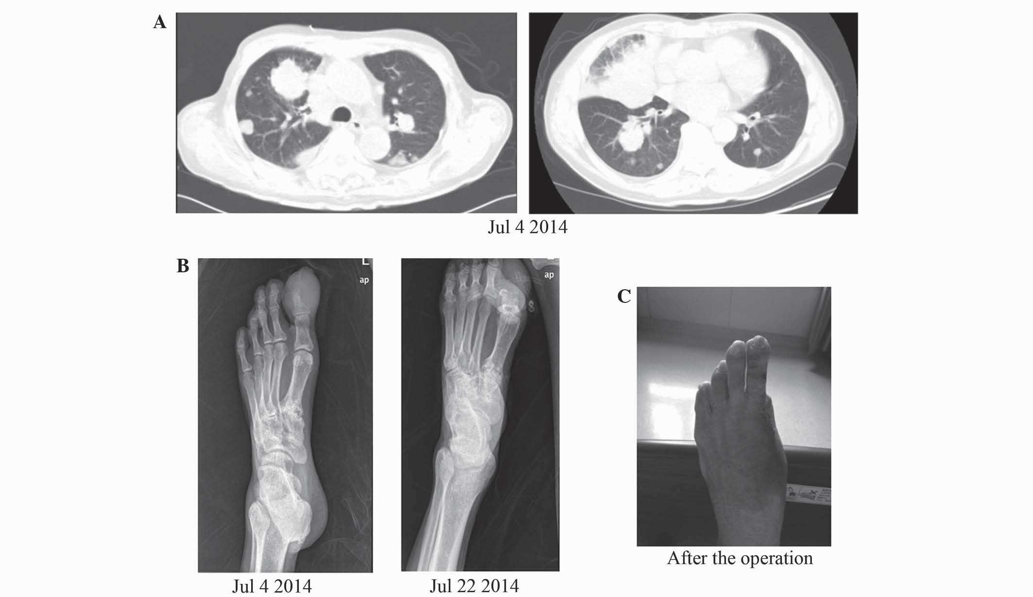
range, <4 ng/ml). An enhanced CT scan of the chest and abdomen
showed a large number of masses in each lung (Fig. 1A). As the patient was admitted due to
a swollen distal phalanx of the left hallux, a bone scan or PET/CT
scan was indicated, however, the patient refused. Therefore, an
X-ray examination was performed (Fig.
1B).
The X-ray results showed morphological changes of
the phalanx and surrounding soft-tissue swelling. Moreover, an
obvious swelling was found on the terminal phalanx of the toe. The
patient was first treated with incision and drainage of the soft
tissue, but the swelling symptom could not be improved. On the
contrary, it became more and more serious during the next 2 weeks,
accompanied with osteolytic destruction (Fig. 1B). After an orthopedic consultation,
an amputation of the phalanx of the left hallux was performed
(Fig. 1C).
The patient was discharged a week after surgery. A
follow-up visit every week was scheduled and no post-operative
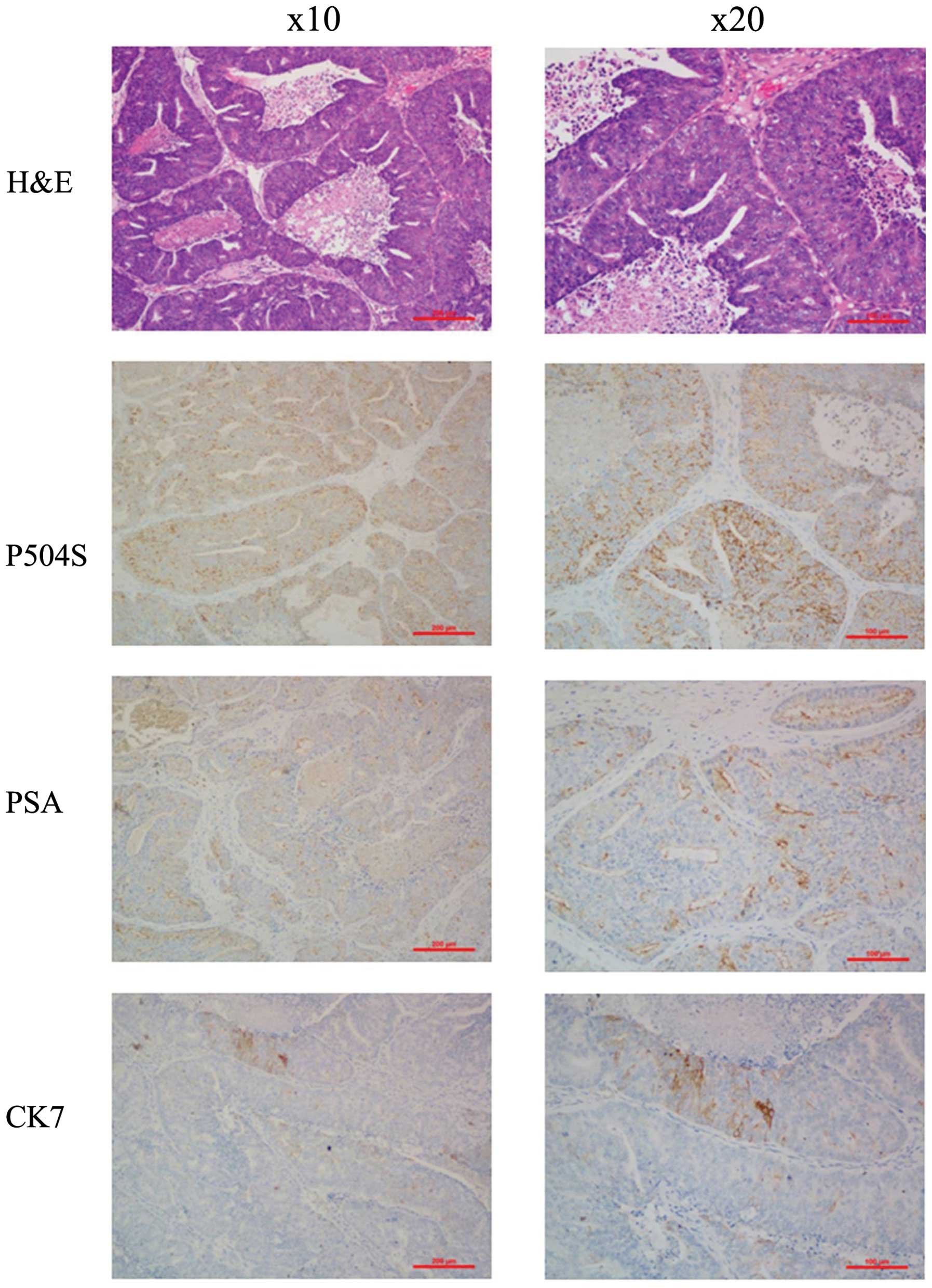
complications occurred. The resected specimen (4 µm) was analyzed
by histological (hematoxylin and eosin) and immunohistochemical
staining. Notably, histological examination revealed a large island
of tumor cells (Fig.2).
Immunohistochemical analysis revealed that the tumor cells were
positive for protein kinase P504S, PSA and cytokeratin (CK)7
(Fig.2), whereas the cells were
negative for cluster of differentiation (CD)56, synaptophysin
(Syn), chromogranin A (CgA) and neuron-specific enolase (NSE). The
following antibodies were used: Mouse monoclonal anti-P504S
(catalog number, sc-81710; dilution, 1:500; Santa Cruz
Biotechnology, Inc., Dallas, TX, USA), mouse monoclonal anti-PSA
(catalog number, sc-7316; dilution, 1:500; Santa Cruz
Biotechnology, Inc.), mouse monoclonal anti-CK7 (catalog number,
ab-9021; dilution, Abcam, Cambridge, MA, USA), mouse monoclonal
anti-CD56 (catalog number, 3576; dilution, 1:100; Cell Signaling
Technology, Inc., Danvers, MA, USA), mouse monoclonal anti-Syn
(catalog number, sc-398849; dilution, 1:500; Santa Cruz
Biotechnology, Inc.), rabbit polyclonal anti-CgA (catalog number,
ab-15160; dilution, 1:1,000; Abcam) and rabbit polyclonal anti-NSE
(catalog number, ab-53025; dilution, 1:1,000; Abcam). Taken
together, these analyses confirmed that the lesion of the distal
phalanx of the left hallux was a metastatic mass from prostate
cancer.
Based on the aforementioned evidence, a diagnosis of
prostate cancer with metastasis to the distal phalanx of the left
hallux was made. The patient succumbed to cardiac and respiratory
failure on February 15, 2015.
Discussion
Prostate cancer is the most common noncutaneous
cancer in men around the world and it preferentially spreads to the
skeleton. Therefore, bone is the dominant and often only metastatic
lesion for the vast majority of men with advanced prostate cancer
(5). Bone metastasis is the main
cause for the morbidity and mortality associated with prostate
cancer, leading to skeletal-related events that include severe
pain, pathological fractures and the risk of spinal cord compromise
(6). The non-axial bone metastasis of
prostate cancer is an extremely rare event. Only phalangeal thumb
metastasis has been reported to date (7). Besides bone, lymph node, lung, liver,
pleura and adrenal gland metastases, several rare metastatic events
have been reported in prostate cancer patients. Although cutaneous
metastasis from prostate cancer is rare in the literature, it has
recently been reported in a large number of the case studies
(Table I) (8–11). In
addition, the iris (12),
gastrointestinal tract (13), parotid
gland (14), proximal ureter
(15), duodenum (16), intramedullary spinal cord (17), larynx (18) and mandibular condyle (19) are also involved metastatic sites of
prostate cancer. Nonetheless, prostate cancer with metastasis to
the phalanx of the toe has been not reported to date. The present
case is therefore the first report on this rare type of
metastasis.
 | Table I.Characteristics of patients with
prostate cancer presenting with rare metastatic events. |
Table I.
Characteristics of patients with
prostate cancer presenting with rare metastatic events.
| First author | Year | Age, years | Site | PSA, ng/ml | (Ref.) |
|---|
| Massraf and Wand | 1998 | 62 | Thumb | 396,000 | (7) |
| Patel et
al | 2015 | 93 | Cutaneous; proximal
right lower trunk | 415 | (8) |
| Ogunmola et
al | 2013 | 62 | Cutaneous; neck,
limbs and trunk | Unknown | (9) |
| Brown et
al | 2014 | 73 | Cutaneous; left
chest | 47 | (10) |
| Keen and Hassan | 2013 | 70 | Cutaneous; left
inguinal region and inner left thigh | 210 | (11) |
| Martin et
al | 2015 | 66 | Iris | 17 | (12) |
| Soe et al | 2014 | 64 | Stomach and
rectum | >10,000 | (13) |
| Hélissey et
al | 2015 | 87 | Parotid gland | 227 | (14) |
| Zhang et
al | 2014 | 76 | Proximal ureter | 18.14 | (15) |
| Kaswala et
al | 2014 | 42 | Duodenum | Unknown | (16) |
| Wu et al | 2014 | 74 | Intramedullary spinal
cord | Unknown | (17) |
| Oliveira et
al | 2012 | 73 | Larynx | 78 | (18) |
| Freudlsperger et
al | 2012 | 75 | Mandibular
condyle | Unknown | (19) |
Several tumor markers are useful in determining the
diagnosis of prostate cancer. PSA is a specific tumor marker for
prostate cancer that commonly occurs at high serum levels in
affected patients (20). PSA
evaluation by immunohistochemistry has been demonstrated to be a
useful tool for determining the diagnosis and prognosis of prostate
cancer (21). However, PSA
immunohistochemistry for the detection of metastatic prostate
cancer can also be of limited utility (22). In the present case, the
immunoreactivity of PSA staining tended to be mildly positive in
the metastatic site.
α-methylacyl-CoA racemase (AMACR/P504S) is a
metabolic enzyme whose overexpression has been shown to be a
diagnostic indicator of prostatic adenocarcinoma in clinical
practice (23,24). Increasing evidence has shown
AMACR/P504S is a diagnostically useful marker for prostatic cancer
(25,26). In the present study,
immunohistochemical examinations revealed positive P504S staining
in the surgical sample. CK7 is commonly used to identify primary
tumor sites and to distinguish invasive prostate cancer from cancer
in situ (27). However, the
immunohistochemical examination of CK is not specific for prostatic
cancer. Focal positivity for CK7 was found in the present
results.
The neuroendocrine antigens (NSE, CgA, Syn and CD56)
are useful in identifying neuroendocrine prostate cancer, but are
usually not expressed in prostatic adenocarcinoma (28). In agreement with this, the present
immunohistochemical staining also showed negative results for CD56,
Syn, CgA and NSE in prostatic adenocarcinoma.
In summary, the present study is the first to report
a case of prostate cancer metastasis to the phalanx of the toe, in
a man that was referred because of persistent pain and swelling of
the distal phalanx of the left hallux, initially misdiagnosed and
treated as abscess. No post-surgery complications and recurrence
occurred following local surgery. The observations made in the
present study contribute to a better understanding of bone
metastasis of prostate cancer.
Acknowledgements
This study was supported by grants from the National
Natural Science Foundation of China (no. 81301891), the Zhejiang
Provincial Natural Science Foundation of China (no. LQ13H160008),
the Zhejiang Province Science and Technology Project of Traditional
Chinese Medicine (no. 2015ZB033) and the Zhengshu Medical Elite
Scholarship Fund.
References
|
1
|
Siegel RL, Miller KD and Jemal A: Cancer
statistics, 2015. CA Cancer J Clin. 65:5–29. 2015. View Article : Google Scholar : PubMed/NCBI
|
|
2
|
Merabishvili VM, Petrova NG, Atroshchenko
AV and Kharitonov MV: Epidemiology of prostate cancer (cohort
study). Vopr Onkol. 60:457–463. 2014.(In Russian). PubMed/NCBI
|
|
3
|
Toomey A and Friedman L: Mortality in
cancer patients after a fall-related injury: The impact of cancer
spread and type. Injury. 45:1710–1716. 2014. View Article : Google Scholar : PubMed/NCBI
|
|
4
|
Cho JY, Shim EJ, Kim IS, Nam EM, Choi MY,
Lee KE, Mun YC, Seoung CM, Lee SN, Song DE and Han WS: Cancer of
unknown primary finally revealed to be a metastatic prostate
cancer: A case report. Cancer Res Treat. 41:45–49. 2009. View Article : Google Scholar : PubMed/NCBI
|
|
5
|
Tu SM, Millikan RE, Mengistu B, Delpassand
ES, Amato RJ, Pagliaro LC, Daliani D, Papandreou CN, Smith TL, Kim
J, et al: Bone-targeted therapy for advanced androgen-independent
carcinoma of the prostate: A randomised phase II trial. Lancet.
357:336–341. 2001. View Article : Google Scholar : PubMed/NCBI
|
|
6
|
Suzman DL, Boikos SA and Carducci MA:
Bone-targeting agents in prostate cancer. Cancer Metastasis Rev.
33:619–628. 2014. View Article : Google Scholar : PubMed/NCBI
|
|
7
|
Massraf AB and Wand JS: Haemorrhagic
secondary prostatic metastasis of the terminal phalanx of the
thumb. Injury. 29:243–245. 1998. View Article : Google Scholar : PubMed/NCBI
|
|
8
|
Patel P, Patel J and Siddiqui S:
Recurrence of prostate cancer with cutaneous metastasis after
radical prostatectomy. Case Rep Urol. 2015:8251752015.PubMed/NCBI
|
|
9
|
Ogunmola AO, Shittu OB and Olapade-Olaopa
EO: Cutaneous metastasis from prostate cancer in a nigerian: A case
report and literature review. Afr J Med Med Sci. 42:283–286.
2013.PubMed/NCBI
|
|
10
|
Brown GT, Patel V and Lee CC: Cutaneous
metastasis of prostate cancer: A case report and review of the
literature with bioinformatics analysis of multiple healthcare
delivery networks. J Cutan Pathol. 41:524–528. 2014. View Article : Google Scholar : PubMed/NCBI
|
|
11
|
Keen MA and Hassan I: Cutaneous metastasis
of an advanced prostate cancer. Indian J Dermatol Venereol Leprol.
79:828–829. 2013. View Article : Google Scholar : PubMed/NCBI
|
|
12
|
Martin V, Cuenca X, Lopez S, Albertini AF,
Lang P, Simon JM, Hémery CG, Feuvret L and Mazeron JJ: Iris
metastasis from prostate carcinoma: A case report and review of the
literature. Cancer Radiother. 19:331–333. 2015. View Article : Google Scholar : PubMed/NCBI
|
|
13
|
Soe AM, Bordia S, Xiao PQ, Lopez-Morra H,
Tejada J, Atluri S and Krishnaiah M: A rare presentation of
metastasis of prostate adenocarcinoma to the stomach and rectum. J
Gastric Cancer. 14:271–274. 2014. View Article : Google Scholar : PubMed/NCBI
|
|
14
|
Hélissey C, Rouanne M, Arnaud FX and Le
Moulec S: Parotid gland metastasis from prostate cancer: Is
docetaxel still the best treatment option? Anticancer Drugs.
26:367–370. 2015. View Article : Google Scholar : PubMed/NCBI
|
|
15
|
Zhang T, Wang Q, Min J, Yu D, Xie D, Wang
Y, Ding D, Chen L, Zou C, Zhang Z and Wang D: Metastasis to the
proximal ureter from prostatic adenocarcinoma: A rare metastatic
pattern. Can Urol Assoc J. 8:E859–E861. 2014. View Article : Google Scholar : PubMed/NCBI
|
|
16
|
Kaswala DH, Patel N, Jadallah S and Wang
W: Metastatic prostate cancer to the duodenum: A rare case. J
Family Med Prim Care. 3:166–168. 2014. View Article : Google Scholar : PubMed/NCBI
|
|
17
|
Wu Z, Xu S, Zhong C, Gao Y, Liu Q, Zheng
Y, Guo Y, Wang Y, Luo Q and Jiang J: Intramedullary conus
medullaris metastasis from prostate carcinoma: A case report and
review of the literature. Oncol Lett. 7:717–720. 2014.PubMed/NCBI
|
|
18
|
Oliveira JA, Said Rde A, Cartaxo Rde S,
Santos JA and Gondim RL: Laryngeal metastasis of a prostate
carcinoma: One rare entity. Braz J Otorhinolaryngol. 78:1352012.(In
Portuguese). View Article : Google Scholar : PubMed/NCBI
|
|
19
|
Freudlsperger C, Kurth R, Werner MK,
Hoffmann J and Reinert S: Condylar metastasis from prostatic
carcinoma mimicking temporomandibular disorder: A case report. Oral
Maxillofac Surg. 16:79–82. 2012. View Article : Google Scholar : PubMed/NCBI
|
|
20
|
Bhavsar T, McCue P and Birbe R: Molecular
diagnosis of prostate cancer: Are we up to age? Semin Oncol.
40:259–275. 2013. View Article : Google Scholar : PubMed/NCBI
|
|
21
|
Garde SV, Sheth AR, Venkatesan VM, Panchal
CJ, Porter AT and Grignon DJ: Prostate inhibin peptide (PIP) in
prostate cancer: A comparative immunohistochemical study with
prostate-specific antigen (PSA) and prostatic acid phosphatase
(PAP). Cancer Lett. 78:11–17. 1994. View Article : Google Scholar : PubMed/NCBI
|
|
22
|
Bernacki KD, Fields KL and Roh MH: The
utility of PSMA and PSA immunohistochemistry in the cytologic
diagnosis of metastatic prostate carcinoma. Diagn Cytopathol.
42:570–575. 2014. View
Article : Google Scholar : PubMed/NCBI
|
|
23
|
Wilson BA, Wang H, Nacev BA, Mease RC, Liu
JO, Pomper MG and Isaacs WB: High-throughput screen identifies
novel inhibitors of cancer biomarker α-methylacyl coenzyme A
racemase (AMACR/P504S). Mol Cancer Ther. 10:825–838. 2011.
View Article : Google Scholar : PubMed/NCBI
|
|
24
|
Kumaresan K, Kakkar N, Verma A, Mandal AK,
Singh SK and Joshi K: Diagnostic utility of α-methylacyl CoA
racemase (P504S) & HMWCK in morphologically difficult prostate
cancer. Diagn Pathol. 5:832010. View Article : Google Scholar : PubMed/NCBI
|
|
25
|
Shen Y, Wang Z, Zhu J, Chen Y, Gu W and
Liu Q: α-Methylacyl-CoA racemase (P504S) is a useful marker for the
differential diagnosis of solid pseudopapillary neoplasm of the
pancreas. Ann Diagn Pathol. 18:146–150. 2014. View Article : Google Scholar : PubMed/NCBI
|
|
26
|
Murray NP, Calaf GM, Badinez L, Dueñas R,
Badinez O, Orellana N, Reyes E and Fuentealba C: P504S expressing
circulating prostate cells as a marker for prostate cancer. Oncol
Rep. 24:687–692. 2010. View Article : Google Scholar : PubMed/NCBI
|
|
27
|
Fichtenbaum EJ, Marsh WL Jr and Zynger DL:
CK5, CK5/6, and double-stains CK7/CK5 and p53/CK5 discriminate in
situ vs. invasive urothelial cancer in the prostate. Am J Clin
Pathol. 138:190–197. 2012. View Article : Google Scholar : PubMed/NCBI
|
|
28
|
Aggarwal R, Zhang T, Small EJ and
Armstrong AJ: Neuroendocrine prostate cancer: Subtypes, biology,
and clinical outcomes. J Natl Compr Canc Netw. 12:719–726.
2014.PubMed/NCBI
|