Introduction
Prostate cancer is the second most common cause of
cancer-associated mortality among men worldwide (1). By the time prostate cancer is diagnosed,
metastasis has occurred in the majority of men, as early prostate
cancer has no symptoms. Metastatic diseases are a significant
public health problem affecting cancer patients and their families
(2). Metastasis occurs when malignant
cells leave the primary tumor, migrate via the circulatory system,
localize in distant areas and lead to the development of secondary
tumors. It is a multistep process, and the steps are similar in all
tumors. Mobilization of tumor cells is an important step in
metastasis (3). Ion channels
regulate, and stimulate numerous behavioral changes in cells that
are associated with cancer and metastasis, including cell movement
(elongation and lateral motility) (4,5),
migration, galvanotaxis (6) and
invasion (7,8). A number of in vitro (9–11) and
in vivo (12) studies
performed using tetrodotoxin (TTX), which specifically blocks
voltage-gated sodium channels (VGSCs) (13), have suggested that the plasma membrane
of prostate cancer cells may gain a more excitable phenotype due to
increased VGSC expression, and thus malignancy is able to progress.
Bennett et al (11)
demonstrated that VGSC expression was ‘necessary’ and ‘enough’ for
the invasiveness of prostate cancer cells. Prostate cancer tends to
invade the bones, lungs and lymph nodes (14). Combating metastasis formation and
growth are important for successfully treating the disease
(2). Various studies have revealed
targeted pharmacological agents aiming to prevent metastasis and
inhibit proliferation (5,6). However, serious side effects of
chemotherapy have encouraged people to request treatment by using
natural agents. Therefore, alternative and complementary treatments
for combating illnesses have increased in popularity. Investigation
of novel and effective therapeutics obtained from natural sources,
including plants and other organisms, is necessary.
The leaves and fruits of Hedera helix L.
(common name, ivy; family, Araliaceae) mainly contain triterpenoid
saponins (15,16). Saponin derivatives obtained from
Hedera spp. have numerous biological activities, including
antiproliferative, cytotoxic (17,18),
antibacterial (19), antifungal
(20), anthelmintic (21), antileishmanial (22), anti-elastase and anti-hyaluronidase
(23) effects. de Medeiros et
al (24) demonstrated that H.
helix spp. canariensis exhibits strong antithrombin
activity, and suggested that there may be a correlation between the
antithrombin activity and a reduction in tumor cell spread.
However, to the best of our knowledge, no investigation into the
potential effects of H. helix on tumor cell migration has
been conducted.
The main aims of the present study were as follows:
i) To investigate whether ethanolic extracts from H. helix
leaves and unripened fruits (HLE and HFE, respectively) have
antiproliferative effects on rat prostate cancer cell lines and,
ii) to investigate how lateral cell motility is affected by these
extracts to reveal the potential effects on cell migration.
Materials and methods
Cell culture
The present study used the highly metastatic
Mat-LyLu cell line and the weakly metastatic AT-2 cell line, which
were derived from the same Dunning rat prostate tumor (25). These cell lines were obtained from
Imperial College, London (UK), and were cultured in RPMI-1640
medium supplemented with 1% heat inactivated fetal bovine serum, 1%
l-glutamine and 0.5% dexamethasone. The cells were maintained under
cell culture conditions of 37°C and 5% CO2 in a
humidified chamber. All chemicals for the cell culture were
purchased from Invitrogen (Thermo Fisher Scientific, Inc., Waltham,
MA, USA).
Preparation of extracts
H. helix L. (family, Araliaceae) was
collected in winter from rural areas of Mersin, located in the
Mediterranean region of Turkey. The plant samples were identified
by Professor Tuna Ekim from Istanbul University (Istanbul, Turkey).
Voucher specimens of the plant were stored in the Herbarium of the
Faculty of Science at the University of Istanbul (ISTF Herbarium
number, 40074). Following identification, shiny, light green H.
helix leaves and unripened fruits were washed and dried in a
chamber at 40°C for 24 h. The dry samples (500 g) were powdered
mechanically and extracted using ethanol (>98%; Honeywell
Riedel-de Haën AG, Seelze, Germany; 1:10 w/v) in an orbital shaker
at room temperature for at least 24 h. These extracts were
filtrated using Whatman filter paper no. 4. Following filtration,
the supernatant was lyophilized at −40°C under a vacuum (Edwards,
Crawley, UK). Following lyophilization, the weight of crude
extracts was 11 and 7 g for H. helix leaves and fruits,
respectively.
Preparation of test medium
Stock solutions (10 mg/ml) of the extracts were
prepared using RPMI medium with the aforementioned supplements.
Stock solutions were filtered using 0.45- or 0.20-µm-diameter
disposable filters. The final concentrations of the HLE were 25, 50
and 75 µg/ml and the final concentrations of the HFE were 18, 20
and 22 µg/ml. The extracts were diluted in conditioned RPMI medium.
The concentrations were selected as result of preliminary toxicity
assays (described below). TTX was prepared according to the
manufacturer's protocol (Alomone Labs, Jerusalem, Israel). Briefly,
1 mg TTX was dissolved in 1 ml sterile distilled water. The stock
solution (3,132 µM) of TTX was frozen in aliquots and stored at
−20°C until use. The test concentration of TTX (200 nM) was diluted
in conditioned RPMI medium.
Toxicity assay
Trypan blue exclusion assays, which are based on
living cells excluding trypan blue dye (4), were performed to determine whether the
ivy extracts and TTX had toxic effects on the cell lines. After
treatment of the cell lines with the ivy extracts or TTX for 48 h,
trypan blue dye (4%; Sigma-Aldrich, St. Louis, MO, USA) was added
into the cell culture dishes (Greiner BioOne GmbH, Frickenhausen,
Germany). The live/dead cell number was counted by using an
inverted microscope (Olympus, Tokyo, Japan) (40X objective).
Proliferation assay
The effects of the HLE and HFE on the proliferation
of Dunning model rat prostate cancer cell lines were assayed
spectrophotometrically using the
3-(4,5-dimethyl-2-thiazolyl)-2,5-diphenyl-2H-tetrazolium bromide
(MTT) reagent (Sigma-Aldrich), as described previously (5). A total of 3×10⁴ cells per
dish were plated into 35-mm-diameter cell culture dishes. The cells
were left to incubate for 24 h undisturbed, and following this, the
medium was replaced daily for 2 days while incubation continued. In
the control dishes, fresh supplemented RPMI medium was used. In the
test dishes, fresh test medium of various concentrations was used.
To determine the cell proliferation, spectrophotometric
measurements were performed using the MTT assay 48 h subsequent to
the addition of the test medium. Absorbance of MTT was measured at
570 nm. The cell counts were normalized using the absorbance values
of the test groups compared with the control group. Additionally,
the cell inhibition rates were calculated proportionally on the
basis of the absorbance value of the control group.
Mitotic activity
The mitotic index (MI) was determined by observing
the effects of the extracts on the mitotic activity of prostate
cancer cell lines. The cells were plated on coverslips and treated
with control or test media for 48 h. The cells were then fixed
using Clarke's fluid (ethanol:acetic acid, 3:1) and stained using
the Feulgen method (26). The number
of cells in the mitotic phases (including the late prophase,
metaphase, anaphase and telophase; n) per total cells (4,000–6,000;
C) was determined by the same person. The MI (%) was calculated
using the following formula: MI=(n/C)×100.
Migration assay
The effect of H. helix extracts on cell
motility was evaluated using wound healing assays (5). Cells were plated in 35-mm-diameter cell
culture dishes at an initial concentration of 15×104
cells per dish. Following 24 h of incubation at 37°C, three
parallel scratches per dish were made using a sterile pipette tip.
The initial widths (mm) of these wounds (W0) were
measured using a graticule on an inverted microscope (Olympus) (10X
objective). A total of 24–48 h after the initial measurement, the
widths of the wounds were measured again (Wt). The
control and test media were replaced at 24 and 48 h following
plating. Motility index (MoI) was calculated using the formula:
MoI=1-(Wt/W0).
Statistical analysis
Data are presented as the mean ± standard error.
Student's t-test, the comparison test of two ratios and the Pearson
correlation test were used to compare the data. P<0.05 was
considered to indicate a statistically significant difference.
Statistical tests were performed using Microsoft Office Excel 2007
(Microsoft Corporation, Redmond, WA, USA) and SPSS version 11.0
(SPSS, Inc., Chicago, IL, USA).
Results
Initial findings
In the present study, an initial evaluation was
performed to determine the non-toxic concentrations of HLE and HFE
on the two cell lines (Mat-LyLu and AT-2) by using a trypan blue
exclusion assay. HLE (25, 50 and 75 µg/ml) and HFE (18, 20 and 22
µg/ml) were not toxic against the cells following 48 h of
incubation. Thus, these non-toxic concentrations were used in the
subsequent experiments. In addition, 200 nM TTX did not demonstrate
toxicity against the two cell lines. Metastatic properties of
Mat-LyLu and AT-2 cells are closely associated with VGSCs, as has
been demonstrated in studies on TTX, which specifically blocks
VGSCs (4,7,8,11). In the present study, 200 nM TTX was
used as the positive control, as it has been previously reported
that TTX does not produce changes in the proliferation of either
cell line even at higher doses (600 nM and 6 µM) than 200 nM
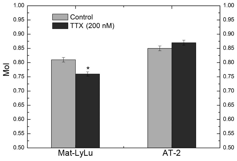
(5,7,27). TTX
reduced the movement distance of Mat-LyLu cells in a lateral
direction, and this effect was found to be statistically
significant. The MoI was 0.81±0.02 and 0.76±0.02 for the control
(non-treated) and test (TTX-treated) groups, respectively
(P<0.01). The MoI for AT-2 cells was 0.85±0.01 in the control
and 0.87±0.03 in the test groups. No significant difference was
observed between the control and TTX-treated groups in AT-2 cells
(P>0.05; Fig. 1). The
aforementioned data revealed that the cell lines maintained their
metastatic properties (Mat-LyLu strongly metastatic and AT-2 weakly
metastatic) associated with VGSCs.
Cell kinetics
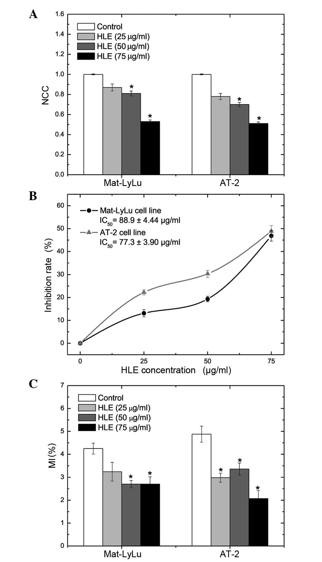
HLE treatment of Mat-LyLu and AT-2 cells for 48 h
resulted in significant inhibition of cell proliferation at 50 and
75 µg/ml (P<0.01). Normalized cell counts (NCCs) of Mat-LyLu
cells were 0.87±0.03, 0.81±0.02 and 0.53±0.02 following treatment
with 25, 50 and 75 µg/ml of HLE for 48 h, respectively. Following
48 h of incubation, the NCCs of AT-2 cells were 0.78±0.03,
0.70±0.02 and 0.51±0.02 for 25, 50 and 75 µg/ml HLE, respectively
(Fig. 2A). The inhibition rates (%)
of Mat-LyLu cell proliferation were 19.3±1.20 for 50 µg/ml and
46.9±2.34 for 75 µg/ml HLE. HLE treatment for 48 h produced
increased rates of inhibition on AT-2 cell proliferation compared
with Mat-LyLu cells. The inhibition rates (%) for AT-2 cells were
30.3±1.51 and 48.9±2.44 for 50 and 75 µg/ml HLE, respectively. The
half-maximal (50%) inhibitory concentration (IC50) of
HLE on cell growth at 48 h was 88.9±4.44 µg/ml for Mat-LyLu cells
and 77.3±3.90 µg/ml for AT-2 cells (Fig.
2B). Following HLE treatment, mitotic activity decreased in
both cell lines. The MI (%) was 4.25±0.24 in the Mat-LyLu cell
control group. The Mat-LyLu cell MI (%) decreased to 3.24±0.41,
2.70±0.16 and 2.70±0.32 as result of treatment with 25, 50 and 75
µg/ml HLE for 48 h, respectively. In a similar manner, the MI (%)
of AT-2 cells decreased from 4.88±0.35 (control group) to 2.07±0.35
(75 µg/ml HLE treatment; Fig.
2C).
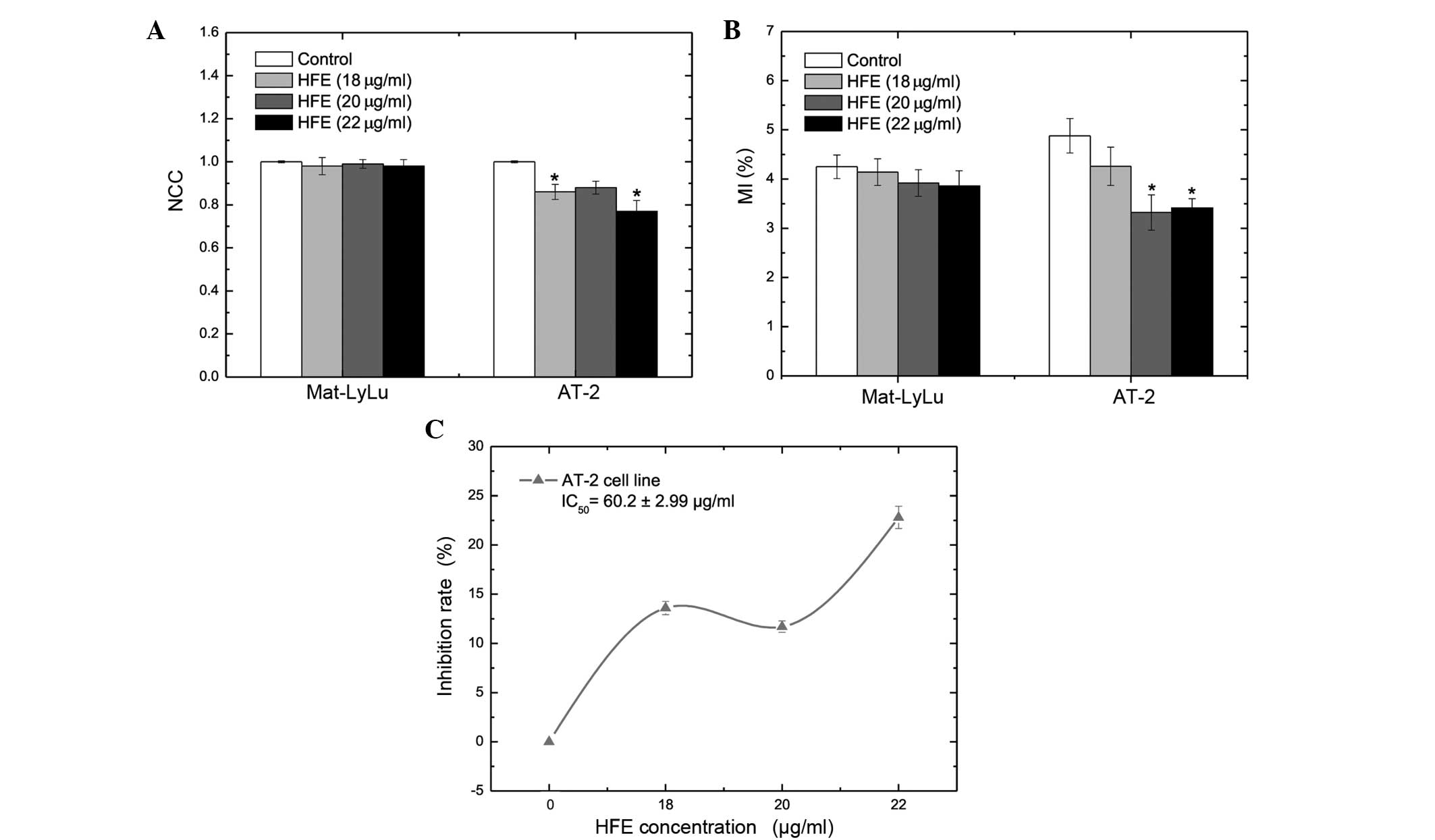
HFE did not produce a significant change in
proliferation (Fig. 3A) or mitotic
activity (Fig. 3B) in the Mat-LyLu
cell line. However, HFE significantly suppressed proliferation and
mitotic activity of AT-2 cells at 18 and 22 µg/ml concentrations,
with inhibition rates (%) of 13.6±0.68 and 22.8±1.14, respectively
(Fig. 3A-C). The MI (%) of AT-2 cells
was 3.41±0.19 following treatment with 22 µg/ml HFE for 48 h, while
the MI (%) was 4.88±0.35 in the control group (Fig. 3B).
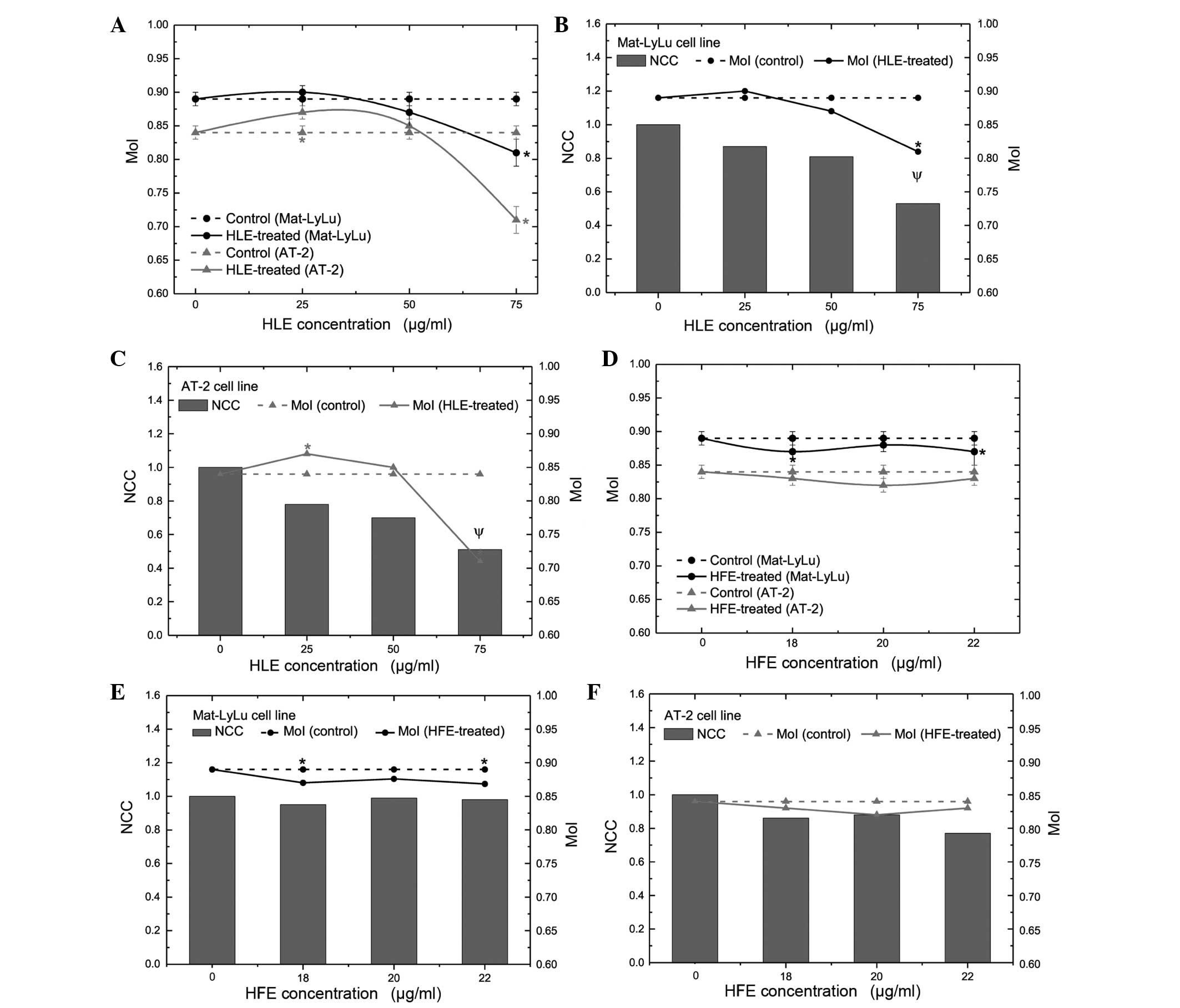
Cell migration
In the present study, the effect of HLE and HFE on
lateral motility of both cell lines was investigated using wound
healing assays. HLE reduced the distance of movement in a lateral
direction in both cell lines (Table
I; Fig. 4A). MoI was 0.89±0.01 in
the Mat-LyLu cell control group. Following 75 µg/ml HLE treatment
for 48 h, the MoI of Mat-LyLu cells reduced to 0.81±0.02
(P<0.01). The MoI of the AT-2 cells was 0.84±0.01 and 0.71±0.02
in the control and 75 µg/ml HLE test groups, respectively. It was
determined that the difference between the control and test groups
was statistically significant (P<0.01). The MoI decreased by
8.83 and 15.7% in the Mat-LyLu and AT-2 cell lines, respectively.
Considering the changing MoI and rate of inhibition of cell
proliferation, it was suggested that the MoI may decrease due to
the inhibition of proliferation in the investigated cell lines
(Fig. 4B and C). A strong positive
correlation was noted between the decrease of MoI and proliferation
(r2=0.97; P<0.01). In addition, HFE treatment reduced
the movement distance of Mat-LyLu cells, but not AT-2 cells
(Table II; Fig. 4D). The MoI for Mat-LyLu cells was
0.89±0.01 and 0.87±0.02 in the control and 22 µg/ml test groups,
respectively (P<0.05). However, no difference in MoI was
observed between the control and test groups for the AT-2 cells.
Notably, HFE treatment did not affect the kinetic parameters
(proliferation and mitotic activity) of Mat-LyLu cells, while
suppressing the growth of AT-2 cells. Additionally, no significant
correlation was observed between the decrease of MoI and
proliferation of Mat-LyLu cells (r2=0.05; P>0.05).
Due to this, the present study concluded that HFE treatment reduced
the motility of Mat-LyLu cells without affecting their
proliferation (Fig. 4E and F).
 | Table I.Initial MoI and change in MoI in
Mat-LyLu and AT-2 cell lines following treatment with ethanolic
extract of HLE for 48 h. |
Table I.
Initial MoI and change in MoI in
Mat-LyLu and AT-2 cell lines following treatment with ethanolic
extract of HLE for 48 h.
|
| MoIa | Change in MoI,
%b |
|---|
|
|
|
|
|---|
| Treatment | Mat-LyLu | AT-2 | Mat-LyLu | AT-2 |
|---|
| Control | 0.89±0.01 | 0.84±0.01 | – | – |
| HLE, 25 µg/ml | 0.90±0.01 |
0.87±0.01c | −0.77 (↑) | −3.96
(↑)c |
| HLE, 50 µg/ml | 0.87±0.02 | 0.85±0.01 | 2.98
(↓) | −0.48 (↑) |
| HLE, 75 µg/ml |
0.81±0.02c |
0.71±0.02c | 8.83
(↓)c | 15.72
(↓)c |
| Table II.Initial MoI and change in MoI of
Mat-LyLu and AT-2 cell lines following treatment with ethanolic
extract of HFE for 48 h. |
Table II.
Initial MoI and change in MoI of
Mat-LyLu and AT-2 cell lines following treatment with ethanolic
extract of HFE for 48 h.
|
| MoIa | Change in MoI,
%b |
|---|
|
|
|
|
|---|
| Treatment | Mat-LyLu | AT-2 | Mat-LyLu | AT-2 |
|---|
| Control | 0.89±0.01 | 0.84±0.01 | – | – |
| HFE, 18 µg/ml |
0.87±0.01c | 0.83±0.01 | 2.51
(↓)c | 1.64 (↓) |
| HFE, 20 µg/ml | 0.88±0.01 | 0.82±0.01 | 1.87 (↓) | 2.38 (↓) |
| HFE, 22 µg/ml |
0.87±0.02c | 0.83±0.01 | 2.69
(↓)c | 1.55 (↓) |
Discussion
To the best of our knowledge, this is the first
study showing the effects of crude H. helix extracts on the
growth and migration of rat prostate cancer cell lines (Mat-LyLu
and AT-2) of different metastatic potential. Previously,
researchers have been focused on in vitro and in vivo
studies using saponins and their derivatives obtained from various
parts (including, leaves or fruits) of herbal sources, and certain
Hedera species (18,28–30).
Triterpenoid saponins such as α-hederin, β-hederin, δ-hederin,
hederacosides A-I, hederacolchisides (A, A1 and B) and
hederagenin are the primary phytochemical components in
Hedera spp. (15,16,18).
Saponin derivatives isolated from Hedera spp. (18,28) and
other plants (29,30) produce antiproliferative, antitumor and
cytotoxic effects (18,28), as well a range of other biological
activities [including, antimicrobial (19), antifungal (20), anthelmintic (21) and antileishmanial (22)]. In the present study, crude extracts
of H. helix leaves and fruits were used. It was determined
that the growth of Mat-LyLu and AT-2 cell lines was inhibited by
HLE. The inhibition of cell proliferation increased with increasing
concentration and application time of HLE. In addition, it was
demonstrated that the mitotic activity of these two cell lines was
suppressed. Danloy et al (17)
reported that α-hederin isolated from H. helix was cytotoxic
against B16 mouse melanoma and 3T3 fibroblast cell lines. α-hederin
isolated from H. helix leaves potentiated the antitumor
activity of 5-fluorouracil in the HT-29 human colon adenocarcinoma
cell line (28). Researchers have
isolated α-hederin or hederagenin from various plants (29,30).
α-Hederin and hederagenin produced weak and strong cytotoxic
effects on human bladder carcinoma cell lines (including, J82 and
T24), respectively (29). Swamy and
Huat (30) reported that α-hederin
induces apoptosis and suppresses DNA, RNA and protein synthesis of
P388 murine leukemia cells. Barthomeuf et al (18) isolated certain saponin derivatives,
including α-hederin, β-hederin, δ-hederin, hederacolchiside (A and
A1) and hederagenin, from H. colchica fruits and used them
for treating 6 human cancer cell lines (PC-3, DLD-1, PA1, A549,
MCF7 and M4Beu) and a human fibroblast cell line. It was observed
that hederacolchiside A1 and β-hederin had the highest
inhibitory activity on cancer cell lines (18). It was also observed that these two
compounds were cytotoxic against fibroblast cells, but that the
concentration producing this cytotoxicity was increased compared
with the dose required for inhibiting the proliferation of cancer
cell lines (18). The present study
revealed that HFE inhibited the proliferation and mitosis of only
the AT-2 cell line and not Mat-LyLu cells.
The most important step for metastatic spread is the
ability to migrate (31). Mat-LyLu
and AT-2, from Dunning model rat prostate cancer cell lines, have
strong and weak metastatic abilities, respectively (32). Therefore, these cell lines are
excellent experimental models for studying the effects of various
agents on cell motility and invasion. No previous studies
investigating the effect of H. helix on the motility of
these cancer cell lines could be identified in the relevant
literature. There are a small number of studies showing the effect
of H. helix extract(s) on cell migration. de Medeiros et
al (24) reported that H.
helix spp. canariensis exhibited antithrombin activity
and that certain compounds from this plant may prevent blood
clotting, thus, blocking tumor cell spread. The results of the
present study have demonstrated that HFE is able to inhibit
Mat-LyLu cell migration, but has no effect on cell proliferation.
By contrast, migration was not affected but proliferation was
inhibited in the AT-2 cells. HLE, however, inhibited cell kinetics
(proliferation and mitotic activity), as well as motility in
Mat-LyLu and AT-2 cell lines.
The initial results indicated that the cell lines
used in the present experiments conserved their metastatic
properties (Mat-LyLu remained strongly metastatic, and AT-2
remained weakly metastatic) that were closely associated with
VGSCs. Therefore, HFE may inhibit cell migration of Mat-LyLu cells
by blocking VGSCs. To clarify the underlying mechanism of
inhibition and to contribute to research on preventing prostate
cancer metastasis, further molecular investigations are required.
Our future studies will focus on explaining the phytochemical
content of H. helix fruits and investigating the underlying
mechanism(s) of their effects on prostate cancer and its
metastasis.
Acknowledgements
The present study was partially supported by The
Scientific and Technological Research Council of Turkey (TUBITAK)
[grant no., TBAG-2422 (104T031)]. The authors would like to thank
Professor Mustafa B.A. Djamgoz (Department of Life Sciences, Sir
Alexander Fleming Building, Imperial College London, South
Kensington Campus, London, UK) for supplying the cell lines and his
contributions.
Glossary
Abbreviations
Abbreviations:
|
HFE
|
ethanolic extract of Hedera
helix fruits
|
|
HLE
|
ethanolic extract of Hedera
helix leaves
|
|
MI
|
mitotic index
|
|
MoI
|
motility index
|
|
MTT
|
3-(4,5-dimethyl-2-thiazolyl)-2,5-diphenyl-2H-tetrazolium
bromide
|
|
NCCs
|
normalized cell counts
|
|
TTX
|
tetrodotoxin
|
|
VGSC
|
voltage-gated sodium channel
|
References
|
1
|
Jemal A, Siegel R, Ward E, Hao Y, Xu J and
Thun MJ: Cancer statistics, 2009. CA Cancer J Clin. 59:225–249.
2009. View Article : Google Scholar : PubMed/NCBI
|
|
2
|
Sleeman J and Steeg PS: Cancer metastasis
as a therapeutic target. Eur J Cancer. 46:1177–1180. 2010.
View Article : Google Scholar : PubMed/NCBI
|
|
3
|
Arya M, Bott RS, Shergill IS, Ahmed HU,
Williamson M and Patel HR: The metastatic cascade in prostate
cancer. Surg Oncol. 15:117–128. 2006. View Article : Google Scholar : PubMed/NCBI
|
|
4
|
Fraser SP, Ding Y, Liu A, Foster CS and
Djamgoz MB: Tetrodotoxin suppresses morphological enhancement of
the metastatic MAT-LyLu rat prostate cancer cell line. Cell Tissue
Res. 295:505–512. 1999. View Article : Google Scholar : PubMed/NCBI
|
|
5
|
Fraser SP, Salvador V, Manning EA, Mizal
J, Altun S, Raza M, Berridge RJ and Djamgoz MB: Contribution of
functional voltage-gated Na+ channel expression to cell behaviors
involved in the metastatic cascade in rat prostate cancer: I.
Lateral motility. J Cell Physiol. 195:479–487. 2003. View Article : Google Scholar : PubMed/NCBI
|
|
6
|
Djamgoz MBA, Mycielska M, Madeja Z, Fraser
SP and Korohoda W: Directional movement of rat prostate cancer
cells in direct-current electric field: Involvement of
voltage-gated Na+ channel activity. J Cell Sci. 114:2697–2705.
2001.PubMed/NCBI
|
|
7
|
Grimes JA, Fraser SP, Stephens GJ, Downing
JE, Laniado ME, Foster CS, Abel PD and Djamgoz MB: Differential
expression of voltage-activated Na+ currents in two prostatic
tumour cell lines: Contribution to invasiveness in vitro. FEBS
Lett. 369:290–294. 1995. View Article : Google Scholar : PubMed/NCBI
|
|
8
|
Laniado ME, Lalani EN, Fraser SP, Grimes
JA, Bhangal G, Djamgoz MB and Abel PD: Expression and functional
analysis of voltage-activated Na+ channels in human prostate cancer
cell lines and their contribution to invasion in vitro. Am J
Pathol. 150:1213–1221. 1997.PubMed/NCBI
|
|
9
|
Smith P, Rhodes NP, Shortland AP, Fraser
SP, Djamgoz MB, Ke Y and Foster CS: Sodium channel protein
expression enhances the invasiveness of rat and human prostate
cancer cells. FEBS Lett. 423:19–24. 1998. View Article : Google Scholar : PubMed/NCBI
|
|
10
|
Abdul M and Hoosein N: Voltage-gated
sodium ion channels in prostate cancer: Expression and activity.
Anticancer Res. 22:1727–1730. 2002.PubMed/NCBI
|
|
11
|
Bennett ES, Smith BA and Harper JM:
Voltage-gated Na+ channels confer invasive properties on human
prostate cancer cells. Pflugers Arch. 447:908–914. 2004. View Article : Google Scholar : PubMed/NCBI
|
|
12
|
Yildirim S, Altun S, Gumushan H, Patel A
and Djamgoz MB: Voltage-gated sodium channel activity promotes
prostate cancer metastasis in vivo. Cancer Lett. 323:58–61. 2012.
View Article : Google Scholar : PubMed/NCBI
|
|
13
|
Cestèle S and Catterall WA: Molecular
mechanisms of neurotoxin action on voltage-gated sodium channels.
Biochimie. 82:883–892. 2000. View Article : Google Scholar : PubMed/NCBI
|
|
14
|
Ritchie CK, Andrews LR, Thomas KG, Tindall
DJ and Fitzpatrick LA: The effects of growth factors associated
with osteoblasts on prostate carcinoma proliferation and
chemotaxis: Implications for the development of metastatic disease.
Endocrinology. 138:1145–1150. 1997. View Article : Google Scholar : PubMed/NCBI
|
|
15
|
Bedir E, Kırmızıpekmez H, Sticher O and
Caliş I: Triterpene saponins from the fruits of Hedera helix.
Phytochemistry. 53:905–909. 2000. View Article : Google Scholar : PubMed/NCBI
|
|
16
|
Demirci B, Goppel M, Demirci F and Franz
G: HPLC profiling and quantification of active principles in leaves
of Hedera helix L. Pharmazie. 59:770–774. 2004.PubMed/NCBI
|
|
17
|
Danloy S, QuetinLeclercq J, Coucke P, De
Pauw-Gillet MC, Elias R, Balansard G, Angenot L and Bassleer R:
Effects of alpha-hederin, a saponin extracted from Hedera helix, on
cells cultured in vitro. Planta Med. 60:45–49. 1994. View Article : Google Scholar : PubMed/NCBI
|
|
18
|
Barthomeuf C, Debiton E, Mshvildadze V,
Kemertelidze E and Balansard G: In vitro activity of
hederacolchisid A1 compared with other saponins from Hedera
colchica against proliferation of human carcinoma and melanoma
cells. Planta Med. 68:672–675. 2002. View Article : Google Scholar : PubMed/NCBI
|
|
19
|
Cioacá C, Margineanu C and Cucu V: The
saponins of Hedera helix with antibacterial activity. Pharmazie.
33:609–610. 1978.
|
|
20
|
MoulinTraffort J, Favel A, Elias R and
Regli P: Study of the action of α-hederin on the ultrastructure of
Candida albicans. Mycoses. 41:411–416. 1998. View Article : Google Scholar : PubMed/NCBI
|
|
21
|
Julien J, Gasquet M, Maillard C, Balansard
G and Timon-David P: Extracts of the ivy plant, Hedera helix, and
their antihelminthic activity on liver flukes. Planta Med.
3:205–208. 1985. View Article : Google Scholar : PubMed/NCBI
|
|
22
|
MajesterSavornin B, Elias R, DiazLanza AM,
Balansard G, Gasquet M and Delmas F: Saponins of the ivy plant,
Hedera helix, and their leishmanicidic activity. Planta Med.
57:260–262. 1991. View Article : Google Scholar : PubMed/NCBI
|
|
23
|
Facino MR, Carini M, Stefani R, Aldini G
and Saibene L: Anti-elastase and anti-hyaluronidase activities of
saponins and sapogenins from Hedera helix, Aesculus hippocastanum,
and Ruscus aculeatus: Factors contributing to their efficacy in the
treatment of venous insufficiency. Arch Pharm (Weinheim).
328:720–724. 1995. View Article : Google Scholar : PubMed/NCBI
|
|
24
|
deMedeiros JM, Macedo M, Contancia JP,
Nguyen C, Cunningham G and Miles DH: Antithrombin activity of
medicinal plants of the Azores. J Ethnopharmacol. 72:157–165. 2000.
View Article : Google Scholar : PubMed/NCBI
|
|
25
|
Tennant TR, Kim H, Sokoloff M and
Rinker-Schaeffer CW: The Dunning model. Prostate. 43:295–302. 2000.
View Article : Google Scholar : PubMed/NCBI
|
|
26
|
Bancroft JD and Stevens A: Proteins and
nucleic acidsTheory and Practice of Histological Techniques. 3rd
edition. Churchill Livingstone; Edinburgh, UK: pp. 148–188.
1990
|
|
27
|
Fraser SP, Grimes JA and Djamgoz MB:
Effects of voltage-gated ion channel modulators on rat protastic
cancer cell proliferation: Comprarison of strongly and weakly
metastic cell lines. Prostate. 44:61–76. 2000. View Article : Google Scholar : PubMed/NCBI
|
|
28
|
Bun SS, Elias R, Baghdikian B, Ciccolini
J, Ollivier E and Balansard G: Alpha-hederin potentiates 5-FU
antitumor activity in human colon adenocarcinoma cells. Phytother
Res. 22:1299–1302. 2008. View Article : Google Scholar : PubMed/NCBI
|
|
29
|
Park HJ, Kwon SH, Lee JH, Lee KH, Miyamoto
KI and Lee KT: Kalopanaxsaponin A is a basic saponin structure for
the anti-tumor activity of hederagenin monodesmosides. Planta Med.
67:118–121. 2001. View Article : Google Scholar : PubMed/NCBI
|
|
30
|
Swamy SM and Huat BT: Intracellular
glutathione depletion and reactive oxygen species generation are
important in alpha-hederin-induced apoptosis of P388 cells. Mol
Cell Biochem. 245:127–139. 2003. View Article : Google Scholar : PubMed/NCBI
|
|
31
|
StetlerStevenson WG, Aznavoorian S and
Liotta LA: Tumor cell interactions with the extracellular matrix
during invasion and metastasis. Annu Rev Cell Dev Biol. 9:541–573.
1993. View Article : Google Scholar
|
|
32
|
Isaacs JT, Isaacs WB, Feitz WF and Scheres
J: Establishment and characterization of seven Dunning rat
prostatic cancer cell lines and their use in developing methods for
predicting metastatic abilities of prostatic cancers. Prostate.
9:261–281. 1986. View Article : Google Scholar : PubMed/NCBI
|