Introduction
Cervical cancer (CC) is the third most common cancer
and the fourth leading cause of cancer-associated mortality in
females worldwide (1). CC is the
leading cause of cancer-associated mortality in young women, and
the most common cause of cancer-associated mortality among women in
the developing world (2). For a long
period of time, focus has been placed on cancer cells, and the
importance of the tumor microenvironment has not been recognized.
However, it is now known that cancer is not merely a local disease,
but a systematic disease, involving a group of rogue cells
(3), in which the tumor
microenvironment plays a pivotal role.
The tumor microenvironment is a complex community
(3), including cancer cells, stromal
tissue with immune cells, fibroblasts, myofibroblasts, cytokines
and vascular tissue, as well as the extracellular matrix
surrounding these components (4). In
the 1880s, Paget reported the ‘seed and soil’ theory (5), which indicated that in order to fully
comprehend cancer initiation and progression, a focus must not only
be placed on the cancer cells, ‘the seed’, but also on ‘the soil’
from which nutrients, oxygen and signals for growth and development
are derived, and within which macrophages and tumor angiogenesis
(6), the most representative factors
in the tumor microenvironment, play crucial roles in the
development of tumors.
Macrophages are important representatives of immune
function during cancer progression process (7), acting as vital components of the host's
defense, and as antigen-presenting cells and effector cells
(8,9).
Macrophages are derived nearly completely from circulating
monocytes, and acquire unique phenotypic characteristics with
diverse functions in response to environmental signals (10). As human solid tumors can often be
anatomically classified into the areas of a cancer nest, the
invading edge and the peritumoral stroma, each with distinct
compositions and functional properties (11,12), the
tumor-associated monocytes/macrophages (TAMs) exhibit different
phenotypes and functions at these distinct tumor sites (13–15).
Macrophages in normal or inflamed tissues exhibit spontaneous
anti-tumor activity, whereas TAMs could be oriented towards
promoting tumor growth, remodeling tissues, promoting angiogenesis
and suppressing adaptive immunity (16–18). Tumor
angiogenesis, another key component in the tumor microenvironment,
also plays an essential role during cancer progression (19). In 1971, Folkman first proposed the
vital role of angiogenesis in the growth of tumors, describing
tumors as ‘hot and bloody’ (20).
Angiogenesis occurs in the normal and vital processes of growth and
development, as well as in wound healing and in granulation tissue
formation (20). However,
angiogenesis is also essential in the tumor transition between
benign and malignant states, resulting in the use of angiogenesis
inhibitors to treat cancer (21).
In addition, studies have shown that TAMs could
promote tumor angiogenesis and metastasis (22). TAMs and cancer cells can secrete
various growth factors, angiogenic factors and a variety of
enzymes, which play important roles in angiogenesis (23). Thus, TAMs and tumor angiogenesis
jointly promote cancer invasion and metastasis. Therefore, it is
urgent to develop a technique, which can image multiple components
in situ so as to study the co-evolution of TAMs and tumor
angiogenesis during cancer invasion. Quantum dots (QDs) are a novel
class of semiconductor nanocrystals of 1–10 nm in diameter
(24,25), which have unique optical and
electrical properties, including narrow and symmetric emission
spectra of light, good light stability, strong fluorescence
intensity, a long fluorescence schedule of 30–100 nsec and a
changeable emission wavelength with the QD size, and are thus of
great potential in the fields of biological imaging and molecular
markers (26,27). Major advances have emerged as a result
of using QDs for highly sensitive cellular imaging over the past
decade (28). In comparison with
organic dyes and fluorescent proteins, QDs exhibit the unique
features of size and composition tunable light emission, enhanced
signal brightness and resistance to photobleaching (29). In this study, the special properties
of QDs were harnessed to better understand the role of tumor
microenvironment during cancer progression, which was also reported
in our previous study (30).
Therefore, the present study aimed to observe the
spatial and temporal distributions of TAMs and tumor angiogenesis
during the progression from chronic cervicitis to cancer through
the use of conventional IHC and QD-based two-component in
situ imaging technology, in order to investigate the
co-evolution between the aforementioned components of the tumor
microenvironment and CC cells.
Materials and methods
Samples
A total of 90 paraffin-embedded cervical samples
were collected from Wuhan Municipality Central Hospital (Wuhan,
Hubei, China) between January 2013 and December 2014, including 30
cases of chronic cervicitis, 30 CIN I–III biopsy samples and 30 CC
resected samples. The study protocol was approved by the Ethics
Committee of Wuhan Municipality Central Hospital. Written informed
consent was obtained from all patients prior to surgery, with
permission to use the specimens for scientific research purposes
and clinicopathological studies.
Reagents
Mouse anti-human monoclonal antibody against
macrophage (catalog no. MA1-38069; dilution, 1/100; ABR Affinity
BioReagents, Golden, CO, USA) and rabbit anti-human monoclonal
antibody against CD105 (catalog no. ab169545; dilution, 1/300;
Abcam, Cambridge, MA, USA) were used. The QDs were goat anti-mouse
secondary antibody QD-525 (dilution, 1/100; Invitrogen; Thermo
Fisher Scientific Inc., Waltham, MA, USA) and goat anti-rabbit
QD-585 (dilution, 1/200; Invitrogen; Thermo Fisher Scientific
Inc.), and the remaining conventional reagents were purchased from
Dako (Glostrup, Denmark).
Immunohistochemistry
The use of IHC followed the instructions of our
previously established technical procedure (31). Briefly, tissue sections (4-µm thick)
were treated by de-paraffinizing, hydration, antigen retrieval and
washing in deionized water prior to proceeding to the following
imaging studies. The main steps of the IHC were as follows: The
endogenous peroxidase activity was blocked by immersion in 3%
H2O2 for 10 min to prevent any non-specific
binding. After being repaired with pH 6.0 citric acid under high
pressure, the slides were first incubated with primary antibody for
30 min, then with the corresponding secondary antibody for 30 min.
Next, they were colored with 3,3′-diaminobenzidine for 10 min,
stained with hematoxylin, dehydrated to transparency and covered
with neutral gum. Phosphate-buffered saline (PBS) was used instead
of a primary antibody for the negative control.
QD-based molecular imaging
For QD-based immune-fluorescent imaging, subsequent
to being blocked with 2% bovine serum antigen (BSA) for 30 min at
37°C, the sections were incubated with the aforementioned primary
antibodies at 37°C for 2 h. Once washed with PBS 3 times, the
sections were incubated with the corresponding secondary antibodies
of the QDs for 2 h at 37°C after another blockade with 2% BSA for
30 min. For the in situ two-component imaging, the primary
antibodies, including mouse anti-human monoclonal antibody against
macrophage (catalog no. MA1-38069; dilution, 1/100) and rabbit
anti-human monoclonal antibody against CD105 (catalog no. ab169545;
dilution, 1/300) were mixed. The sections were incubated with
corresponding mixed secondary antibodies of QD-525 and QD-585. The
sections were assessed after washing.
Image acquisition and processing
All sections were observed under an Olympus BX51
fluorescence microscope (Olympus, Tokyo, Japan). Images of routine
IHC and QDs were collected under bright field and obtained by
ultraviolet (UV) excitation (UV, 330–385 nm), respectively, and all
images were obtained using a CRi Nuance multispectral imaging
system (Cambridge Research and Instrumentation, Inc., Woburn, MA,
USA). Each tissue section was observed by two double-blind
randomized observations, first at a low magnification (×100) in
regions with the highest density of TAMs and tumor neo-vessels
(‘hot spots’), and then at a high magnification (×200) as capture
images in three selected hot spots. After using the aforementioned
spectral separation system, the density of the TAMs and the tumor
neo-vessel number were calculated, and the mean values were
recorded for the cases.
Statistical analysis
The Mann-Whitney U test or the Kruskal-Wallis H test
was analyzed using the SPSS 19.0 statistical software (IBM SPSS,
Armonk, NY, USA), and two-tailed P<0.05 was considered to
indicate a statistically significant difference.
Results
Establishment of QD-based imaging of
CC
IHC is a significant technology for clinical
application, which utilizes the basic principles of immunology and
the antigen-antibody reaction (32).
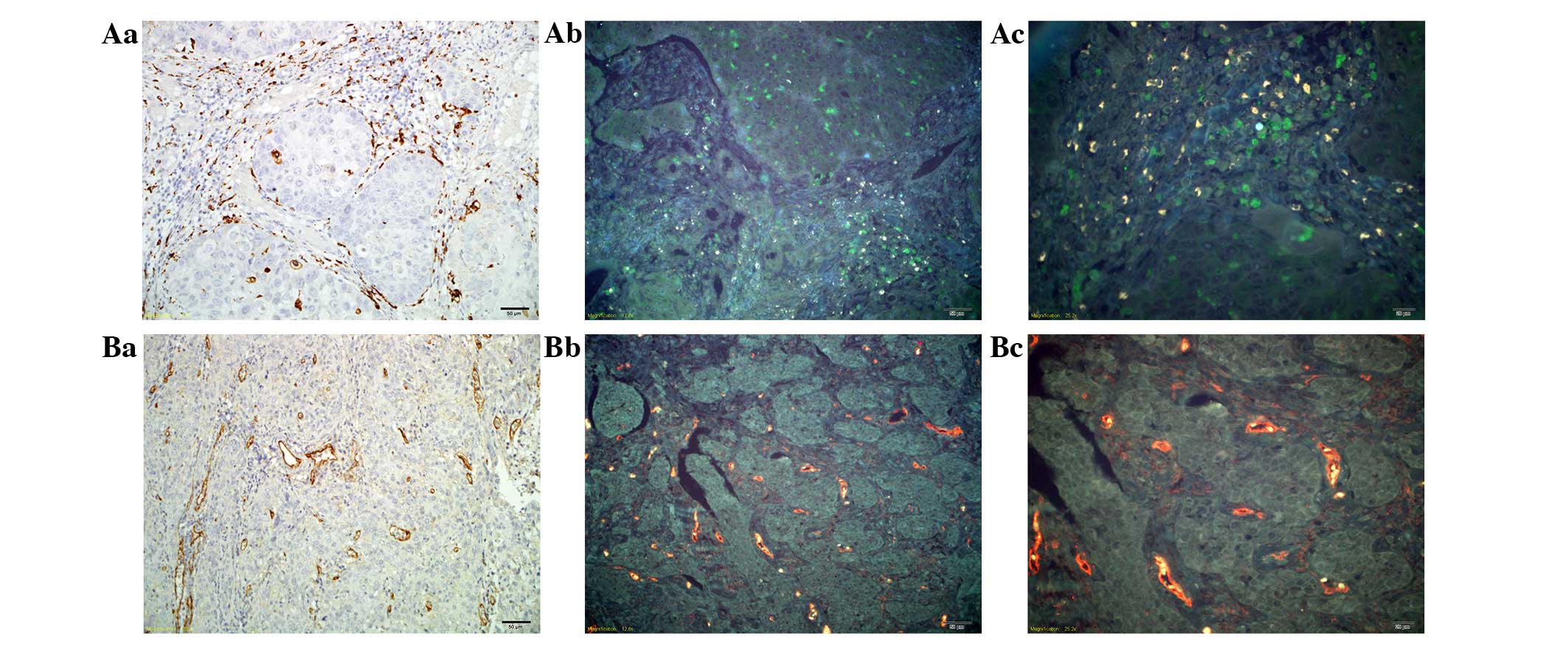
The present study first examined the expression of TAMs and tumor
neo-vessels using traditional IHC in CC tissue. Macrophages marked
by CD68 were stained brown on the cell membrane and in the
cytoplasm (Fig. 1Aa). Tumor
neo-vessels marked by CD105 were stained brown in tumor stroma, and
were mainly located around the cancer nests (Fig. 1Ba). Then, we used QDs-based probes to
image TAMs (Fig. 1Ab and Ac) and
tumor neo-vessels (Fig. 1Bb and Bc).
The QDs fluorescence specifically labeled TAMs with green color and
tumor neo-vessels with yellow color, without non-specific binding.
The background was tissue auto-fluorescence, which did not
interfere with the targets, but could assist in identifying tissue
morphology as a contrasting background. Compared with the results
of the IHC, the success of QD-based imaging was confirmed, as the
same regions of the tissue sections were marked.
Dynamic changes in the tumor
microenvironment during CC progression
The imaging of multiple molecular markers in tumor
and peritumoral tissues in situ can demonstrate the key
events of cancer invasion. Focus is placed on two major components
of cancer invasion: Monocyte infiltration represented by
macrophages (CD68) and tumor vascularization represented by CD105
(30). From normal tissue to invasive
CC tissue, the tumor microenvironment was presented as
significantly heterogeneous in terms of TAMs and tumor neo-vessels
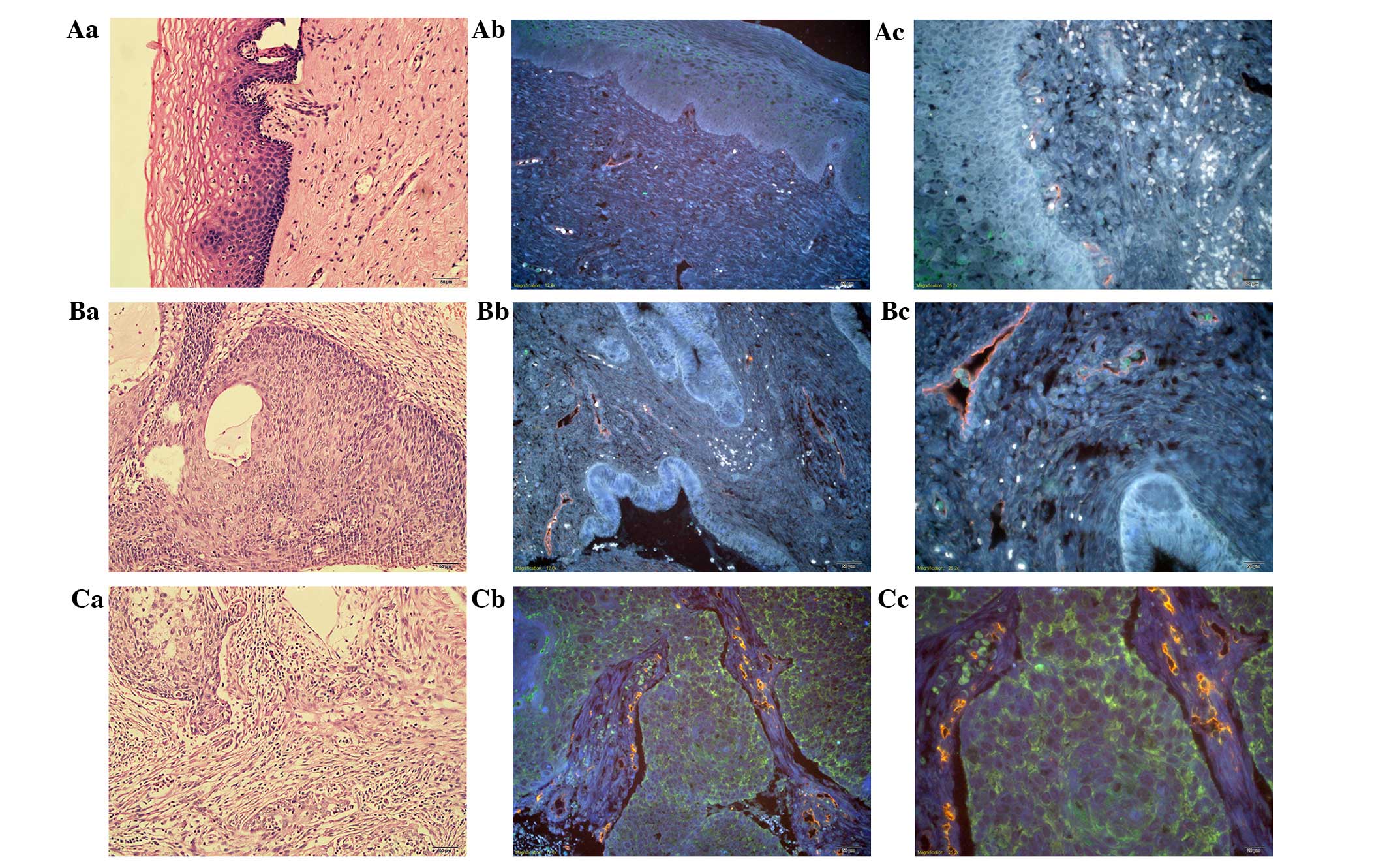
when assessed with QD-based nanoprobes. In normal tissue, there
were few macrophages under the basement membrane (BM), and few
regular and intact vessels with an intact and linear BM (Fig. 2Aa-Ac). In CC in situ tissue,
the cancer cells broke through the BM to form a micro-invasive
front; the TAMs presented mainly at the invasive front in the
stroma together with irregular and incomplete tumor neo-vessels
(Fig. 2Ba-Bc). In well-differentiated
invasive CC, the TAMs were extensively distributed among the cancer
cells and in the stroma, with prominent tumor neo-vessels
surrounding tumor nests (Fig.
2Ca-Cc). Compared with the well-differentiated invasive CC, in
the poorly-differentiated invasive CC, the TAMs were abundant with
a clustered morphology. Meanwhile, tumor neo-vessels were greater
in number compared with in the well-differentiated invasive CC and
were distributed in a scattered manner with an irregular diameter
and generally small morphology (Fig.
2Da-Dc). Overall, during the CC progression process from normal
tissue to invasive CC, it could be clearly observed that the TAMs
(green color) increased gradually in density in Fig. 2Ab→Bb→Cb→Db, as did the tumor
neo-vessels (yellow color) in Fig.
2Ac→Bc→Cc→Dc.
 | Figure 2.Dynamic changes in the tumor
microenvironment during CC progression. (Aa) H&E staining, (Ab)
TAM staining with QDs-525 and (Ac) tumor neo-vessel staining with
QD-585, in normal tissue. (Ba) H&E staining, (Bb) TAM staining
with QDs-525 and (Bc) tumor neo-vessel staining with QD-585, in CC
in situ tissue. (Ca) H&E staining, (Cb) TAM staining
with QD-525 and (Cc) tumor neo-vessel staining with QD-585, in
well-differentiated CC. (Da) H&E staining, (Db) TAM staining
with QD-525 and (Dc) tumor neo-vessel staining with QD-585, in
poorly-differentiated CC. Aa-Ac, Ba and Bb, Ca-Cc and Da-Dc:
Magnification, ×200; and Bc: Magnfication, ×400. TAMs,
tumor-associated macrophages; QD, quantum dot; CC, cervical cancer;
H&E, hematoxylin and eosin. |
QD-based double staining of TAMs and
tumor neo-vessels in situ
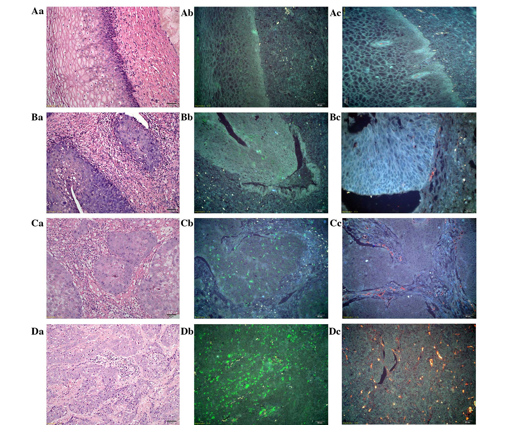
With the advantages of the multiplexed imaging of
QDs, TAMs and tumor neo-vessels were simultaneously visualized in
the tumor tissues. This method breaks the traditional technological
barriers to acquire multiple bio-information in situ and
made it possible to analyze the temporal-spatial process and
morphological characteristics of cancer invasion. In chronic
cervicitis tissue, TAMs were barely expressed and few vessels were
distributed at the BM with regular and conventional morphology
(Fig. 3Aa-Ac). In CC in situ
tissues, the density of the TAMs increased and they were
distributed in close vicinity to tumor neo-vessels or were found
lodging in vessels (Fig. 3Ba-Bc). In
invasive CC, the TAMs were scattered all around the tumor nests and
the stroma, with cluster aggregation, accompanied with rich tumor
angiogenesis (Fig. 3Ca-Cc). Based on
the simultaneous double staining of TAMs and tumor neo-vessels
in situ, it was clearly noted that the TAMs and tumor
angiogenesis increased simultaneously and were distributed in close
proximity during CC progression.
Quantitative analysis of TAMs and
tumor neo-vessels
A set of 90 patients with the three most common
pathological types of cervical disease, namely chronic cervicitis,
CIN I–III and CC, were selected, with 30 cases in each group. The
tissue slides from each patient were quantitatively analyzed for
TAM density and tumor neo-vessel number. According to the
quantitative analysis results, the median value of TAM density was
higher in the CC group (5,540.14) than in the CIN I–III group
(2,502.17) and the chronic cervicitis group (1,403.31), with
statistical significance in all three groups (P<0.001, for
between-group comparisons). The number of neo-vessels was also much
higher in the CC group (n=27) than in the CIN I–III group (n=17) or
the chronic cervicitis group (n=6.5), with statistical significance
in all three groups (P<0.001, for between-group comparisons). In
addition, during the progression process from chronic cervicitis to
CIN I–III to CC, the TAM density and the number of tumor
neo-vessels gradually increased and were distributed in close
proximity.
Discussion
Tumor invasion and metastasis is a multi-factor,
multi-stage, gradually developing and complex dynamic process
(33,34), involving not only the abnormal
expression of a variety of genes of the cancer cells themselves and
the activation of multiple signaling pathways, but also the
interaction between tumor cells and the surrounding
microenvironment (35). The
components of the tumor microenvironment are numerous, and are
composed of stromal cells and interstitial cells, including
fibroblasts, endothelial cells, infiltrating immune cells and
structural support collagen (36).
Tumor inflammation and tumor angiogenesis are considered
indispensible for cancer invasion. The present study indicated that
the tumor microenvironment plays a significant role in cancer
progression, involving immune cell infiltration and tumor
angiogenesis. Endothelial cells and immunocytes in the
microenvironment around the tumor cells can promote the progression
of cancer (4,37). The infiltration of macrophages
distributed in the surrounding tumor tissues is the most important
ingredients of immune infiltration research (38). Therefore, they are also known as
‘tumor-associated macrophages’, infiltrating the peripheral
vasculature or intravasculature, which are important contact media
between inflammation and cancer (39). In addition, the process of tumor
angiogenesis is another crucial hallmark of cancer progression.
Tumor tissue must be supplied with nutrients from the blood vessels
in order to grow (40). Typically,
tumor neovessels are disordered with high permeability, which
results in hypoperfusion. Therefore, tumor angiogenesis is usually
incomplete and of inferior maturity, which could provide strong
conditions for tumor invasion and metastasis (41). Recent studies have showed that
macrophages express a series of factors to promote tumor
progression through the occurrence of hypoxia, angiogenesis, immune
suppression and metastasis (42). It
was shown that the macrophages infiltrated into tumor tissues and
adjacent tissues, mainly in the junctions between cancer nests and
interstitial cells, particularly in the places where the blood
vessels were rich, which is consistent with the present findings.
During the CC invasion progress, the TAMs and tumor neo-vessels
simultaneously increased surrounding the cancer nests and were
located in close proximity. Macrophages can also secrete vascular
endothelial growth factor and other factors to promote the
formation of vascular endothelial cells (43) and the sprouting of blood vessels
through structural support, so as to increase the tumor vascular
network (44). Thus, the
combinational analysis of TAMs and tumor neo-vessels could better
reveal the cancer invasion biology and guide clinical strategy.
However, current methods for tumor invasion and
metastasis research, including polymerase chain reaction, western
blotting and IHC, could be used to perform qualitative and
semi-quantitative research. However, these methods cannot show the
spatial distribution of the key components during the development
in situ within cancer cells (45,46). In
addition, given the complex compositions and functions of the tumor
microenvironment, the single-molecule research techniques, such as
conventional IHC, are difficult to use to accurately reflect the
interactions between cancer cells and the tumor microenvironment.
While one component such as the TAMs could be observed directly at
one time, there must be another imaging procedure to produce the
image for tumor neo-vessels. The shortcoming of these conventional
techniques is that subtle essential information on the major
components of the tumor microenvironment cannot be obtained
simultaneously, thus leading to information loss and low resolution
of different components in situ (34). Therefore, in order to solve this
problem, there is an urgent requirement to develop multi-situ
molecular imaging techniques so as to better guide the clinical
treatment.
QD-based molecular imaging and multispectral
analysis could make a unique contribution in this regard, as the
current study demonstrated. QDs are engineered nanoparticles with
unique optical properties and have shown great potential for
biomedical application. Compared with organic dyes and fluorescent
proteins, due to its unique size and surface features, QDs have
many advantages such as enhanced fluorescence brightness and strong
resistance to photobleaching. In addition, different QDs can be
excited simultaneously by a single light source, which provides
significant advantages for the multiplexed detection of targets,
suitable for investigating the several key components in tumor
microenvironment (27). In the
present study, with the aid of QD-based in situ
two-component imaging of the tumor microenvironment, the TAM and
CD105 levels gradually and significantly increased after
quantitative analysis, and the TAMs were found to be scattered all
around the tumor nests and the stroma with cluster aggregation,
accompanied with rich tumor angiogenesis during the progression
process from chronic cervicitis and CIN I–III to invasive CC.
Therefore, instead of producing artificial overlay images by
conventional imaging techniques, this QD-based molecular imaging
technique could provide authentic multicolor images to better
reveal the complex tumor microenvironment in cancer tissues.
In conclusion, the present study combined the
advantages of QD-based multiplexed imaging in situ to detect
macrophages and tumor angiogenesis in chronic cervicitis, cervical
CIN I–III and CC tissues, emphasizing the importance of the overall
analysis of the tumor microenvironment during cancer invasion and
metastasis, and enhancing the understanding of the process of tumor
invasion. The findings demonstrated that the TAMs and tumor
neo-vessels together promoted cancer invasion. It was also
suggested that a simple one-component study based on cancer cells
or the tumor microenvironment is insufficient to fully reflect the
cancer invasion process. Therefore, there is an urgent requirement
for the development of in situ multi-component imaging
technology and an image analysis method.
Acknowledgements
The authors would like to acknowledge the financial
support provided by the Natural Science Foundation of Hubei
Province (grant no. 2013CFB374).
References
|
1
|
Siegel R, Naishadham D and Jernal A:
Cancer statistics, 2013. CA Cancer J Clin. 63:11–30. 2013.
View Article : Google Scholar : PubMed/NCBI
|
|
2
|
Limmer K, LoBiondo-Wood G and Dains J:
Predictors of cervical cancer screening adherence in the United
States: A systematic review. J Adv Pract Oncol. 5:31–41.
2014.PubMed/NCBI
|
|
3
|
Bizzarri M and Cucina A: Tumor and the
microenvironment: A Chance to reframe the paradigm of
carcinogenesis? Biomed Res Int 2014. 9340382014.
|
|
4
|
Polyak K, Haviv I and Campbell IG:
Co-evolution of tumor cells and their microenvironment. Trends
Genet. 25:30–38. 2009. View Article : Google Scholar : PubMed/NCBI
|
|
5
|
Paget S: The distribution of secondary
growths in cancer of the breast. Lancet. 133:571–573. 1889.
View Article : Google Scholar
|
|
6
|
Sun T, Yang Y, Luo X, Cheng Y, Zhang M,
Wang K and Ge C: Inhibition of tumor angiogenesis by interferon-γ
by suppression of tumor-associated macrophage differentiation.
Oncol Res. 21:227–235. 2014. View Article : Google Scholar : PubMed/NCBI
|
|
7
|
Liu Q, Zhang A, Xu W and Dong J: A new
view of the roles of blood flow dynamics and Kupffer cell in
intra-hepatic metastasis of hepatocellular carcinoma. Med
Hypotheses. 77:87–90. 2011. View Article : Google Scholar : PubMed/NCBI
|
|
8
|
Lewis CE and Pollard JW: Distinct role of
macrophages in different tumor microenvironments. Cancer Res.
66:605–612. 2006. View Article : Google Scholar : PubMed/NCBI
|
|
9
|
Gordon S and Taylor PR: Monocyte and
macrophage heterogeneity. Nat Rev Immunol. 5:953–964. 2005.
View Article : Google Scholar : PubMed/NCBI
|
|
10
|
Biswas SK and Mantovani A: Macrophage
plasticity and interaction with lymphocyte subsets: Cancer as a
paradigm. Nat Immunol. 11:889–896. 2010. View Article : Google Scholar : PubMed/NCBI
|
|
11
|
Budhu A, Forgues M, Ye QH, Jia HL, He P,
Zanetti KA, Kammula US, Chen Y, Qin LX, Tang ZY and Wang XW:
Prediction of venous metastases, recurrence, and prognosis in
hepatocellular carcinoma based on a unique immune response
signature of the liver microenvironment. Cancer Cell. 10:99–111.
2006. View Article : Google Scholar : PubMed/NCBI
|
|
12
|
Vakkila J and Lotze MT: Inflammation and
necrosis promote tumour growth. Nat Rev Immunol. 4:641–648. 2004.
View Article : Google Scholar : PubMed/NCBI
|
|
13
|
Qian BZ and Pollard JW: Macrophage
diversity enhances tumor progression and metastasis. Cell.
141:39–51. 2010. View Article : Google Scholar : PubMed/NCBI
|
|
14
|
Mantovani A, Sozzani S, Locati M, Allavena
P and Sica A: Macrophage polarization: Tumor-associated macrophages
as a paradigm for polarized M2 mononuclear phagocytes. Trends
Immunol. 23:549–555. 2002. View Article : Google Scholar : PubMed/NCBI
|
|
15
|
Gao B, Jeong WI and Tian Z: Liver: An
organ with predominant innate immunity. Hepatology. 47:729–736.
2008. View Article : Google Scholar : PubMed/NCBI
|
|
16
|
Qian BZ and Pollard JW: New tricks for
metastasis-associated macrophages. Breast Cancer Res. 14:3162012.
View Article : Google Scholar : PubMed/NCBI
|
|
17
|
Kuang DM, Zhao Q, Peng C, Xu J, Zhang JP,
Wu C and Zheng L: Activated monocytes in peritumoral stroma of
hepatocellular carcinoma foster immune privilege and disease
progression through PD-L1. J Exp Med. 206:1327–1337. 2009.
View Article : Google Scholar : PubMed/NCBI
|
|
18
|
Zhang JP, Yan J, Xu J, Pang XH, Chen MS,
Li L, Wu C, Li SP and Zheng L: Increased intratumoral
IL-17-producing cells correlate with poor survival in
hepatocellular carcinoma patients. J Hepatol. 50:980–989. 2009.
View Article : Google Scholar : PubMed/NCBI
|
|
19
|
Yu D, Zhuang L, Sun X, Chen J, Yao Y, Meng
K and Ding Y: Particular distribution and expression pattern of
endoglin (CD105) in the liver of patients with hepatocellular
carcinoma. BMC Cancer. 7:1222007. View Article : Google Scholar : PubMed/NCBI
|
|
20
|
Folkman J: Tumor angiogenesis: Therapeutic
implications. N Engl J Med. 285:1182–1186. 1971. View Article : Google Scholar : PubMed/NCBI
|
|
21
|
Devy L, Huang L, Naa L, Yanamandra N,
Pieters H, Frans N, Chang E, Tao Q, Vanhove M, Lejeune A, et al:
Selective inhibition of matrix metalloproteinase-14 blocks tumor
growth, invasion, and angiogenesis. Cancer Res. 69:1517–1526. 2009.
View Article : Google Scholar : PubMed/NCBI
|
|
22
|
Guzmán-Medrano R, Arreola-Rosales RL,
Shibayama M, Silva-Olivares DA, Bologna-Molina R and Rodríguez MA:
Tumor-associated macrophages and angiogenesis: A statistical
correlation that could reflect a critical relationship in
ameloblastoma. Pathol Res Pract. 208:672–676. 2012. View Article : Google Scholar : PubMed/NCBI
|
|
23
|
Zhu JY, Sun QK, Wang W and Jia WD:
High-level expression of HOXB13 is closely associated with tumor
angiogenesis and poor prognosis of hepatocellular carcinoma. Int J
Clin Exp Pathol. 7:2925–2933. 2014.PubMed/NCBI
|
|
24
|
Resch-Genger U, Grabolle M,
Cavaliere-Jaricot S, Nitschke R and Nann T: Quantum dots versus
organic dyes as fluorescent labels. Nat Methods. 5:763–775. 2008.
View Article : Google Scholar : PubMed/NCBI
|
|
25
|
Madani SY, Shabani F, Dwek MV and
Seifalian AM: Conjugation of quantum dots on carbon nanotubes for
medical diagnosis and treatment. Int J Nanomedicine. 8:941–950.
2013.PubMed/NCBI
|
|
26
|
Chen C, Peng J, Sun SR, Peng CW, Li Y and
Pang DW: Tapping the potential of quantum dots for personalized
oncology: Current status and future perspectives. Nanomedicine
(Lond). 7:411–428. 2012. View
Article : Google Scholar : PubMed/NCBI
|
|
27
|
Fang M, Peng CW, Pang DW and Li Y: Quantum
dots for cancer research: Current status, remaining issues, and
future perspectives. Cancer Biol Med. 9:151–163. 2012.PubMed/NCBI
|
|
28
|
Hoshino A, Manabe N, Fujioka K, Suzuki K,
Yasuhara M and Yamamoto K: Use of fluorescent quantum dot
bioconjugates for cellular imaging of immune cells, cell organelle
labeling, and nanomedicine: Surface modification regulates
biological function, including cytotoxicity. J Artif Organs.
10:149–157. 2007. View Article : Google Scholar : PubMed/NCBI
|
|
29
|
Chen LD, Liu J, Yu XF, He M, Pei XF, Tang
ZY, Wang QQ, Pang DW and Li Y: The biocompatibility of quantum dot
probes used for the targeted imaging of hepatocellular carcinoma
metastasis. Biomaterials. 29:4170–4176. 2008. View Article : Google Scholar : PubMed/NCBI
|
|
30
|
Fang M, Peng CW, Yuan JP, Zhang ZL, Pang
DW and Li Y: Coevolution of the tumor microenvironment revealed by
quantum dot-based multiplexed imaging of hepatocellular carcinoma.
Future Oncol. 9:1029–1037. 2013. View Article : Google Scholar : PubMed/NCBI
|
|
31
|
Chen C, Peng J, Xia HS, Yang GF, Wu QS,
Chen LD, Zeng LB, Zhang ZL, Pang DW and Li Y: Quantum dots-based
immunofluorescence technology for the quantitative determination of
HER2 expression in breast cancer. Biomaterials. 30:2912–2918. 2009.
View Article : Google Scholar : PubMed/NCBI
|
|
32
|
Peuscher A, Gassler N, Schneider U, Thom
P, Rasche S, Spiegel H and Schillberg S: An immunohistochemical
assay on human tissue using a human primary antibody. J Immunoassay
Immunochem. 35:322–334. 2014. View Article : Google Scholar : PubMed/NCBI
|
|
33
|
Sulzmaier FJ and Ramos JW: RSK isoforms in
cancer cell invasion and metastasis. Cancer Res. 73:6099–6105.
2013. View Article : Google Scholar : PubMed/NCBI
|
|
34
|
Fang M, Yuan JP, Peng CW, Pang DW and Li
Y: Quantum dots-based in situ molecular imaging of dynamic changes
of collagen IV during cancer invasion. Biomaterials. 34:8708–8717.
2013. View Article : Google Scholar : PubMed/NCBI
|
|
35
|
Friedl P and Alexander S: Cancer invasion
and the microenvironment: Plasticity and reciprocity. Cell.
147:992–1009. 2011. View Article : Google Scholar : PubMed/NCBI
|
|
36
|
Nguyen-Ngoc KV, Cheung KJ, Brenot A,
Shamir ER, Gray RS, Hines WC, Yaswen P, Werb Z and Ewald AJ: ECM
microenvironment regulates collective migration and local
dissemination in normal and malignant mammary epithelium. Proc Natl
Acad Sci USA. 109:E2595–E2604. 2012. View Article : Google Scholar : PubMed/NCBI
|
|
37
|
Zou W and Restifo NP: T(H)17 cells in
tumour immunity and immunotherapy. Nat Rev Immunol. 10:248–256.
2010. View
Article : Google Scholar : PubMed/NCBI
|
|
38
|
Emri E, Egervari K, Varvolgyi T, Rozsa D,
Miko E, Dezso B, Veres I, Mehes G, Emri G and Remenyik E:
Correlation among metallothionein expression, intratumoural
macrophage infiltration and the risk of metastasis in human
cutaneous malignant melanoma. J Eur Acad Dermatol Venereol.
27:e320–e327. 2013. View Article : Google Scholar : PubMed/NCBI
|
|
39
|
Mantovani A and Locati M: Tumor-associated
macrophages as a paradigm of macrophage plasticity, diversity, and
polarization: Lessons and open questions. Arterioscler Thromb Vasc
Biol. 33:1478–1483. 2013. View Article : Google Scholar : PubMed/NCBI
|
|
40
|
Folkman J: Angiogenesis. Annu Rev Med.
57:1–18. 2006. View Article : Google Scholar : PubMed/NCBI
|
|
41
|
Rolny C, Mazzone M, Tugues S, Laoui D,
Johansson I, Coulon C, Squadrito ML, Segura I, Li X, Knevels E, et
al: HRG inhibits tumor growth and metastasis by inducing macrophage
polarization and vessel normalization through downregulation of
PlGF. Cancer Cell. 19:31–44. 2011. View Article : Google Scholar : PubMed/NCBI
|
|
42
|
Luo Y, Zhou H, Krueger J, Kaplan C, Lee
SH, Dolman C, Markowitz D, Wu W, Liu C, Reisfeld RA and Xiang R:
Targeting tumor-associated macrophages as a novel strategy against
breast cancer. J Clin Invest. 116:2132–2141. 2006. View Article : Google Scholar : PubMed/NCBI
|
|
43
|
Stockmann C, Doedens A, Weidemann A, Zhang
N, Takeda N, Greenberg JI, Cheresh DA and Johnson RS: Deletion of
vascular endothelial growth factor in myeloid cells accelerates
tumorigenesis. Nature. 456:814–818. 2008. View Article : Google Scholar : PubMed/NCBI
|
|
44
|
Fantin A, Vieira JM, Gestri G, Denti L,
Schwarz Q, Prykhozhij S, Peri F, Wilson SW and Ruhrberg C: Tissue
macrophages act as cellular chaperones for vascular anastomosis
downstream of VEGF-mediated endothelial tip cell induction. Blood.
116:829–840. 2010. View Article : Google Scholar : PubMed/NCBI
|
|
45
|
Chen C, Sun SR, Gong YP, Qi CB, Peng CW,
Yang XQ, Liu SP, Peng J, Zhu S, Hu MB, et al: Quantum dots-based
molecular classification of breast cancer by quantitative
spectroanalysis of hormone receptors and HER2. Biomaterials.
32:7592–7599. 2011. View Article : Google Scholar : PubMed/NCBI
|
|
46
|
Peng CW, Tian Q, Yang GF, Fang M, Zhang
ZL, Peng J, Li Y and Pang DW: Quantum-dots based simultaneous
detection of multiple biomarkers of tumor stromal features to
predict clinical outcomes in gastric cancer. Biomaterials.
33:5742–5752. 2012. View Article : Google Scholar : PubMed/NCBI
|