Introduction
Lung carcinoma is the leading malignant carcinoma
with the highest mortality rate worldwide (1). The main pathological types of lung
carcinoma are small cell lung carcinoma (~15%) and non-small cell
lung carcinoma (NSCLC) (~85%). The common pathological types of
NSCLC are lung squamous cell carcinoma (~40%), lung adenocarcinoma
(~40%) and lung large-cell carcinoma (~10%) (2). Although the use of targeted agents in
recent years has increased the survival rate of the NSCLC, it
remains the severest malignant carcinoma with the poorest treatment
effects (3).
Determination of the molecular mechanism underlying
the occurrence and development of NSCLC, and the development of new
therapeutic targets are important means for improving its poor
therapeutic effect and low survival rate. Stem cell core
transcription factor SOX2 is a member of the SOX (SRY-like HMG box)
gene family, which includes HMG in the DNA-binding zone and plays
an important role in maintaining the self-renewal,
multi-differentiation and reprogramming of stem cells (4–6). The
abnormal expression of SOX2 is associated with the occurrence and
development of many malignant carcinomas, such as esophageal,
breast, gastric and colon carcinoma (7–10).
Previous findings showed that SOX2 is overexpressed in NSCLC, and
its positive rate in lung squamous cell carcinoma is significantly
higher than that in lung adenocarcinoma. However, the correlation
of the overexpression of SOX2 and the prognosis of NSCLC remains
controversial (11–14).
By means of immunohistochemical analysis, the aim of
the present study was to examine the expression of SOX2 in the
NSCLC of Chinese individuals and analyze the correlation of SOX2
expression, clinicopathological factors and prognosis.
Patients and methods
Patients and samples
A total of 127 patients with NSCLC that accepted
radical resection of pulmonary carcinoma from January, 2007 to
December, 2008 were included in the present study. None of the
patients received neoadjuvant chemotherapy. Of the 127 patients,
there were 101 males and 26 females of age between 39 and 75 years,
with an average age of 57 years. All 127 patients were followed-up
for >60 months. Paraffin specimens of carcinoma and
para-carcinoma tissues were collected from the patients for tissue
microarray. Each piece of tissue was set with three 2 mm repeat
points. After hematoxylin and eosin staining, the specimens were
analyzed and diagnosed by two chief physicians of the pathology
department. In addition to tissue microarray, lung adenosquamous
carcinoma tissues were also subjected to paraffin section and
immunohistochemical staining. In the course of
immunohistochemistry, no carcinoma tissue shed and 5 cases of
carcinoma adjacent tissues shed.
Immunohistochemistry
The SOX2 antibody was purchased from Bethyl
Laboratories, Inc. (A301-741A; Montgomery, TX, USA).
Immunohistochemical reagent (PV 9000 and DAB) coloring solution was
purchased from Beijing Zhongshan Golden Bridge Biotechnology Co.,
Ltd. (Beijing, China). Tissue sections were dewaxed in xylene,
hydrated in gradient ethanol (100, 95, 90, 85, 80 and 75%), placed
in 3% hydrogen peroxide and incubated for 10 min to block
endogenous peroxidase. Subsequently, the sections were rinsed with
phosphate-buffered saline 3 times, and incubated with 5% goat serum
at 25°C for 30 min, followed by incubation with rabbit polyclonal
SOX2 primary antibody (Abcam, Cambridge, MA, USA; catalog no.
ab97959; dilution of 1:500) at 4°C overnight. The following day,
the sections were incubated with PV 9000 at room temperature, and
color was developed using DAB solution. After rinsing the slides in
deionized water, the sections were counterstained with hematoxylin
and decolorized with 1% hydrochloric acid, returned to blue
following 1% ammonia and dehydrated with gradient ethanol before
sealing.
Interpretation of immunohistochemical
results
Tissue microarray staining results were read by two
chief physicians from the Department of Pathology, Fujian
Provincial Cancer Hospital (Fuzhou, China), independently.
According to the staining degree of the nucleus, the sections were
assigned a score of 0–4. According to the percentage of positive
cells, the sections were scored as 0 (≤5%), 1 (6–25%), 2 (26–50%),
3 (5l-75%), and 4 (76–100%). According to the comprehensive scores
of staining intensity and positive cell number, the sections were
numbered as ‘0’, indicating negative (−); ‘1–4’, indicating
positive (+); ‘5–7’, indicating positive (++); ≥8, indicating
positive (+++).
Statistical analysis
SPSS 18.0 statistical software (SPSS, Inc., Chicago,
IL, USA) was used for statistical analysis. SOX2 expression and
analysis of the clinicopathological factors were performed using
the χ2 test. Survival analysis was performed using the
Kaplan-Meier method and log-rank test. COX model was used to
determine the multi-factor regression analysis of prognosis-related
factors. P<0.05 was considered to indicate a statistically
significant difference.
Results
Expression of SOX2 in NSCLC tissues
was higher than that in para-carcinoma tissues
The expression of SOX2 in NSCLC tissues showed 29
cases (22.8%) of negative (−) expression, 53 cases (41.7%) of
positive (+) expression, 28 cases (22.8%) of positive (++)
expression, and 16 cases (12.6%) of positive (+++) expression. The
expression of SOX2 in para-carcinoma tissues showed 74 cases
(60.7%) of negative (−) expression, 45 cases (36.9%) of positive
(+) expression, 3 cases (2.5%) of positive (++) expression, and 0
case of positive (+++) expression. Positive (++) and (+++) was
considered a high SOX2 expression, and negative (−) and positive
(+) were considered a low SOX2 expression. The high expression rate
of SOX2 in NSCLC tissues was significantly higher than that in
para-carcinoma tissues (Table I). The
difference was statistically significant (P<0.001).
 | Table I.Staining degree of SPX3 in non-small
cell lung carcinoma and para-carcinoma tissues [case (%)]. |
Table I.
Staining degree of SPX3 in non-small
cell lung carcinoma and para-carcinoma tissues [case (%)].
| Group | Case | Weakly positive | Strongly
positive | P-value |
|---|
| Carcinoma | 127 | 82 (64.6%) | 45 (35.4%) | <0.001 |
| Normal mucosa | 122 | 119 (97.6%) | 3 (2.5%) |
|
Expression of SOX2 in NSCLC tissues
was significantly correlated with the pathological type of
carcinoma
The expression level of SOX2 was not associated with
gender, age, smoking history or TNM stage, but was significantly
associated with the pathological type of carcinoma. The high
expression rate of SOX2 in lung squamous cell carcinoma was 50%
(25/50) and in lung adenocarcinoma was 20.3% (12/59) (Table II), indicating that SOX2 high
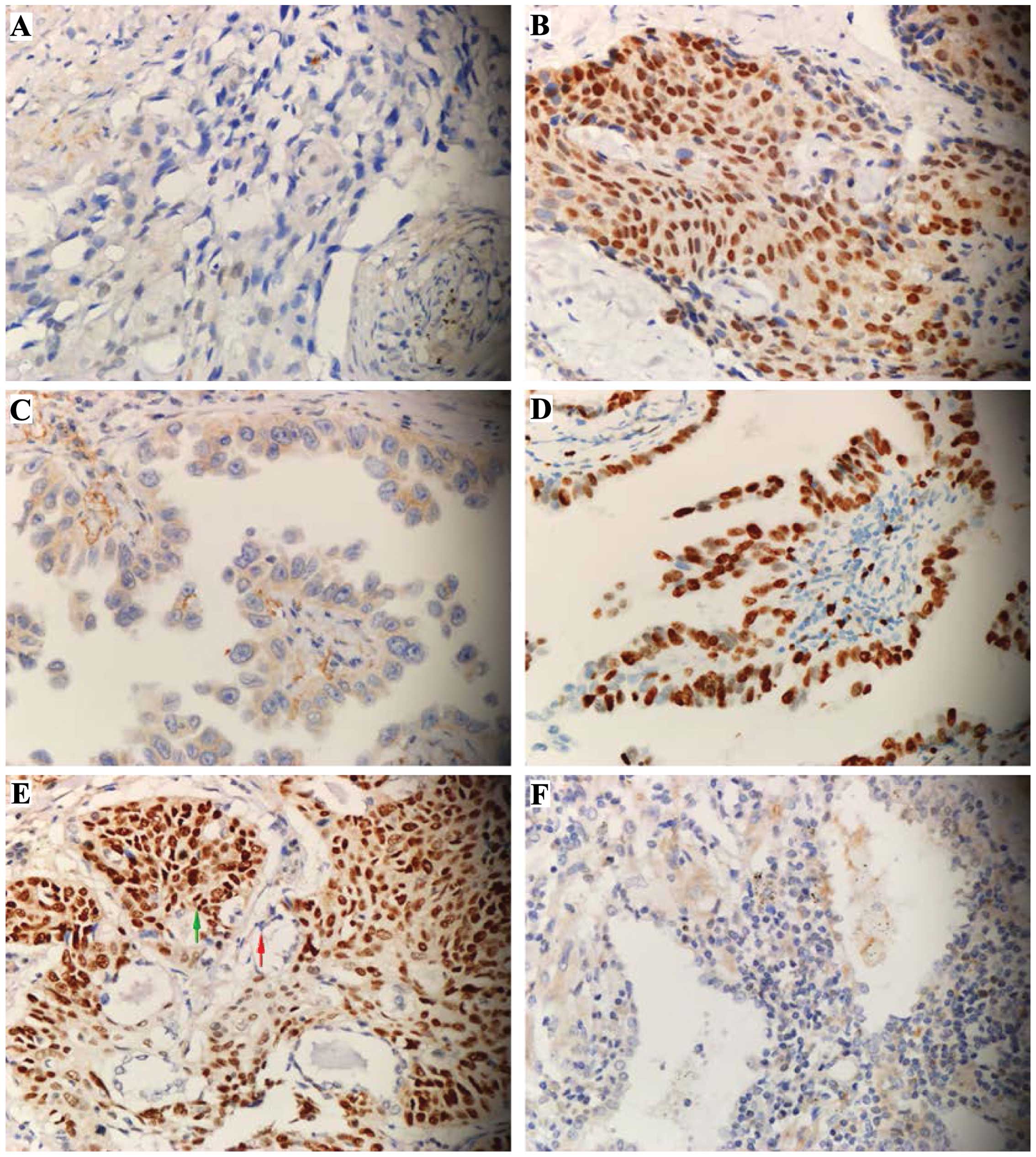
expression mainly occurred in lung squamous cell carcinoma. Of the
18 cases of high expression of lung adenosquamous carcinoma, SOX2
showed a high expression in the adenocarcinoma and squamous cell
carcinoma cells of 1 adenosquamous carcinoma tissue. SOX2 also
showed a high expression in squamous carcinoma cells and low
expression in adenocarcinoma cells of 7 tissues. By contrast, SOX2
showed a low expression in adenocarcinoma and squamous cell
carcinoma cells of 10 tissues (Fig.
1), confirming that the expression level of SOX2 was
significantly associated with the pathological type of carcinoma.
Additionally, the high expression rate of squamous cell carcinoma
cells was significantly higher than that of adenocarcinoma
cells.
| Table II.Correlation between SOX2 expression
and clinicopathological factors in patients with non-small cell
lung carcinoma [case (%)]. |
Table II.
Correlation between SOX2 expression
and clinicopathological factors in patients with non-small cell
lung carcinoma [case (%)].
| Variables | Case | SOX2 low
expression | SOX2 high
expression | P-value |
|---|
| Gender |
|
|
|
|
| Male | 101 | 61 (74.4) | 40 (88.9) | 0.066 |
|
Female | 26 | 21 (25.6) | 5
(11.1) |
|
| Age |
|
|
|
|
| ≥57
years | 66 | 40 (48.8) | 26 (57.8) | 0.358 |
| <57
years | 61 | 42 (51.2) | 19 (42.2) |
|
| Smoking history |
|
|
|
|
| Yes | 56 | 32 (39.0) | 24 (53.3) | 0.138 |
| No | 71 | 50 (61.0) | 21 (46.7) |
|
| Pathological
type |
|
|
|
|
|
Adenocarcinoma | 59 | 47 (57.3) | 12 (26.7) | 0.004 |
| Squamous
carcinoma | 50 | 25 (30.5) | 25 (55.6) |
|
|
Adenosquamous carcinoma | 18 | 10 (12.2) | 8
(17.8) |
|
| T staging |
|
|
|
|
| T1 | 8 | 4 (4.9) | 4 (8.9) | 0.273 |
| T2 | 90 | 62 (75.6) | 28 (62.2) |
|
| T3 | 29 | 16 (19.5) | 13 (28.9) |
|
| N staging |
|
|
|
|
| N0 | 47 | 31 (37.8) | 16 (35.6) | 0.403 |
| N1 | 31 | 17 (20.7) | 14 (31.1) |
|
| N2 | 49 | 34 (41.5) | 15 (33.3) |
|
| TNM staging |
|
|
|
|
| I | 38 | 26 (31.7) | 12 (26.7) | 0.570 |
| II | 30 | 17 (20.7) | 13 (28.9) |
|
| III | 59 | 39 (47.6) | 20 (44.4) |
|
Expression of SOX2 in NSCLC was
correlated with the survival of patients
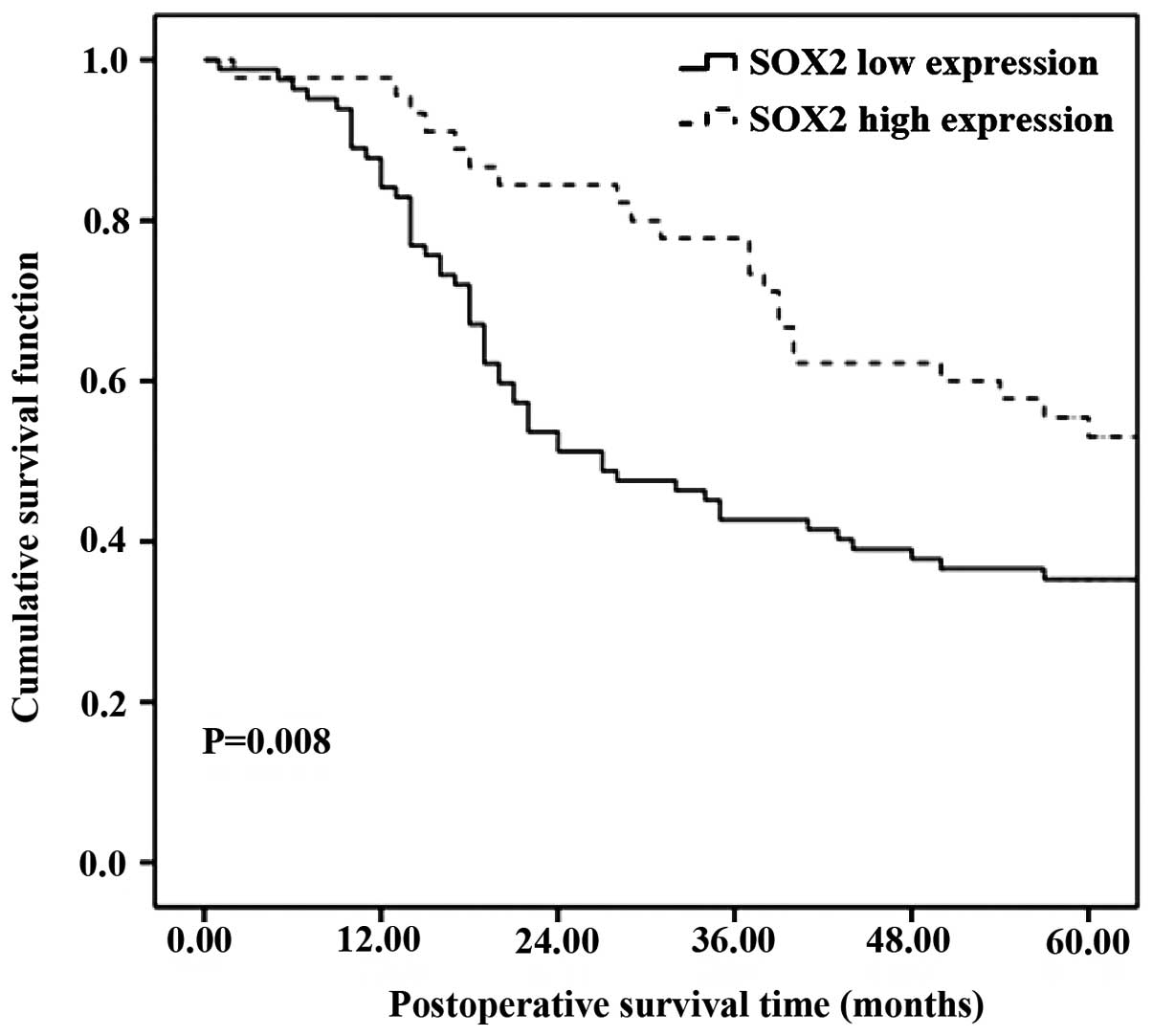
The Kaplan-Meier method was applied to plot the
survival curve, and the log-rank test and COX multiple regression
model were applied for estimation of the survival analysis. The
prognosis of patients with a high SOX2 expression was significantly
better than those with a low SOX2 expression (Fig 2). The COX multiple regression analysis
revealed that T and N staging of the carcinoma was the same,
indicating that the expression level of SOX2 was an independent
prognostic factor of patients with NSCLC (P<0.001) (Table III).
| Table III.SOX2 expression level was an
independent prognostic factor (COX multivariate regression
analysis). |
Table III.
SOX2 expression level was an
independent prognostic factor (COX multivariate regression
analysis).
| Factors | Relative risk (95%
CI) | P-value |
|---|
| Gender
(male/female) | 0.795
(0.416–1.519) |
0.487 |
| Age (≥57/<57
years) | 1.158
(0.668–2.006) |
0.601 |
| Pathological type
(adenocarcinoma/adenosquamous carcinoma)a | 1.325
(0.945–1.859) |
0.102 |
| T staging
(T3/T2/T1) | 1.610
(1.004–2.584) |
0.048 |
| N staging
(N2/N1/N0) | 2.036
(1.527–2.714) | <0.001 |
| SOX2 expression (high
expression/low expression) | 0.380
(0.223–0.649) | <0.001 |
Discussion
SOX2 protein is an important role in cell cycle
regulation, DNA damage repair and maintaining the totipotency of
stem cells. In many malignant carcinomas, the high expression of
SOX2 is associated with the invasion and poor prognosis of
carcinoma (7,8,16).
However, at present, detailed information on the expression of SOX2
in the Chinese population of NSCLC is lacking. Previous studies
have suggested that the occurrence and therapeutic targets of lung
carcinoma in different ethnic groups are varied (17,18). In
this study, we mainly focused on examining the association of SOX2
expression and clinicopathological factors and prognosis of NSCLC
of Chinese patients. The results of the present study showed that
the expression of SOX2 in NSCLC tissues was higher than that in the
para-carcinoma tissues and a high SOX2 expression mainly occurred
in lung squamous cell carcinoma cells. The study results were
consistent with those of European and Japanese investigators
(11,13).
The results of our study also show that of the 18
cases of adenosquamous carcinoma, the positive rate of SOX2
expression in adenocarcinoma cells was higher than that in squamous
cell carcinoma cells. The 7 cases showed a high SOX2 expression in
squamous carcinoma cells and a low expression in adenocarcinoma
cells, further confirming that the expression level of SOX2 was
significantly associated with the pathological type of carcinoma.
Additionally, SOX2 is a potential molecular marker for the
diagnosis of lung squamous cell carcinoma. The study of Sasaki
et al showed that the DNA copy number of SOX2 gene
was increased significantly in lung squamous cell carcinoma, but no
significant changes were found in lung adenocarcinoma (11), which indicated that the overexpression
of SOX2 in lung squamous cell carcinoma may be caused by the
increase of DNA copy number and the overexpression of SOX2 in some
lung adenocarcinoma may be caused by other mechanisms, such as
transcription level and post-ßtranscriptional modifications.
At present, the regulatory mechanism of SOX2 in
carcinoma cells is not entirely clear. Existing research results
have shown that as a nuclear transcription factor, SOX2 can induce
the expression of squamous carcinoma labeled carcinoma-associated
factor p63 and keratin 6, and activate the downstream epidermal
growth factor receptor and insulin-like growth factor-1 signals, to
promote the proliferation, invasion and migration of carcinoma
cells (7,8,16).
Consequently, the high expression of SOX2 protein may lead to poor
prognosis. However, consensus on the correlation of the expression
of SOX2 and the prognosis of NSCLC is lacking. Sholl et al
(12) suggested that SOX2
overexpression was associated with the poor prognosis of stage I
lung adenocarcinoma, whereas the study of Sasaki et al
showed that there was no significant correlation of the SOX2
expression and prognosis of lung squamous cell carcinoma (11). Wilbertz et al (14) suggested that the expression of SOX2
was associated with the prognosis of lung squamous cell carcinoma,
and this expression suggested an improved prognosis. In a study
using a large sample, Velcheti et al (13) found that SOX2 overexpression in NSCLC
(squamous cell carcinoma and adenocarcinoma) was more favorable for
prognosis than a low SOX2 expression, a finding which was
consistent with the results of the present study. Nevertheless,
since the regulation mechanism of SOX2 in carcinoma cells is not
very clear, this result lacks a convincing explanation. In the
present study as well as that of Velcheti et al (13), the majority of the subjects were in
stage II and III and underwent chemotherapy and/or radiotherapy
after surgery. In the study of Sholl et al (12), where patients were primarily in stage
I and did not undergo chemotherapy and/or radiotherapy after
surgery, the opposite result was obtained. Thus, a better prognosis
caused by SOX2 overexpression may be associated with the
sensitivity of lung carcinoma cells to chemotherapy. The present
study and that of Velcheti et al are retrospective studies,
the postoperative adjuvant treatment plan was not unified,
information obtained was not complete, and we cannot exclude the
influence of postoperative adjuvant therapy on prognosis.
Therefore, the sensitivity of SOX2 overexpression and carcinoma
cells to radiotherapy and chemotherapy remains to be
investigated.
There are some shortcomings to the present study, as
we only used immunohistochemistry for detection and did not study
the DNA copy number, or mRNA level of the genes. Most of the
subjects were in stage II and III and required chemotherapy and/or
radiotherapy after surgery. Since the present study was a
retrospective one, adjuvant therapy after surgery was not strictly
controlled, which may affect the analysis of prognosis.
Additionally, the sample size was insufficient. These factors
should be considered in future studies.
References
|
1
|
Jemal A, Bray F, Center MM, Ferlay J, Ward
E and Forman D: Global cancer statistics. CA cancer J Clin.
61:69–90. 2011. View Article : Google Scholar : PubMed/NCBI
|
|
2
|
Hoffman PC, Mauer AM and Vokes EE: Lung
cancer. Lancet Oncol. 355:479–485. 2000. View Article : Google Scholar
|
|
3
|
Bruera S, Shete E, McDevitt J, Kelly M,
Wang LK, Hong TM and Chen HY: Surgery for non-small cell lung
cancer. Asian Pac J Surg Oncol. 1:157–170. 2015.
|
|
4
|
Que J, Luo X, Schwartz RJ and Hogan BL:
Multiple roles for Sox2 in the developing and adult mouse trachea.
Development. 136:1899–1907. 2009. View Article : Google Scholar : PubMed/NCBI
|
|
5
|
Gontan C, de Munck A, Vermeij M, Grosveld
F, Tibboel D and Rottier R: Sox2 is important for two crucial
processes in lung development: Branching morphogenesis and
epithelial cell differentiation. Dev Biol. 317:296–309. 2008.
View Article : Google Scholar : PubMed/NCBI
|
|
6
|
Graham V, Khudyakov J, Ellis P and Pevny
L: SOX2 functions to maintain neural progenitor identity. Neuron.
39:749–765. 2003. View Article : Google Scholar : PubMed/NCBI
|
|
7
|
Long KB and Hornick JL: SOX2 is highly
expressed in squamous cell carcinomas of the gastrointestinal
tract. Hum Pathol. 40:1768–1773. 2009. View Article : Google Scholar : PubMed/NCBI
|
|
8
|
Maier S, Wilbertz T, Braun M, Scheble V,
Reischl M, Mikut R, Menon R, Nikolov P, Petersen K, Beschorner C,
et al: SOX2 amplification is a common event in squamous cell
carcinomas of different organ sites. Hum Pathol. 42:1078–1088.
2011. View Article : Google Scholar : PubMed/NCBI
|
|
9
|
Lengerke C, Fehm T, Kurth R, Neubauer H,
Scheble V, Müller F, Schneider F, Petersen K, Wallwiener D, Kanz L,
et al: Expression of the embryonic stem cell marker SOX2 in
early-stage breast carcinoma. BMC cancer. 11:422011. View Article : Google Scholar : PubMed/NCBI
|
|
10
|
Neumann J, Bahr F, Horst D, Kriegl L,
Engel J, Luque RM, Gerhard M, Kirchner T and Jung A: SOX2
expression correlates with lymph-node metastases and distant spread
in right-sided colon cancer. BMC Cancer. 11:5182011. View Article : Google Scholar : PubMed/NCBI
|
|
11
|
Sasaki H, Yokota K, Hikosaka Y, Moriyama
S, Yano M and Fujii Y: Increased Sox2 copy number in lung squamous
cell carcinomas. Exp Ther Med. 3:44–48. 2012.PubMed/NCBI
|
|
12
|
Sholl LM, Barletta JA, Yeap BY, Chirieac
LR and Hornick JL: Sox2 protein expression is an independent poor
prognostic indicator in stage I lung adenocarcinoma. Am J Surg
Pathol. 34:1193–1198. 2010. View Article : Google Scholar : PubMed/NCBI
|
|
13
|
Velcheti V, Schalper K, Yao X, Cheng H,
Kocoglu M, Dhodapkar K, Deng Y, Gettinger S and Rimm DL: High SOX2
levels predict better outcome in non-small cell lung carcinomas.
PLoS One. 8:e614272013. View Article : Google Scholar : PubMed/NCBI
|
|
14
|
Wilbertz T, Wagner P, Petersen K, Stiedl
AC, Scheble VJ, Maier S, Reischl M, Mikut R, Altorki NK, Moch H, et
al: SOX2 gene amplification and protein overexpression are
associated with better outcome in squamous cell lung cancer. Mod
Pathol. 24:944–953. 2011. View Article : Google Scholar : PubMed/NCBI
|
|
15
|
Brock MV, Hooker CM, Ota-Machida E, Han Y,
Guo M, Ames S, Glöckner S, Piantadosi S, Gabrielson E, Pridham G,
et al: DNA methylation markers and early recurrence in stage I lung
cancer. N Engl J Med. 358:1118–1128. 2008. View Article : Google Scholar : PubMed/NCBI
|
|
16
|
Ge N, Lin HX, Xiao XS, Guo L, Xu HM, Wang
X, Jin T, Cai XY, Liang Y, Hu WH, et al: Prognostic significance of
Oct4 and Sox2 expression in hypopharyngeal squamous cell carcinoma.
J Transl Med. 8:942010. View Article : Google Scholar : PubMed/NCBI
|
|
17
|
Cataldo VD, Gibbons DL, Pérez-Soler R and
Quintás-Cardama A: Treatment of non-small-cell lung cancer with
erlotinib or gefitinib. N Engl J Med. 364:947–955. 2011. View Article : Google Scholar : PubMed/NCBI
|
|
18
|
Shaw AT, Kim DW, Nakagawa K, Seto T, Crinó
L, Ahn MJ, De Pas T, Besse B, Solomon BJ, Blackhall F, et al:
Crizotinib versus chemotherapy in advanced ALK-positive lung
cancer. N Engl J Med. 368:2385–2394. 2013. View Article : Google Scholar : PubMed/NCBI
|