Introduction
Matrine (Mat) is one of the main active ingredients
of Chinese herbal medicine (1), such
as Sophora alopecuroides, Sophora tonkinensis, and Radix
Sophorae Flavescentis. Its pharmacological effects are quite
extensive. Previous studies have focused on Mat in depth and its
antitumor effect. It was previously identified that Mat is capable
of inhibiting tumor cell proliferation and inducing apoptosis
(1). It can also inhibit tumor
angiogenesis, invasion, and metastasis as well as reverse tumor
cell resistance to pharmacological agents. In addition, combined
medication can enhance its efficacy (2). A large body of studies worldwide has
reported that Mat treatment results in different degrees of
inhibition on melanoma, lung, gastric, breast, ovarian and bladder
cancers as well as others (3–9). However, studies on the effects of Mat on
rhabdomyosarcoma (RMS), as well as those on the combined effects of
Mat with other chemotherapeutic drugs on RMS are limited. The
present study examined cisplatin, a chemotherapeutic drug commonly
used to treat clinical cases of RMS, and whether its effectiveness
can be improved when used in combination with Mat, to produce an
antitumor effect.
X-linked inhibitor of apoptosis protein (XIAP) is a
new member of the IAP family and one of the most powerful
inhibitors of apoptosis (10,11). At present, XIAP has been shown to be a
protein of universal expression. It is expressed in various human
tissues (12,13), but at low levels in normal cells and
is increased in cancer cells (14–16). XIAP
can inhibit tumor cell apoptosis and reduce tumor cell sensitivity,
such as in ovarian cancer, glioma, prostate cancer, rectal cancer
and childhood acute leukemia cells (17–20).
However, to the best of our knowledge, there are few relevant
studies reporting its role in children's RMS.
In the present study, XIAP was used as a reporter to
examine the effect of Mat combined with cisplatin on human RMS
cells, in order to identify a new option for the clinical treatment
of RMS.
Materials and methods
Reagents
The RMS RD cell line was purchased from the Chinese
Academy of Sciences Cell Bank (Shanghai, China). Mat (purity ≥98%)
was purchased from Shaanxi Zhongxin Biotechnology Co., Ltd.
(Shaanxi, China); the methyl hiazolyl tetrazolium (MTT) assay was
purchased from Sigma (St. Louis, MO, USA); the flow cytometry kit
(Annexin V-FITC kit) was purchased from Beijing Biosea
Biotechnology Co., Ltd. (Beijing, China); and the total RNA
extraction kit, and reverse transcription (RT)-PCR two-step kit
were purchased from Tiangen Biotech (Beijing) Co., Ltd. (Beijing,
China). The PCR primers were designed and produced by Sangon
Biotech Co., Ltd. (Shanghai, China).
Cell culture
RD cells were grown in RPMI-1640 supplemented with
100 g/l fetal bovine serum and 10 g/l penicillin/streptomycin and
maintained in a constant temperature incubator at 37°C, and 50 ml/l
CO2. The medium was changed once every 3–4 days, and
cells were passaged once every 5–7 days. The cells were collected
and used during the logarithmic phase of growth for all the
experiments.
Experimental grouping and
treatments
Grouping and treatments used were: i) Control group,
only RMS RD cells, without any other processing; ii) 5 mg/l
cisplatin group, 5 mg/l cisplatin was added to RD cells and
processed for 16 h; iii) 0.5 g/l Mat group, 0.5 g/l Mat was added
to RD cells and processed for 24 h; iv) 1.0 g/l Mat group, 1.0 g/l
Mat was added to RD cells and processed for 24 h; v) 1.5 g/l Mat
group, 1.5 g/l Mat was added to RD cells and processed for 24 h;
vi) 0.5 g/l Mat combined with cisplatin group, 0.5 g/l Mat was
added to RD cells and processed for 24 h, followed by the addition
of 5 mg/l cisplatin and processed for 16 h; vii) 1.0 g/l Mat
combined with cisplatin group, 1.0 g/l Mat was added to RD cells
and processed for 24 h, followed by the addition of 5 mg/l
cisplatin and processed for 16 h; and viii) 1.5 g/l Mat combined
with cisplatin group, 1.5 g/l Mat was added to RD cells and
processed for 24 h, followed by the addition of 5 mg/l cisplatin
and processed for 16 h.
MTT detection of RD cell proliferation
inhibitory rate
RD cells were collected in the logarithmic phase of
growth, and adjusted to a concentration of 1×10⁵/l. The
cell suspension (100 µl) was then inoculated into appropriate wells
of a 96-well plate. Five repeats of each condition were prepared.
After the cells were allowed to adhere to the plate for 24 h, they
were treated in accordance with the above groupings. After 24 h,
the assay was performed as previously described (21). Absorbance of each group was taken at
570 nm using a microplate reader (Bio-Rad, Hercules, CA, USA), and
the cell proliferation inhibition rate of each group was
calculated. The experiment was repeated three times.
Flow cytometry mediated detection of
RD cell apoptotic rate
RD cells were collected in the logarithmic phase and
adjusted to a concentration of 1×10⁵/l. The cells were
subsequently inoculated into 6-well plates, with each well
containing 2 ml RPMI-1640 medium. After the cells were allowed to
adhere to the plate, they were treated in accordance with the above
groupings. After 24 h, the apoptotic rate of each group was
assessed using an Annexin V-FITC kit according to the
manufacturer's instructions. The experiment was repeated three
times.
RT-PCR detection of XIAP mRNA
Total RNA extraction was then carried out. Briefly,
harvested cells in suspension were adjusted to a concentration of
1×10⁶/l, and inoculated into cell culture flasks. The
cells were then treated in accordance with the above groupings.
After 24 h, the cells were collected again and total RNA was
extracted. Subsequently, 5 µl of RNA was added to 2% agarose gel
for electrophoresis for detection of its quality, and the RNA
concentration and purity were detected using ultraviolet
spectrophotometry using GeneQuant Pro (Amersham Biosciences, Little
Chalfont, UK). RT was then performed. For PCR amplification, XIAP
primer sequences and internal reference sequences (GAPDH) were
identified (Table I). Amplification
conditions used were: Pre-degenerated under 94°C for 2 min,
degenerated under 94°C for 30 sec, annealed under 55°C for 45 sec,
extended under 72°C for 30 sec, for a total 32 cycles, and then
extended under 72°C for 10 min. This was followed by agarose gel
electrophoresis on the amplification products. The ImageJ software
(National Institutes of Health, Bethesda, Maryland, USA) was used
to analyze the gray level of the bands after electrophoresis, and
the gene expression levels were calculated by the ratio of target
gene to the level of the internal amplification product. The
experiment was repeated three times.
 | Table I.Primer sequence and annealing
temperature of the XIAP and reference genes. |
Table I.
Primer sequence and annealing
temperature of the XIAP and reference genes.
| Gene | Primer (5′→3′) | PCR amplification
products size (bp) | Annealing temperature
(°C) |
|---|
| XIAP | Sense: |
ATGGGATTTGATTTCAAGGA | 256 | 55 |
|
| Antisense: |
GTCCACAAGGAATAAAAACA |
|
|
| GAPDH | Sense: |
ACCACAGTCCATGCCATCAC | 452 | 55 |
|
| Antisense: |
TCCACCACCCTGTTGCTGTA |
|
|
Statistical analysis
SPSS 16.0 statistical software (Chicago, IL, USA)
was used for statistical analysis. Data were presented as mean ±
standard deviation (SD). A normality and homogeneity test of
variance was performed on the experimental data and variance
analysis of two-factor factorial design was applied to make
comparisons among groups. The Student-Newman-Keuls q-test was used
in performing pairwise comparison. P<0.05 was considered to be
statistically significant.
Results
Mat and cisplatin act synergistically
to inhibit the proliferation of RD cells
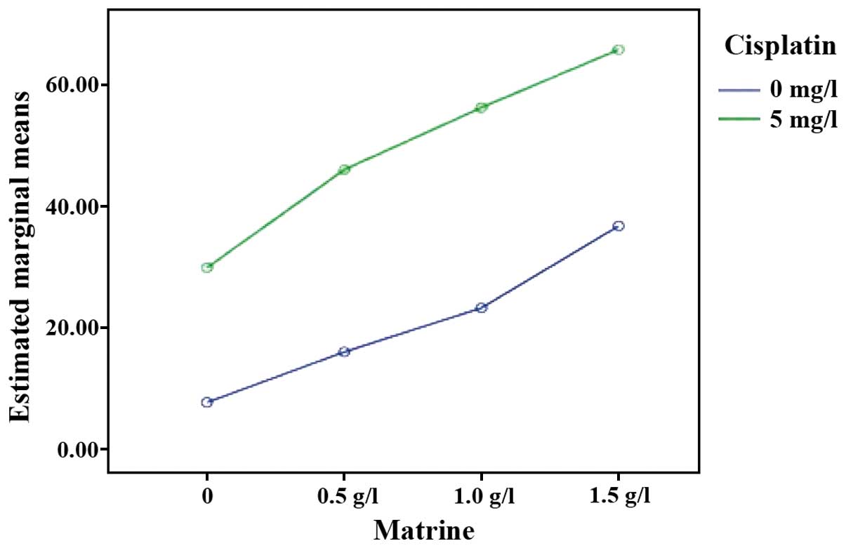
Using an MTT assay, it was observed that the
proliferation of RD cells in each experimental group was inhibited
to different degrees, compared with the control group. As the drug
concentration increased, the proliferation inhibition effect was
gradually increased (P<0.05). The proliferation inhibition rate
of Mat combined with the cisplatin groups was significantly higher
than those of the single Mat and single cisplatin groups under the
same concentration (P<0.01). These data suggested that Mat and
cisplatin act synergistically to inhibit the proliferation of RD
cells (Table II and Fig. 1).
| Table II.Proliferation inhibition rate,
apoptosis rate and XIAP mRNA expression level (n=3, mean ± SD) of
each group. |
Table II.
Proliferation inhibition rate,
apoptosis rate and XIAP mRNA expression level (n=3, mean ± SD) of
each group.
| Group | Proliferation
inhibition rate (%) | Apoptosis rate
(%) | XIAP expression
(FI) |
|---|
| Control | 7.60±6.22 | 7.80±0.85 | 0.90±0.57 |
| Cisplatin |
29.83±1.60a |
22.33±0.97a |
0.36±0.04a |
| Mat |
|
|
|
| 0.5
g/l |
15.93±2.50a |
12.63±0.85a |
0.67±0.06a |
| 1.0
g/l |
23.20±2.33a, b |
16.93±0.81a, b |
0.50±0.03a, b |
| 1.5
g/l |
36.73±2.48a, b |
27.63±0.70a, b |
0.28±0.04a, b |
| Mat +
cisplatin |
|
|
|
| 0.5
g/l |
46.00±1.45a, c |
36.47±3.00a, c |
0.21±0.02a, c |
| 1.0
g/l |
56.27±2.24a,c,d |
44.03±2.06a,c,d |
0.14±0.02a,c,d |
| 1.5
g/l |
68.83±1.35a,c,d |
69.00±5.48a,c,d |
0.07±0.03a,c,d |
| F-value | 141.581 | 203.905 | 162.600 |
| P-value | 0.01 | <0.0001 | <0.0001 |
Mat and cisplatin act cooperatively to
induce apoptosis of RD cells
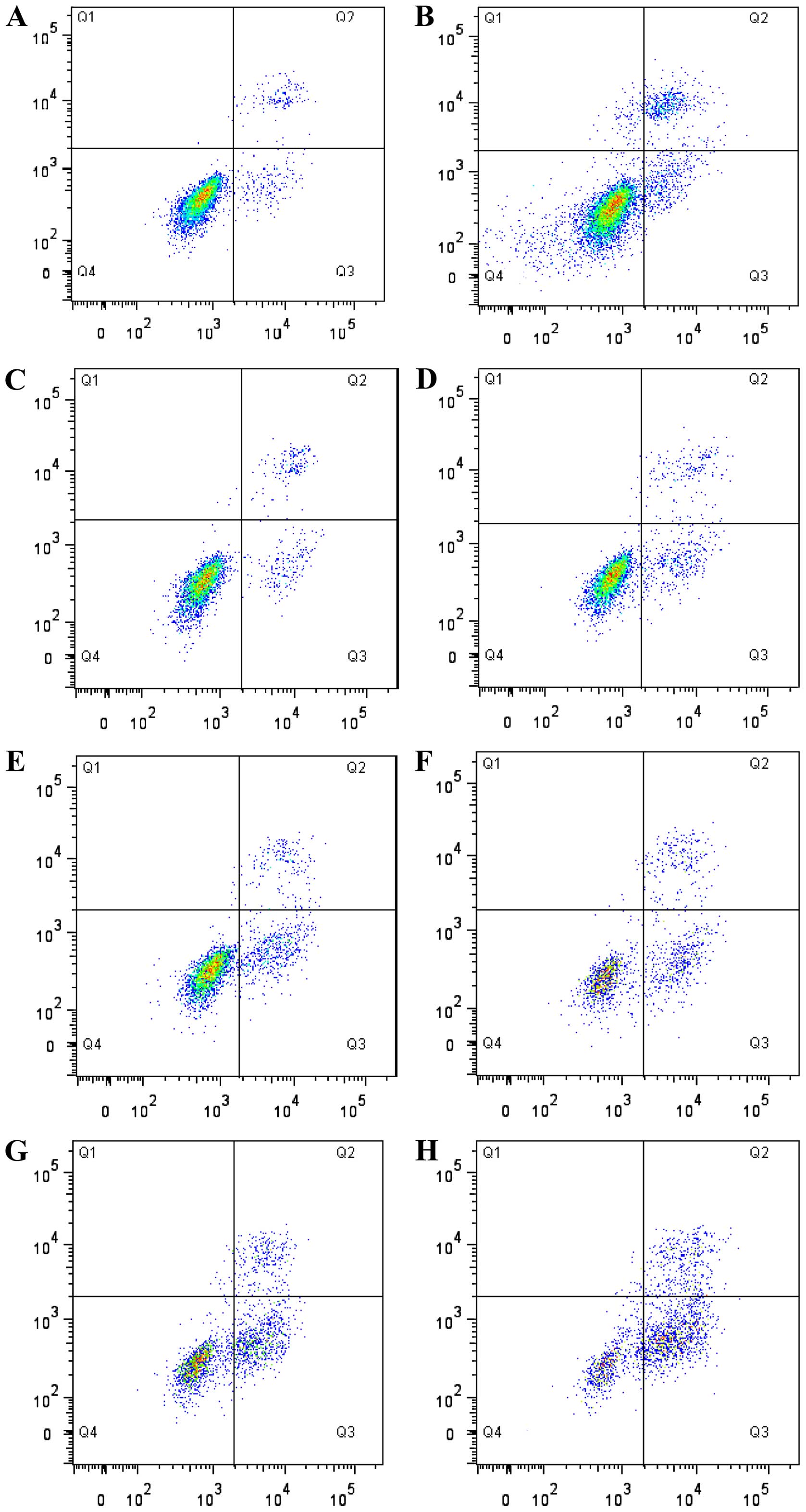
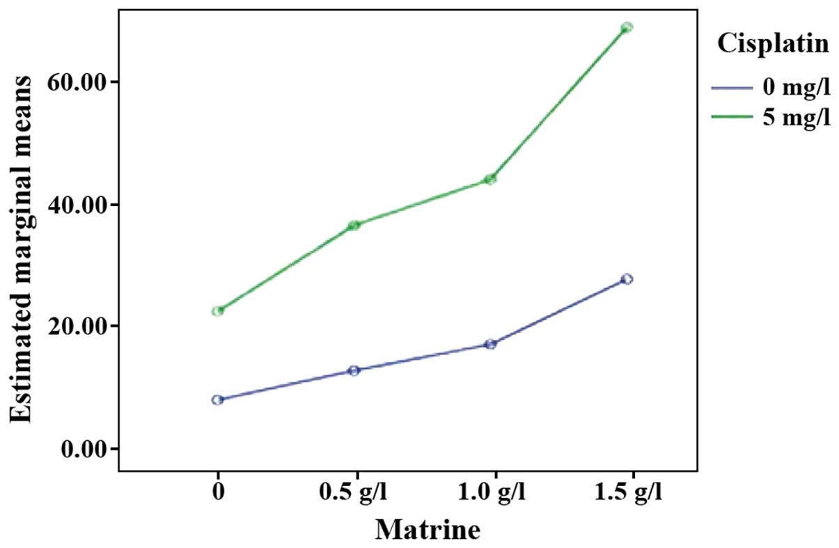
Flow cytometry results showed that, compared with
the control group, the apoptotic rate of RD cells in each
experimental group was increased to varying degrees (P<0.05). An
increase in drug concentration, led to the apoptotic rate of RD
cells in single Mat groups being gradually increased (P<0.05).
The apoptotic rate of Mat combined with the cisplatin groups was
significantly higher than those of the single cisplatin and single
Mat groups under the same concentration (P<0.05). Consistent
with the observation on the inhibition of cell proliferation, Mat
and cisplatin acted cooperatively to induce the apoptosis of RD
cells (Table II; Figs. 2 and 3).
Mat and cisplatin act synergistically
to reduce the XIAP mRNA expression levels
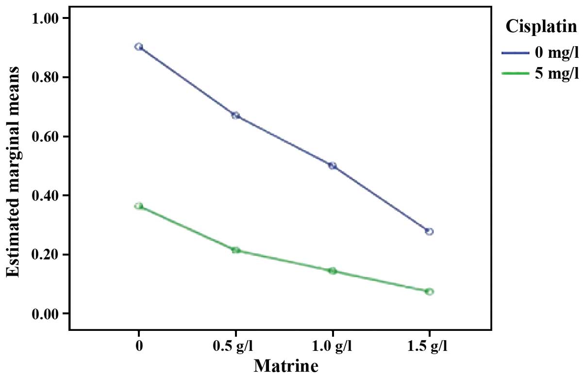
XIAP mRNA was expressed in cells of the control and
each experimental group. The expression levels of XIAP mRNA in RD
cells of each experimental group were lower than those in the
control group (P<0.05). Compared with the single cisplatin
group, the expression levels of XIAP mRNA in Mat combined with the
cisplatin group were significantly reduced (P<0.05). Compared
with the single Mat groups under the same concentration, the
expression levels of XIAP mRNA in the Mat combined with cisplatin
group were significantly reduced (P<0.01). These data showed
that Mat and cisplatin act synergistically to reduce the expression
levels of XIAP mRNA (Table II;
Figs. 4 and 5).
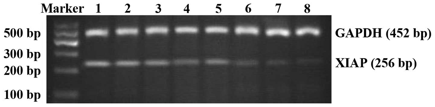 | Figure 5.XIAP mRNA expression in each group of
RD cells. The upper band is the internal reference (GAPDH). The
lower band is the target band (XIAP). Lane 1, control group; lane
2, 0.5 g/l Mat group; lane 3, 1.0 g/l Mat group; lane 4, 1.5 g/l
Mat group; lane 5, mg/l cisplatin group; lane 6, 0.5 g/l Mat
combined with cisplatin group; lane 7, 1.0 g/l Mat combined with
cisplatin group; and lane 8, 1.5 g/l Mat combined with cisplatin
group. XIAP, X-linked inhibitor of apoptosis protein; Mat,
matrine. |
Discussion
RMS is the most common soft tissue sarcoma in
children, accounting for 4–8% of all malignant pediatric solid
tumors (22), and 53% of soft tissue
sarcomas (23). RMS can appear in any
part of the body. It is most commonly seen in the head and neck of
children and is characterized by high malignancy and rapid
progression (24). It is challenging
to diagnose the disease in its early stages. When patients arrive
under hospital care, the disease is at an advanced stage and the
tumor cannot be removed by surgery. At this point, comprehensive
therapy by combining surgery with chemo- and radiotherapy should be
utilized. Drug resistance to chemotherapy often results in
treatment failure. Therefore, the need to improve the curative
effect of chemotherapy drugs, and reverse the drug resistance of
tumor cells has become a research hotspot.
In recent years, the traditional Chinese medicine,
Mat, has increasingly aroused the interest of scholars due to its
physicochemical properties of low toxicity and high efficacy.
Previous findings have shown that Mat had a significant inhibitory
effect on a variety of tumor cells, such as neuroblastoma,
medulloblastoma, colorectal cancer, nasopharyngeal carcinoma,
leukemia cells and RMS (25–31). In addition, the antitumor mechanism of
Mat mainly included obstructing tumor cell cycle progression,
inhibiting tumor cell proliferation, inducing tumor cell apoptosis
and reversing chemotherapy drug resistance (32).
Guo et al (33)
and Li et al (34)
investigated the 24 and 48 h effects of different concentrations of
Mat on RMS. The results have confirmed that Mat inhibited the
proliferation of RMS, inducing cell apoptosis and maintaining the
cell cycle in the G0/G1 phase. The results of the present study
have shown that the proliferation inhibition rate and apoptosis
rate of RD cells treated at different concentrations of Mat (0.5,
1.0 and 1.5 g/l) were higher than those in the control group.
Additionally, as the concentration of Mat increased, the apoptosis
rate of RD cells also increased, confirming that Mat suppressed the
proliferation and induced the apoptosis of RMS cells. The effect
was dose-dependent to a certain degree. Our experimental results
were consistent with those of Guo et al and Li et al
(33,34).
Thanks to its broad anticancer properties, cisplatin
has occupied an extremely significant and unique position in the
field of cancer treatment since 1970 (35). It is commonly regarded as an
anticancer drug with epoch-making significance, particularly
effective for urogenital tumors and malignant carcinomas of the
neck (35). However, as the use of
cisplatin became increasingly widespread, it became apparent that
it had toxic side effects while killing cancer cells and that most
of these side effects were related to the dose of cisplatin. The
toxic side effects of cisplatin mainly manifested as a result of
its application in the clinic. However, approaches regarding how to
reduce the toxicity and side effects of cisplatin have become an
area of interest in clinical research. Further studies on cisplatin
revealed that it had synergistic effects with multiple other drugs.
Drug combinations can enhance their antitumor effects, reduce the
required dose of cisplatin, and reduce the toxic side effect of
cisplatin. Sárosi and Lénárt showed that the combination of
gemcitabine and cisplatin was effective in the treatment of
advanced non-small cell lung cancer, and that the side effects were
relatively minor (36). Solár et
al have shown that geldanamycin enhanced the cytotoxic effect
of cisplatin on tumor cells (37).
In recent years, as research on the antitumor
effects of Chinese medicine progressed, investigators examined how
to enhance the anticancer effect of antitumor drugs while lowering
their toxicity and side effects. Several studies have shown that
rhubarb, bufalin, Radix Astragali, Shengmai, Mat and other
traditional Chinese medicines may enhance the anticancer effects of
cisplatin to varying degrees (38–41).
However, to the best of our knowledge, few studies focused on the
effects of Mat combined with cisplatin on RMS. In the present
study, we used Mat at different concentration (final concentrations
were 0.5, 1.0 and 1.5 g/l) in combination with cisplatin (final
concentration was 5 mg/l) to treat RMS RD cells cultured in
vitro. We then used the MTT assay, flow cytometry and RT-PCR to
measure the effects on proliferation inhibition and apoptosis of RD
cells. The results of the present study show that the proliferation
inhibition rate and apoptosis rate of RD cells in Mat (different
concentrations) combined with cisplatin groups was significantly
higher than that in the single Mat or cisplatin groups. Our study
also confirmed that Mat and cisplatin produced a synergistic
effect, and that the combination therapy can significantly kill RMS
RD cells.
In recent years, with the extensive clinical
application of cisplatin, the phenomenon of cisplatin resistance
has become increasingly widespread, which has generated interest
among scholars to explore the mechanisms of drug resistance.
Amantana et al have shown that DU145 prostate cancer cells
were resistant to cisplatin and that high expression levels of XIAP
were a cause of DU145 resistance to cisplatin. The study also
confirmed that antisense nucleic acid therapy of XIAP can enhance
the antitumor effect of cisplatin and increase the sensitivity of
the DU145 cell line to cisplatin (42). XIAP is a new member of the IAPs family
and also one of the most powerful inhibitors of apoptosis (23,24). At
present, XIAP has been shown to be a protein of universal
expression. It is expressed in various human tissues (25,26), but
at low levels in normal tissue cells and is increased in cancer
cells (27–29). We have studied the effects of
different concentrations of Mat in combination with cisplatin on
the expression of XIAP in human RMS RD cells using RT-PCR analysis.
The results showed that XIAP was expressed in RMS RD cells.
Following treatment with Mat and/or cisplatin, the expression
levels of XIAP in the cells were decreased. In the dose-response
experiments, XIAP mRNA decreased as a function of increasing Mat
concentration. The expression of XIAP mRNA was significantly
reduced under the combined effect of Mat and cisplatin versus Mat
or cisplatin alone.
In conclusion, our experimental results show that
Mat combined with cisplatin inhibited the expression of XIAP in RMS
RD cells and induced apoptosis in these tumor cells. The results
also indicate that Mat may lower the tolerance of RMS RD cells to
cisplatin by inhibiting the expression of XIAP, thus improving the
curative effect of cisplatin. We believe this study reveals a
potential new target for the study of drug-resistant RMS cell lines
and may also lead to new therapeutic options for the clinical
treatment of RMS.
References
|
1
|
Song YQ, Liu SP, Liu ZF and Hu XL:
Determination of matrine and oxymatrine in Radix Sophorae
Flavescentis by resonance rayleigh scattering, second-order
scattering and frequency doubling scattering technique. Chem Res
Chin Univ. 27:746–749. 2011.
|
|
2
|
Zhaowu Z, Xiaoli W, Yangde Z and Nianfeng
L: Preparation of matrine ethosome, its percutaneous permeation in
vitro and anti-inflammatory activity in vivo in rats. J Liposome
Res. 19:155–162. 2009. View Article : Google Scholar : PubMed/NCBI
|
|
3
|
Niu H, Zhang Y, Wu B, Zhang Y, Jiang H and
He P: Matrine induces the apoptosis of lung cancer cells through
downregulation of inhibitor of apoptosis proteins and the Akt
signaling pathway. Oncol Rep. 32:1087–1093. 2014.PubMed/NCBI
|
|
4
|
Liu YQ, Li Y, Qin J, Wang Q, She YL, Luo
YL, He JX, Li JY and Xie XD: Matrine reduces proliferation of human
lung cancer cells by inducing apoptosis and changing miRNA
expression profiles. Asian Pac J Cancer Prev. 15:2169–2177. 2014.
View Article : Google Scholar : PubMed/NCBI
|
|
5
|
Li H, Li X, Bai M, Suo Y, Zhang G and Cao
X: Matrine inhibited proliferation and increased apoptosis in human
breast cancer MCF-7 cells via upregulation of Bax and
downregulation of Bcl-2. Int J Clin Exp Pathol. 8:14793–14799.
2015.PubMed/NCBI
|
|
6
|
Gao H, Guo Y, Deng N, Fei P, Qiu X, Zheng
P, Feng J and Dai G: Suppressive effect of matrine on tumor
invasion in N-butyl-N-4-hydroxybutyl)nitrosamine-induced urinary
bladder carcinogenesis. Chemotherapy. 60:119–128. 2014. View Article : Google Scholar : PubMed/NCBI
|
|
7
|
Li Q, Lai Y, Wang C, Xu G, He Z, Shang X,
Sun Y, Zhang F, Liu L and Huang H: Matrine inhibits the
proliferation, invasion and migration of castration-resistant
prostate cancer cells through regulation of the NF-κB signaling
pathway. Oncol Rep. 35:375–381. 2016.PubMed/NCBI
|
|
8
|
Rong B, Zhao C, Gao W and Yang S: Matrine
promotes the efficacy and safety of platinum-based doublet
chemotherapy for advanced non-small cell lung cancer. Int J Clin
Exp Med. 8:14701–14717. 2015.PubMed/NCBI
|
|
9
|
Wang HQ, Jin JJ and Wang J: Matrine
induces mitochondrial apoptosis in cisplatin-resistant non-small
cell lung cancer cells via suppression of β-catenin/survivin
signaling. Oncol Rep. 33:2561–2566. 2015.PubMed/NCBI
|
|
10
|
Flanagan L, Sebastià J, Tuffy LP, Spring
A, Lichawska A, Devocelle M, Prehn JH and Rehm M: XIAP impairs Smac
release from the mitochondria during apoptosis. Cell Death Dis.
1:e492010. View Article : Google Scholar : PubMed/NCBI
|
|
11
|
Kempkensteffen C, Jäger T, Bub J, Weikert
S, Hinz S, Christoph F, Krause H, Schostak M, Miller K and Schrader
M: The equilibrium of XIAP and Smac/DIABLO expression is gradually
deranged during the development and progression of testicular germ
cell tumours. Int J Androl. 30:476–483. 2007. View Article : Google Scholar : PubMed/NCBI
|
|
12
|
Ramp U, Krieg T, Caliskan E, Mahotka C,
Ebert T, Willers R, Gabbert HE and Gerharz CD: XIAP expression is
an independent prognostic marker in clear-cell renal carcinomas.
Hum Pathol. 35:1022–1028. 2004. View Article : Google Scholar : PubMed/NCBI
|
|
13
|
Hanson AJ, Wallace HA, Freeman TJ,
Beauchamp RD, Lee LA and Lee E: XIAP monoubiquitylates Groucho/TLE
to promote canonical Wnt signaling. Mol Cell. 45:619–628. 2012.
View Article : Google Scholar : PubMed/NCBI
|
|
14
|
Zheng ZL, Tan LZ, Yu YP, Michalopoulos G
and Luo JH: Interaction of CSR1 with XIAP reverses inhibition of
caspases and accelerates cell death. Am J Pathol. 181:463–471.
2012. View Article : Google Scholar : PubMed/NCBI
|
|
15
|
Berezovskaya O, Schimmer AD, Glinskii AB,
Pinilla C, Hoffman RM, Reed JC and Glinsky GV: Increased expression
of apoptosis inhibitor protein XIAP contributes to anoikis
resistance of circulating human prostate cancer metastasis
precursor cells. Cancer Res. 65:2378–2386. 2005. View Article : Google Scholar : PubMed/NCBI
|
|
16
|
Doudican NA, Byron SA, Pollock PM and
Orlow SJ: XIAP downregulation accompanies mebendazole growth
inhibition in melanoma xenografts. Anticancer Drugs. 24:181–188.
2013. View Article : Google Scholar : PubMed/NCBI
|
|
17
|
Castells M, Milhas D, Gandy C, Thibault B,
Rafii A, Delord JP and Couderc B: Microenvironment mesenchymal
cells protect ovarian cancer cell lines from apoptosis by
inhibiting XIAP inactivation. Cell Death Dis. 4:e8872013.
View Article : Google Scholar : PubMed/NCBI
|
|
18
|
Cao Z, Li X, Li J, Luo W, Huang C and Chen
J: X-linked inhibitor of apoptosis protein (XIAP) lacking RING
domain localizes to the nuclear and promotes cancer cell
anchorage-independent growth by targeting the E2F1/Cyclin E axis.
Oncotarget. 5:7126–7137. 2014. View Article : Google Scholar : PubMed/NCBI
|
|
19
|
Liew JC, Tan WS, Alitheen NB, Chan ES and
Tey BT: Over-expression of the X-linked inhibitor of apoptosis
protein (XIAP) delays serum deprivation-induced apoptosis in CHO-K1
cells. J Biosci Bioeng. 110:338–344. 2010. View Article : Google Scholar : PubMed/NCBI
|
|
20
|
Katragadda L, Carter BZ and Borthakur G:
XIAP antisense therapy with AEG 35156 in acute myeloid leukemia.
Expert Opin Investig Drugs. 22:663–670. 2013. View Article : Google Scholar : PubMed/NCBI
|
|
21
|
Yang CL, Liu SS, Ma YG, Liu YY, Xue YX and
Huang B: The influence of intraoperative pleural perfusion with
matrine- cisplatin or cisplatin on stromal cell-derived factor-1 in
non-small cell lung cancer patients with subclinical pleural
metastasis. Med Oncol. 29:574–581. 2012. View Article : Google Scholar : PubMed/NCBI
|
|
22
|
Rossi S, Stoppani E, Puri PL and Fanzani
A: Differentiation of human rhabdomyosarcoma RD cells is regulated
by reciprocal, functional interactions between myostatin, p38 and
extracellular regulated kinase signalling pathways. Eur J Cancer.
47:1095–1105. 2011. View Article : Google Scholar : PubMed/NCBI
|
|
23
|
Paulino AC and Okcu MF: Rhabdomyosarcoma.
Curr Probl Cancer. 32:7–34. 2008. View Article : Google Scholar : PubMed/NCBI
|
|
24
|
Panta P and Felix D: Rhabdomyosarcoma: a
rapidly growing malignancy. Pan Afr Med J. 22:1212015. View Article : Google Scholar : PubMed/NCBI
|
|
25
|
Li H, Xie S, Liu X, Wu H, Lin X, Gu J,
Wang H and Duan Y: Matrine alters microRNA expression profiles in
SGC-7901 human gastric cancer cells. Oncol Rep. 32:2118–2126.
2014.PubMed/NCBI
|
|
26
|
Meng F, Zhang ZX, Xie J, Huang CB, Liu Y
and Liao YG: Effects of matrine on the apoptosis and the expression
of PEG10 in human hepatocarcinoma cell Line HepG2. J Pract Med.
10:1523–1526. 2014.(In Chinese).
|
|
27
|
Gao XH, Xu W, Li WJ, Ma HW and Bai YL:
Anti-proliferative effects of matrine on human neuroblastoma
SK-N-SH cells Chin. J Exp Trad Med Formulae. 23:196–199. 2013.(In
Chinese).
|
|
28
|
Yu Q, Chen B, Zhang X, Qian W, Ye B and
Zhou Y: Arsenic trioxide-enhanced, matrine-induced apoptosis in
multiple myeloma cell lines. Planta Med. 79:775–781. 2013.
View Article : Google Scholar : PubMed/NCBI
|
|
29
|
Yan F, Liu Y and Wang W: Matrine inhibited
the growth of rat osteosarcoma UMR-108 cells by inducing apoptosis
in a mitochondrial-caspase-dependent pathway. Tumour Biol.
34:2135–2140. 2013. View Article : Google Scholar : PubMed/NCBI
|
|
30
|
Guo L, Xue TY, Xu W and Gao JZ: Matrine
promotes G0/G1 arrest and down-regulates cyclin D1 expression in
human rhabdomyosarcoma cells. Panminerva Med. 55:291–296.
2013.PubMed/NCBI
|
|
31
|
Tan C, Qian X, Jia R, Wu M and Liang Z:
Matrine induction of reactive oxygen species activates p38 leading
to caspase-dependent cell apoptosis in non-small cell lung cancer
cells. Oncol Rep. 30:2529–2535. 2013.PubMed/NCBI
|
|
32
|
Liu Y, Xu Y, Ji W, Li X, Sun B, Gao Q and
Su C: Anti-tumor activities of matrine and oxymatrine: literature
review. Tumour Biol. 35:5111–5119. 2014. View Article : Google Scholar : PubMed/NCBI
|
|
33
|
Guo L, Xue TY, Xu W and Gao JZ: Effects of
matrine on the proliferation and apoptosis of human
rhabdomyosarcoma RD cells. Zhongguo Dang Dai Er Ke Za Zhi.
14:780–784. 2012.(In Chinese). PubMed/NCBI
|
|
34
|
Li YX: Effects and mechanisms of matrine
on proliferation and apoptosis of human rhabdomyosarcoma cell RD.
Seek Med Ask Med:. 10:295–296. 2012.
|
|
35
|
Miao L, Guo S, Zhang J, Kim WY and Huang
L: Nanoparticles with precise ratiometric co-loading and
co-delivery of gemcitabine monophosphate and cisplatin for
treatment of bladder cancer. Adv Funct Mater. 24:6601–6611. 2014.
View Article : Google Scholar : PubMed/NCBI
|
|
36
|
Sárosi V and Lénárt T:
Gemcitabine-cisplatin combination in the first line treatment of
non-small cell lung cancer. Our experience and analysis of safety.
Magy Onkol. 47:189–193. 2003.(In Hungarian). PubMed/NCBI
|
|
37
|
Solár P, Horváth V, Kleban J, Koval' J,
Solárová Z, Kozubík A and Fedorocko P: Hsp90 inhibitor geldanamycin
increases the sensitivity of resistant ovarian adenocarcinoma cell
line A2780cis to cisplatin. Neoplasma. 54:127–130. 2007.PubMed/NCBI
|
|
38
|
Huang Q, Lu G, Shen HM, Chung MC and Ong
CN: Anti-cancer properties of anthraquinones from rhubarb. Med Res
Rev. 27:609–630. 2007. View Article : Google Scholar : PubMed/NCBI
|
|
39
|
Yin PH, Liu X, Qiu YY, Cai JF, Qin JM, Zhu
HR and Li Q: Anti-tumor activity and apoptosis-regulation
mechanisms of bufalin in various cancers: new hope for cancer
patients. Asian Pac J Cancer Prev. 13:5339–5343. 2012. View Article : Google Scholar : PubMed/NCBI
|
|
40
|
Tang MX: Reduction of metastasis of lung
cancer by ‘Fufang Shengmai-compound pulse inducer’ - a preliminary
report on experimental, clinical and pathologic study with
reference to the ultrastructure. Zhonghua Zhong Liu Za Zhi.
5:176–179. 1983.(In Chinese). PubMed/NCBI
|
|
41
|
Xiao WL, Motley TJ, Unachukwu UJ, Lau CB,
Jiang B, Hong F, Leung PC, Wang QF, Livingston PO, Cassileth BR, et
al: Chemical and genetic assessment of variability in commercial
Radix Astragali (Astragalus spp.) by ion trap LC-MS and nuclear
ribosomal DNA barcoding sequence analyses. J Agric Food Chem.
59:1548–1556. 2011. View Article : Google Scholar : PubMed/NCBI
|
|
42
|
Amantana A, London CA, Iversen PL and Devi
GR: X-linked inhibitor of apoptosis protein inhibition induces
apoptosis and enhances chemotherapy sensitivity in human prostate
cancer cells. Mol Cancer Ther. 3:699–707. 2004.PubMed/NCBI
|