Open Access

Evaluation by quantitative image analysis of anticancer drug activity on multicellular spheroids grown in 3D matrices

  • Authors:
    • Aurélie Gomes
    • Adrien Russo
    • Guillaume Vidal
    • Elise Demange
    • Pauline Pannetier
    • Zied Souguir
    • Jean‑Michel Lagarde
    • Bernard Ducommun
    • Valérie Lobjois
  • View Affiliations

  • Published online on: October 4, 2016     https://doi.org/10.3892/ol.2016.5221
  • Pages: 4371-4376
  • Copyright: © Gomes et al. This is an open access article distributed under the terms of Creative Commons Attribution License.

Metrics: Total Views: 0 (Spandidos Publications: | PMC Statistics: )
Total PDF Downloads: 0 (Spandidos Publications: | PMC Statistics: )


Abstract

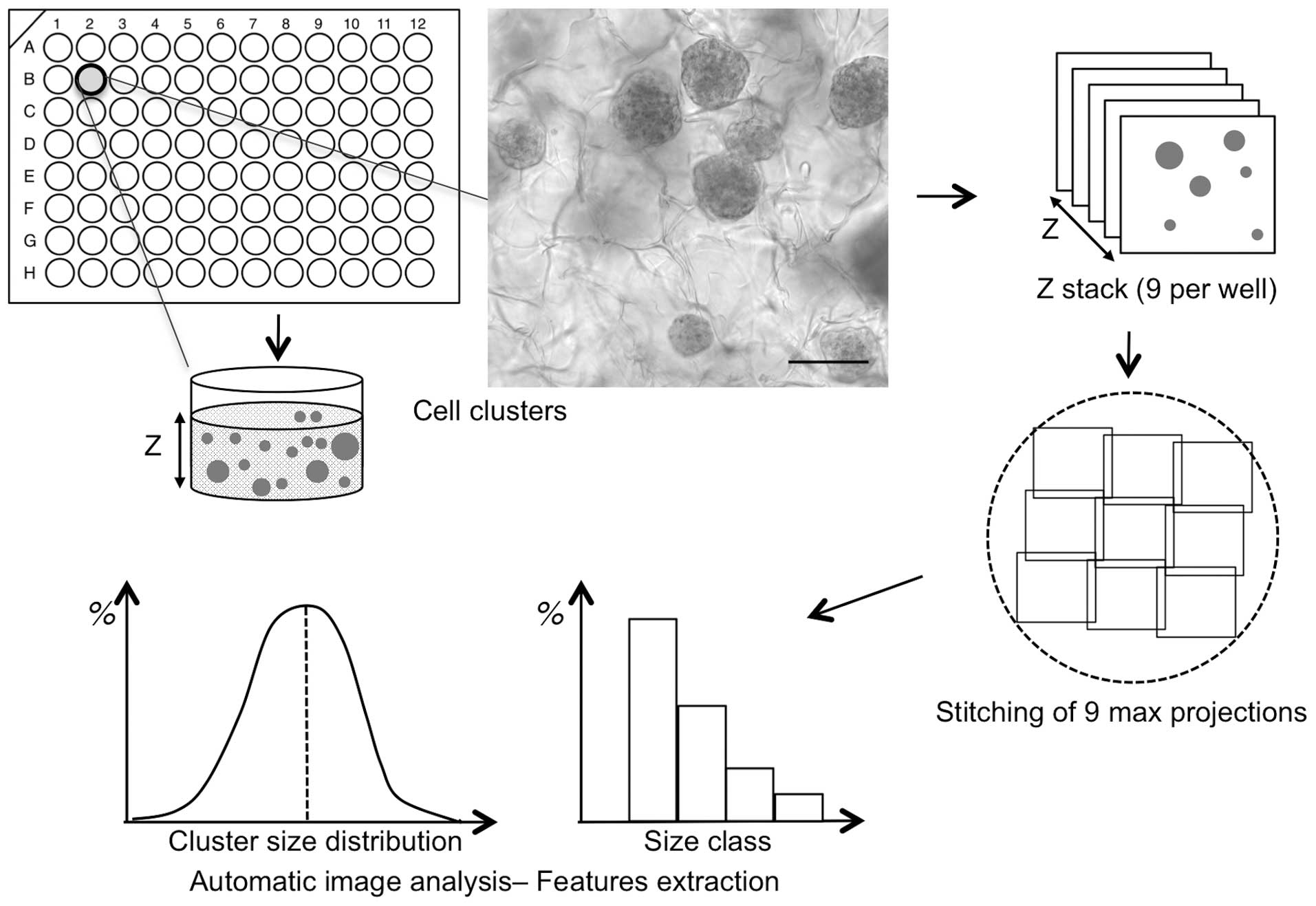
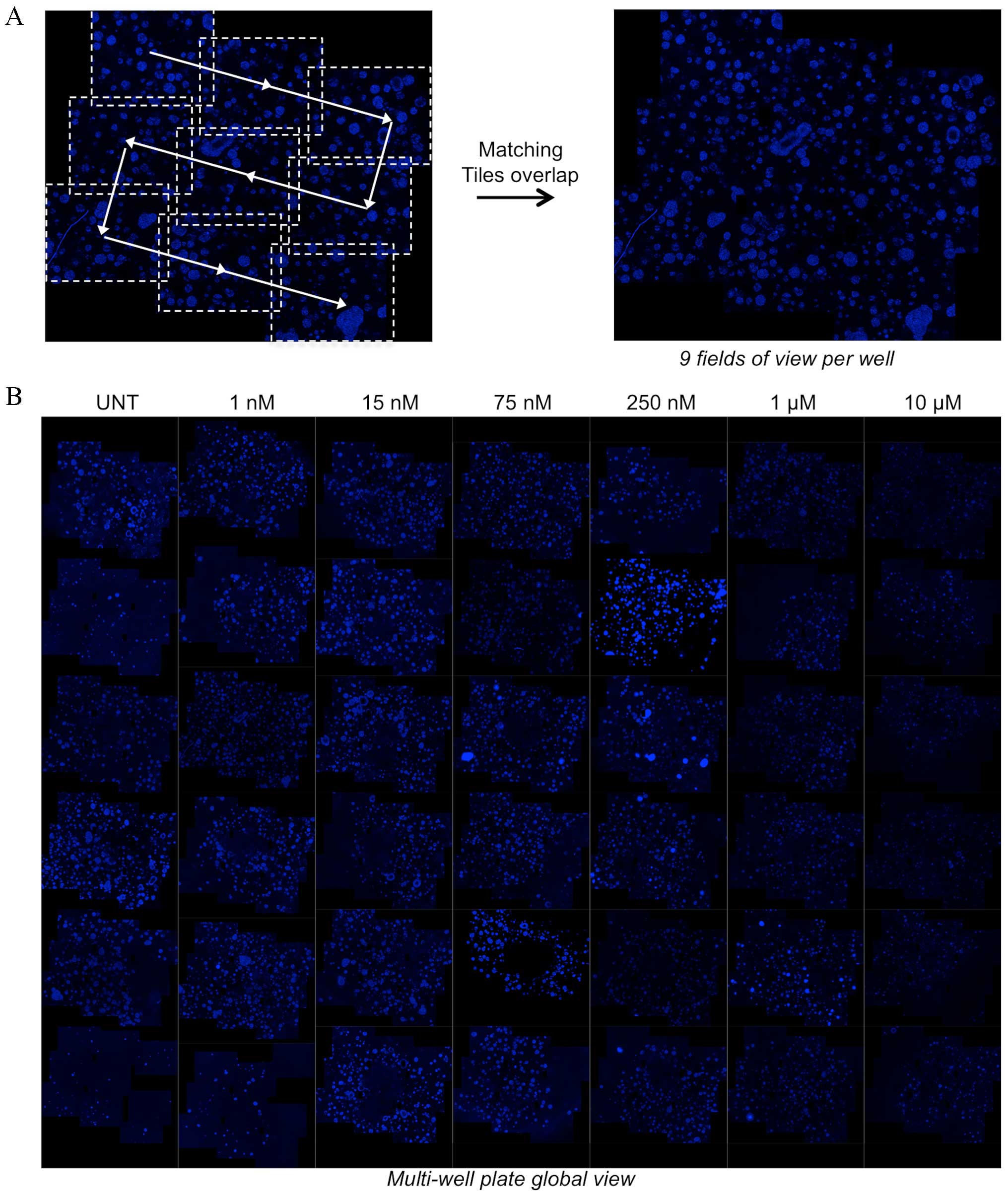
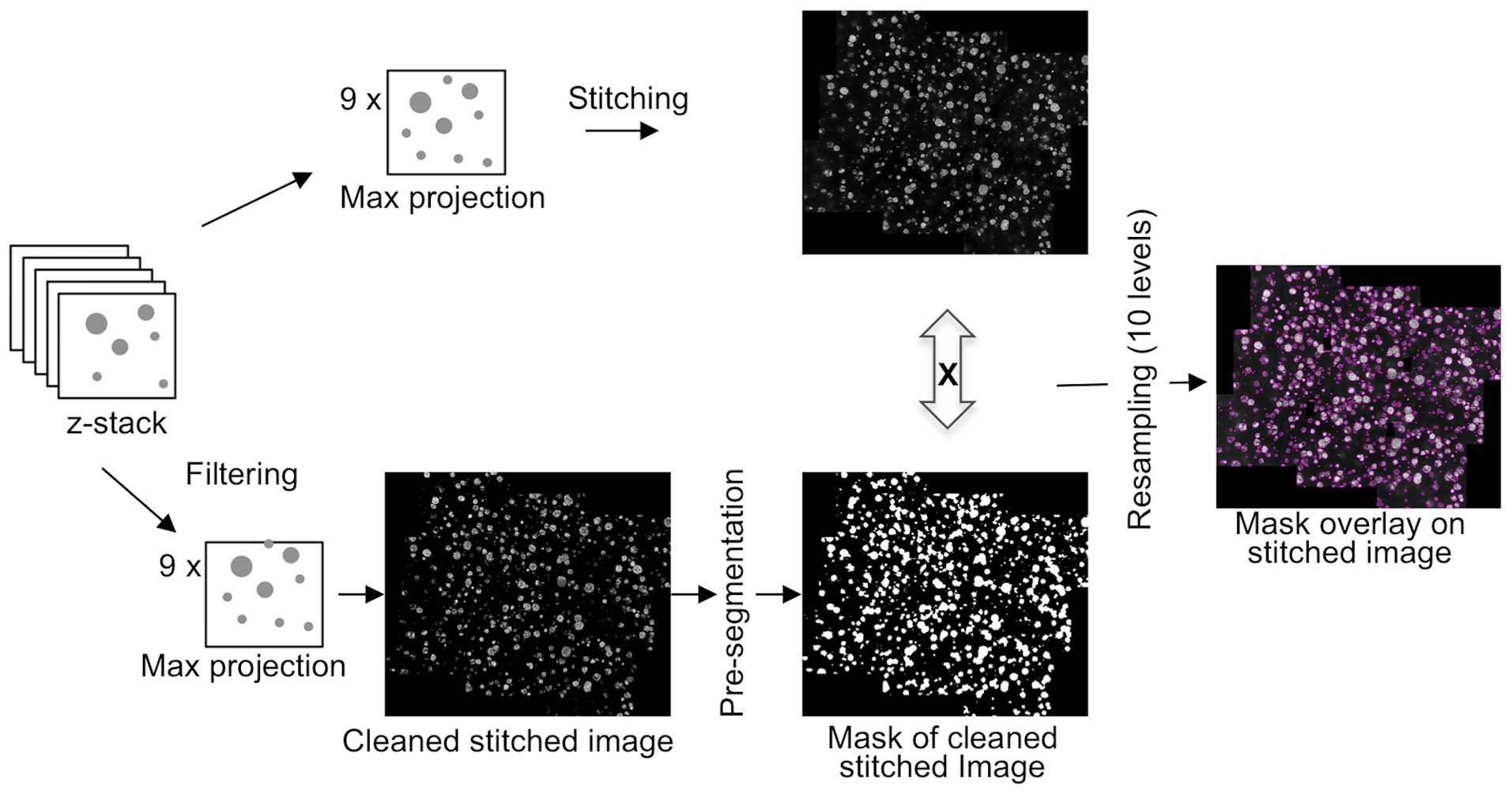
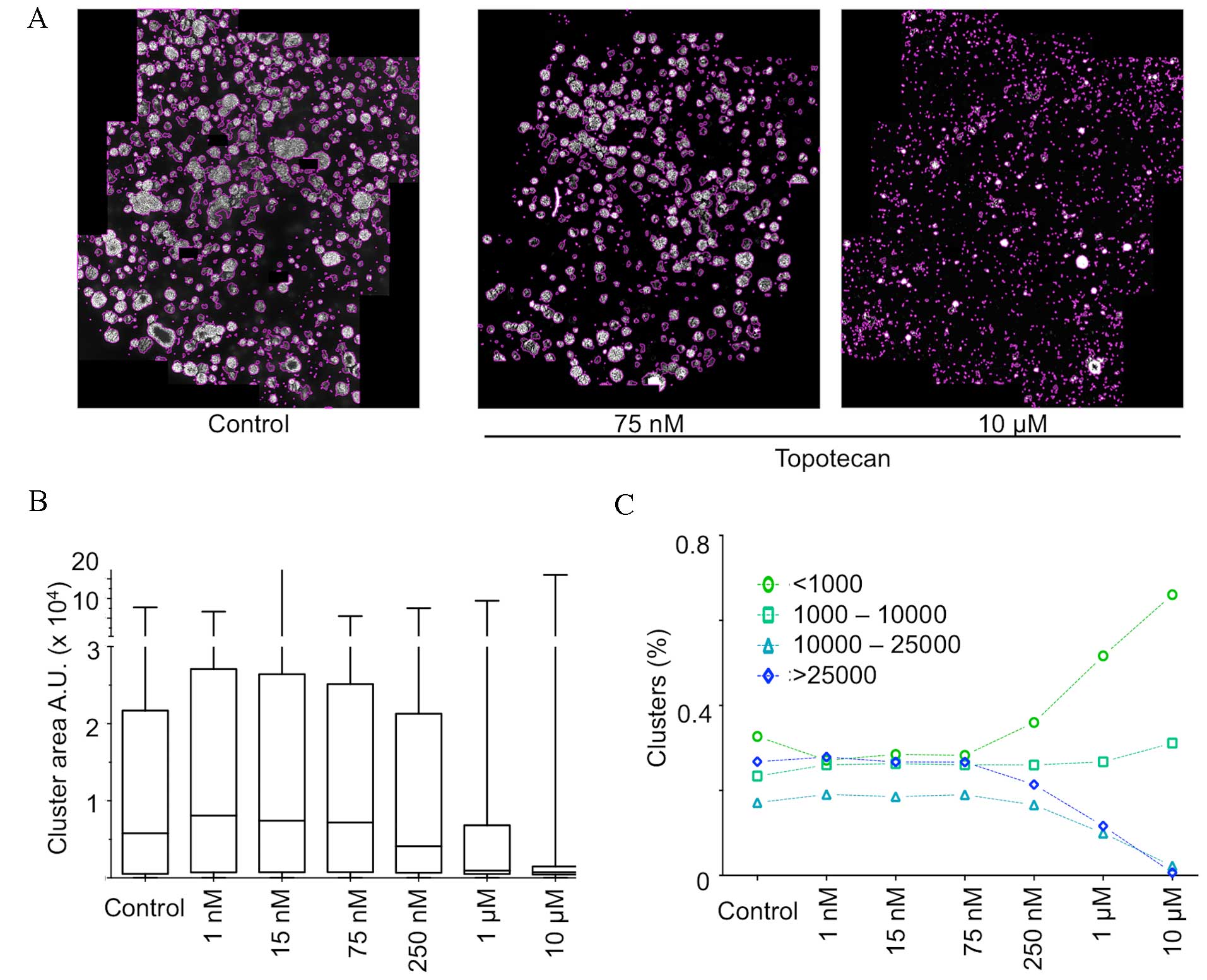
Pharmacological evaluation of anticancer drugs using 3D in vitro models provides invaluable information for predicting in vivo activity. Artificial matrices are currently available that scale up and increase the power of such 3D models. The aim of the present study was to propose an efficient and robust imaging and analysis pipeline to assess with quantitative parameters the efficacy of a particular cytotoxic drug. HCT116 colorectal adenocarcinoma tumor cell multispheres were grown in a 3D physiological hyaluronic acid matrix. 3D microscopy was performed with structured illumination, whereas image processing and feature extraction were performed with custom analysis tools. This procedure makes it possible to automatically detect spheres in a large volume of matrix in 96‑well plates. It was used to evaluate drug efficacy in HCT116 spheres treated with different concentrations of topotecan, a DNA topoisomerase inhibitor. Following automatic detection and quantification, changes in cluster size distribution with a topotecan concentration‑dependent increase of small clusters according to drug cytotoxicity were observed. Quantitative image analysis is thus an effective means to evaluate and quantify the cytotoxic and cytostatic activities of anticancer drugs on 3D multicellular models grown in a physiological matrix.
View Figures
View References

Related Articles

Journal Cover

December-2016
Volume 12 Issue 6

Print ISSN: 1792-1074
Online ISSN:1792-1082

Sign up for eToc alerts

Recommend to Library

Copy and paste a formatted citation
x
Spandidos Publications style
Gomes A, Russo A, Vidal G, Demange E, Pannetier P, Souguir Z, Lagarde JM, Ducommun B and Lobjois V: Evaluation by quantitative image analysis of anticancer drug activity on multicellular spheroids grown in 3D matrices. Oncol Lett 12: 4371-4376, 2016
APA
Gomes, A., Russo, A., Vidal, G., Demange, E., Pannetier, P., Souguir, Z. ... Lobjois, V. (2016). Evaluation by quantitative image analysis of anticancer drug activity on multicellular spheroids grown in 3D matrices. Oncology Letters, 12, 4371-4376. https://doi.org/10.3892/ol.2016.5221
MLA
Gomes, A., Russo, A., Vidal, G., Demange, E., Pannetier, P., Souguir, Z., Lagarde, J., Ducommun, B., Lobjois, V."Evaluation by quantitative image analysis of anticancer drug activity on multicellular spheroids grown in 3D matrices". Oncology Letters 12.6 (2016): 4371-4376.
Chicago
Gomes, A., Russo, A., Vidal, G., Demange, E., Pannetier, P., Souguir, Z., Lagarde, J., Ducommun, B., Lobjois, V."Evaluation by quantitative image analysis of anticancer drug activity on multicellular spheroids grown in 3D matrices". Oncology Letters 12, no. 6 (2016): 4371-4376. https://doi.org/10.3892/ol.2016.5221