Introduction
Laryngeal squamous cell carcinoma (LSCC) is one of
the most common malignant tumors identified in the head and neck.
LSCCs account for 90% of all larynx carcinomas, ~90% of which are
in males. Furthermore, the majority of patients are aged between 40
and 60-years-old (1). At present,
there is no specific biomarker for LSCC diagnosis and prognosis.
Thus, the current study aimed to examine potential biomarkers that
could improve the early diagnosis of LSCC.
The cell adhesion molecule 1 (CADM1) gene
encodes a membrane protein, which is silenced in ~44% of patients
with non-small cell lung cancer and 30–60% of those with other
types of cancer (2). Previous studies
have identified two mechanisms of CADM1 inactivation:
Promoter hypermethylation and loss of heterozygosity (3–5).
Furthermore, it has been demonstrated that the CADM1 protein
participates in a number of biological functions, including synapse
formation, cell adhesion and tumor suppression (6). In several types of tumors, the absence
of CADM1 expression is closely related to invasive tumor behavior
(6–9).
However, the role of CADM1 in LSCC remains largely unknown. To
investigate the association between CADM1 protein expression and
the development of LSCC, 60 surgically resected specimens were
collected and assessed for loss of the CADM1 protein using
immunohistochemistry. Subsequently, the association of CADM1
protein expression with LSCC clinicopathological parameters and the
histological growth pattern of the tumors were investigated.
Folate is a water-soluble B vitamin that mediates
one-carbon metabolism in vivo and serves multiple functions
in physiological processes. Folate provides one-carbon groups for
DNA replication, epigenetic modifications and DNA mutation
prevention, thus protecting against tumorigenesis (10). Additionally, tetrahydrofolate
synthesis in vivo requires folate for blood cell development
and maturation (11). Folate
deficiency is closely associated with glossitis, anemia and, in
pregnancy, fetal neural tube defects (12). Previous studies have suggested that
folate may aid cancer prevention (13,14), and
several studies have demonstrated that a reduction in serum folate
levels may be closely related to several types of cancer, including
colorectal cancer (15–17). However, no similar report in LSCC was
found in the literature. Therefore, it is necessary to investigate
the relationship between serum folate levels and cancer progression
and prognosis. The present study aimed to investigate the
underlying association between folate deficiency and CADM1 protein
expression in LSCC.
Patients and methods
Patients and tissue specimens
A total of 60 pairs of LSCC and precancerous
specimens were collected from patients treated at the Department of
Otolaryngology, Head and Neck Surgery at the First Affiliated
Hospital of Henan University of Science and Technology (Luoyang,
China) between September 2011 and September 2014. The control group
consisted of 30 healthy volunteers whose blood was collected during
routine health examinations. The ratio of patients to healthy
volunteers was 1:2. Precancerous tissues were defined as the
laryngeal mucosa 2 cm from the edge of the cancerous site. Patients
had undergone preoperative partial or total laryngectomy without
radical and chemical therapies. Patients with megaloblastic anemia
or other types of anemia, upper gastrointestinal tract diseases,
and those treated with any other surgical procedures, were
excluded. Patients with a history of smoking, oral contraceptive
use or those who had taken B vitamins in the past 6 months were
also excluded from the current study. The Ethics Committee of the
First Affiliated Hospital of Henan University of Science and
Technology (Luoyang, China) approved the use of human tissues for
the current study. All patients and volunteers provided written
informed consent to participate.
Immunohistochemistry
Expression of the CADM1 protein was tested using an
Immunohistochemical S-P kit against CADM1 (Kangwei Bio, Ltd.,
Beijing, China), according to the manufacturer's protocol. A rabbit
polyclonal antibody against CADM1 (1:100 dilution, cat. no.
Rs-1147R) was purchased from Kangwei Bio, Ltd. Tissues were fixed
with 10% formaldehyde solution, cut into 3-µm sections, embedded in
paraffin blocks and subsequently incubated with the polyclonal
antibody at 4°C overnight (>14 h). Sections were subsequently
incubated with biotinylated goat anti-rabbit immunoglobulin G
(1:100 dilution, cat. no. CW2035S; Kangwei Bio, Ltd.) for 40 min at
room temperature. Phosphate-buffered saline was used as a negative
control. 3,3′-diaminobenzidine was used to stain the sections and
hematoxylin was used as a counterstain to highlight the cytoblasts
and background color. Each section was observed in 10 random fields
under ×200 magnification (18). The
score was calculated by adding the scores for intensity (weak=1,
moderate=2, intense=3) and proportion (5%=1, 6–24%=2, 25–49%=3,
50–74%=4, ≥75%=5) of positively stained cells (19). Scores of ≥2 were considered positive.
Two independent pathologists assessed each slide.
Radioimmunoassay
Whole blood samples (6 ml) were drawn from a
peripheral vein of each participant after overnight fasting.
Following the centrifugation of whole blood samples at 300 ×
g for 15 min, serum samples were collected and stored at
−80°C until analysis. Serum folate levels were tested using
radioimmunoassay quantification with a commercial kit for
measurement of folate (Shanghai Ruiqi Bio-Technology, Co., Ltd.,
Shanghai, China), according to the manufacturer's instructions.
Statistical analysis
Data were analyzed using Student's t-test,
χ2 test, one-way analysis of variance (ANOVA),
Student-Newman-Keuls test, Fisher's exact test and Spearman's rank
correlation coefficient. SPSS version 16.0 for Windows (SPSS Inc.,
Chicago, IL, USA) was used to analyze all results. P<0.05 was
considered to indicate a statistically significant difference.
Results
Expression of the CADM1 protein in
LSCC
CADM1 protein expression was detected via
immunohistochemical analysis of 60 malignant squamous cells of
larynx tumors and 60 corresponding healthy adjacent non-neoplastic
tissues, which were examined as a comparison. The results
demonstrated that 47 specimens (78%) of adjacent normal tissues
expressed CADM1 protein. By contrast, only 12 tumor specimens (20%)
expressed the CADM1 protein (P<0.001; Table I). The associations among CADM1
expression patterns in the tumors, serum folate levels and patient
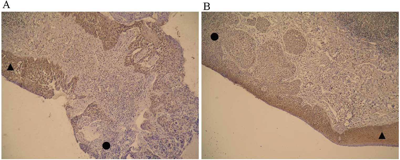
clinical characteristics are presented in Table II. Positive staining indicated that
CADM1 was primarily located in the cytoplasm in the adjacent normal
tissues, but was absent in the tumor tissues (Fig. 1A and B, indicated by shapes).
Expression of the CADM1 protein was not correlated with patient age
or anatomic localization (P=1.000 and 0.386, respectively), but was
correlated with histological differentiation and clinical stage
(P=0.010 and 0.020, respectively; Table
II).
 | Table I.Expression of the CADM1 protein in
laryngeal squamous cell carcinoma and adjacent normal tissues. |
Table I.
Expression of the CADM1 protein in
laryngeal squamous cell carcinoma and adjacent normal tissues.
|
|
| CADM1 |
|
|
|---|
|
|
|
|
|
|
|---|
| Group | n | Positive | Negative | χ2 | P-value |
|---|
| Tumors | 60 | 12 | 48 | 28.229 | 0.001a |
| Adjacent normal
tissues | 60 | 47 | 13 |
|
|
| Table II.Associations among patient clinical
characteristics, protein expression of CADM1 and serum folate
levels. |
Table II.
Associations among patient clinical
characteristics, protein expression of CADM1 and serum folate
levels.
| Characteristic | n | P | N | Folic acid
(ng/ml) | P-value: Fisher's
exact test | P-value: One-way
ANOVA |
|---|
| Age (years) |
|
|
|
|
|
|
| ≥50 | 49 | 15 | 34 | 3.53±1.53 |
|
|
|
<50 | 11 | 3 | 8 | 2.36±0.98 | 1.000 | 0.058 |
| TNM stage |
|
|
|
|
|
|
| I | 27 | 12 | 15 | 3.69±1.45 |
|
|
| II | 17 | 6 | 11 | 3.65±1.57 |
|
|
| III | 11 | 0 | 11 | 2.44±1.32 |
|
|
| IV | 5 | 0 | 5 | 2.46±1.07 | 0.010a | 0.001b |
| Histological
differentiation |
|
|
|
|
|
|
| Well | 34 | 15 | 19 | 3.85±1.33 |
|
|
|
Moderately | 16 | 3 | 13 | 3.18±1.66 |
|
|
|
Poorly | 10 | 0 | 10 | 1.95±0.86 | 0.010a | 0.001b |
| Anatomic
localization |
|
|
|
|
|
|
|
Supraglottic | 8 | 2 | 6 | 2.08±0.96 |
|
|
|
Glottic | 39 | 14 | 25 | 3.86±1.45 |
|
|
|
Subglottic | 13 | 2 | 11 | 2.94±1.23 | 0.386 | 0.001b |
Associations among patient
characteristics, expression of CADM1 and serum folate levels
One-way ANOVA was used to determine differences in
the serum folate levels among LSCC tissues with different clinical
characteristics, including age, clinical stage, histological
differentiation and anatomic localization. The differences were
statistically significant among tumor tissues with different
clinical stages, histological differentiation and anatomic
localizations (P=0.001, 0.001 and 0.001, respectively; Table II). To further clarify the
relationship between serum folate levels and stage of LSCC, a
Student-Newman-Keuls test was used for multiple comparisons of
clinical stage, histological differentiation and anatomic
localization. The results indicated that serum folate levels
decreased as tumor malignancy increased (Table III). Additionally, serum folate
levels were significantly lower in patients with LSCC compared with
healthy control subjects (P=0.002; Table
IV). The spearman correlation analysis identified a significant
correlation between CADM1 protein expression and serum folate
levels (r=0.642, P=0.001). Therefore, folate deficiency may
decrease the methylation of CADM1 promotor and regulate
CADM1 expression. However, further studies are required to
confirm this.
| Table III.Associations between serum folic acid
levels in patients with laryngeal squamous cell carcinoma and
clinical stage, histological differentiation and anatomic
localization. |
Table III.
Associations between serum folic acid
levels in patients with laryngeal squamous cell carcinoma and
clinical stage, histological differentiation and anatomic
localization.
| Clinical stage
(TNM) | P-value | Histological
differentiation and anatomic localization | P-value |
|---|
| I and II | 0.610 | 1 and 2 | 0.110 |
| I and III | 0.002a | 1 and 3 | 0.001b |
| I and IV | 0.001b | 2 and 3 | 0.020a |
| II and III | 0.014a | A and B | 0.001b |
| II and IV | 0.004b | A and C | 0.160 |
| III and IV | 0.320 | B and C | 0.080 |
| Table IV.Serum folic acid levels in patients
with LSCC and healthy controls. |
Table IV.
Serum folic acid levels in patients
with LSCC and healthy controls.
| Group | n | Folic acid
(ng/ml) |
|---|
| Healthy
controls | 30 |
4.40±1.47a |
| Patients with
LSCC | 60 | 3.35±1.51 |
Discussion
In the present study, all included patients were
male and most patients aged <50 years were TNM stage III or IV
with higher serum folate levels, whereas patients aged >50 years
were stages I–II with lower serum folate levels. The critical
factor in the prognosis of LSCC is tumor metastasis, however, the
etiopathogenesis of tumor metastasis remains largely unknown and
may involve immune dysregulation. CADM1, located on
chromosome 11q23.2, encodes a transmembrane protein from the
immunoglobulin superfamily (20).
Previous studies have demonstrated that CADM1 is a human tumor
suppressor and mutations in its cytoplasmic domain have been linked
to lung tumor cell metastasis, aggravated histological
differentiation, clinical stage classification and a poor prognosis
(6,21,22). The
results of the present study were consistent with previous studies
(23,24) and established that, as compared with
adjacent non-neoplastic tissue, tumor tissues express lower levels
of CADM1. This is important as CADM1 may serve a potential role in
the diagnosis and prognosis of LSCC.
Duthie et al (25) affirmed the effect of folate deficiency
on chromosome breaks and the risk of carcinogenesis. Furthermore,
it has been determined that folate deficiency may decrease
thymidine synthesis and stimulate uracil misincorporation into DNA
(26,27). Homeostasis of folate is critical to
DNA stability and integrity, as well as the repair of damaged DNA,
and folate supplementation may have a protective effect against
cancer (15,16,28,29). The
results of the current study demonstrated that serum folate levels
in patients with LSCC were markedly lower than those in healthy
individuals and decreased with the deterioration in the degree of
tumor malignancy. This indicates that low serum folate levels may
be associated with the increased malignancy of LSCC.
The results of the present study suggested that
there was a significant association between CADM1 protein
expression and serum levels of serum folate in patients with LSCC
(r=0.642, P=0.001). CADM1 expression varied among different
anatomic localizations; therefore, this relationship may result
from the different malignant degrees and carcinoma progression.
However, the unique microenvironments of different anatomical
localizations may also contribute to the difference. Therefore,
further studies are required to understand why these differences
occur.
The analysis demonstrated that the decline of serum
folate levels may be the cause of low CADM1 expression in LSCC
tumor tissue. It was observed that a significant proportion of LSCC
patients had low serum folate levels. Furthermore, as the severity
of LSCC increased, serum folic acid levels decreased. Therefore,
folate deficiency may be associated with the excessive consumption
of folic acid in the body (due to the increased energy needs of
patients with cancer) during repair of the damaged CADM1
gene (17).
Folate is a critical factor of DNA methylation
(29) and methylation of promoter DNA
has been regarded as an important mechanism of CADM1 gene
silencing (30). In the present
study, CADM1 downregulation in LSCC tumor tissue was observed;
however, the underlying mechanism was not investigated. Therefore,
further clinical studies are necessary. In future studies, we
intend to evaluate the effectiveness of serum folic acid
supplementation in patients with LSCC and to identify the intrinsic
relationship between folate and promoter methylation of the
CADM1 gene.
Acknowledgements
This study was supported by the Provincial Science
and Technology Foundation of Henan Province, China (grant no.
132102310029).
References
|
1
|
Huang XZ, Wang JB and Kong WJ: Practice of
Otorhinolaryngology-Head and Neck Surgery. 2nd. People's Medical
Publishing House; Bei Jing, China: 2007, View Article : Google Scholar
|
|
2
|
Murakami Y, Nobukuni T, Tamura K, Maruyama
T, Sekiya T, Arai Y, Gomyou H, Tanigami A, Ohki M, Cabin D, et al:
Localization of tumor suppressor activity important in nonsmall
cell lung carcinoma on chromosome 11q. Proc Natl Acad Sci USA.
95:8153–8158. 1998. View Article : Google Scholar : PubMed/NCBI
|
|
3
|
Allinen M, Peri L, Kujala S,
Lahti-Domenici J, Outila K, Karppinen SM, Launonen V and Winqvist
R: Analysis of 11q21-24 loss of heterozygosity candidate target
genes in breast cancer: Indications of TSLC1 promoter
hypermethylation. Genes Chromosomes Cancer. 34:384–389. 2002.
View Article : Google Scholar : PubMed/NCBI
|
|
4
|
Fong KM, Kida Y, Zimmerman PV, Ikenaga M
and Smith PJ: Loss of heterozygosity frequently affects chromosome
17q in non-small cell lung cancer. Cancer Res. 55:4268–4272.
1995.PubMed/NCBI
|
|
5
|
Zhou L, Jiang W, Ren C, Yin Z, Feng X, Liu
W, Tao Q and Yao K: Frequent hypermethylation of RASSF1A and TSLC1
and high viral load of Epstein-Barr Virus DNA in nasopharyngeal
carcinoma and matched tumor-adjacent tissues. Neoplasia. 7:809–815.
2005. View Article : Google Scholar : PubMed/NCBI
|
|
6
|
Kuramochi M, Fukuhara H, Nobukuni T, Kanbe
T, Maruyama T, Ghosh HP, Pletcher M, Isomura M, Onizuka M, Kitamura
T, et al: TSLC1 is a tumor suppressor gene in human non-small cell
lung cancer. Nat Genet. 27:427–430. 2001. View Article : Google Scholar : PubMed/NCBI
|
|
7
|
Murakami Y: Involvement of a cell adhesion
molecule, TSLC1/IGSF4, in human oncogenesis. Cancer Sci.
96:543–552. 2005. View Article : Google Scholar : PubMed/NCBI
|
|
8
|
Fukuhara H, Kuramochi M, Fukami T,
Kasahara K, Furuhata M, Nobukuni T, Maruyama T, Isogai K, Sekiya T,
Shuin T, et al: Promoter methylation of TSLC1 and tumor suppression
by its gene product in human prostate cancer. Jpn J Cancer Res Jun.
93:605–609. 2002. View Article : Google Scholar
|
|
9
|
Jansen M, Fukushima N, Rosty C, Walter K,
Altink R, Heek TV, Hruban R, Offerhaus JG and Goggins M: Aberrant
methylation of the 5′ CpG island of TSLC1 is common in pancreatic
ductal adenocarcinoma and is first manifest in high-grade PanlNs.
Cancer Biol Ther. 1:293–296. 2002. View
Article : Google Scholar : PubMed/NCBI
|
|
10
|
Kamen B: Folate and antifolate
pharmacology. Semin Oncol. 24:(5 Suppl 18). S18-30–S18-39.
1997.
|
|
11
|
Friso S and Choi SW: Gene-nutrient
interactions and DNA methylation. J Nutr. 132:(8 Suppl).
2382S–2387S. 2002.PubMed/NCBI
|
|
12
|
Allen RH, Stabler SP, Savage DG and
Lindenbaum J: Metabolic abnormalities in cobalamin (vitamin B12)
and folate deficiency. FASEB J. 7:1344–1353. 1993.PubMed/NCBI
|
|
13
|
Sie KK, Medline A, van Weel J, Sohn KJ,
Choi SW, Croxford R and Kim YI: Effect of maternal and postweaning
folic acid supplementation on colorectal cancer risk in the
offspring. Gut. 60:1687–1694. 2011. View Article : Google Scholar : PubMed/NCBI
|
|
14
|
Jennings BA and Willis G: How folate
metabolism affects colorectal cancer development and treatment; a
story of heterogeneity and pleiotropy. Cancer Lett. 356:224–230.
2015. View Article : Google Scholar : PubMed/NCBI
|
|
15
|
Kim YI: Role of folate in colon cancer
development and progression. J Nutr. 133(11): Suppl 1. 3731S–3739S.
2003.PubMed/NCBI
|
|
16
|
Choi SW and Mason JB: Folate status:
Effects on pathways of colorectal carcinogenesis. J Nutr. 132:(8
Suppl). 2413S–2418S. 2002.PubMed/NCBI
|
|
17
|
Kim YI: Folate and carcinogenesis:
Evidence, mechanisms, and implications. J Nutr Biochem. 10:66–88.
1999. View Article : Google Scholar : PubMed/NCBI
|
|
18
|
Kuramochi M, Fukuhara H, Nobukuni T, Kanbe
T, Maruyama T, Ghosh HP, Pletcher M, Isomura M, Onizuka M, Kitamura
T, et al: TSLC1 is a tumor-suppressor gene in human non-small-cell
lung cancer. Nat Genet. 27:427–430. 2001. View Article : Google Scholar : PubMed/NCBI
|
|
19
|
Yong M, Yang L, Suyila Q, Han W, Yuan H,
Zhao C and Su X: Expression and clinical implications of P53, P63,
and P73 protein in malignant tumor of the parotid gland. Turk J Med
Sci. 44:875–882. 2014. View Article : Google Scholar : PubMed/NCBI
|
|
20
|
Yurdakul A, Akyurek N, Yilmaz Ş, Karakaya
J, Memİş L and Ozturk C: Prognostic impact of matrix
metalloproteinases (MMP-9 and MMP-2) and vascular endothelial
growth factor expression in non-small cell lung cancer. Turk J Med
Sci. 42:281–288. 2012.
|
|
21
|
Uchino K, Ito A, Wakayama T, Koma Y, Okada
T, Ohbayashi C, Iseki S, Kitamura Y, Tsubota N, Okita Y and Okada
M: Clinical implication and prognostic significance of the tumor
suppressor TSLC1 gene detected in adenocarcinoma of the lung.
Cancer. 98:1002–1007. 2003. View Article : Google Scholar : PubMed/NCBI
|
|
22
|
Blount BC, Mack MM, Wehr CM, MacGregor JT,
Hiatt RA, Wang G, Wickramasinghe SN, Everson RB and Ames BN: Folate
deficiency causes uracil misincorporation into human DNA and
chromosome breakage: Implications for cancer and neuronal damage.
Proc Natl Acad Sci USA. 94:3290–3295. 1997. View Article : Google Scholar : PubMed/NCBI
|
|
23
|
Surace EI, Lusis E, Murakami Y,
Scheithauer BW, Perry A and Gutmann DH: Loss of tumor suppressor in
lung cancer-1 (TSLC1) expression in meningioma correlates with
increased malignancy grade and reduced patient survival. J
Neuropathol Exp Neurol. 63:1015–1027. 2004. View Article : Google Scholar : PubMed/NCBI
|
|
24
|
Steenbergen RD, Kramer D, Braakhuis BJ,
Stern PL, Verheijen RH, Meijer CJ and Snijders PJ: TSLC1 gene
silencing in cervical cancer cell lines and cervical neoplasia. J
Natl Cancer Inst. 96:294–305. 2004. View Article : Google Scholar : PubMed/NCBI
|
|
25
|
Duthie SJ, Grant G and Narayanan S:
Increased uracil misincorporation in lymphocytes from
folate-deficient rats. Br J Cancer. 83:1532–1537. 2000. View Article : Google Scholar : PubMed/NCBI
|
|
26
|
Duthie SJ, Narayanan S, Brand GM, Pirie L
and Grant G: Impact of folate deficiency on DNA stability. J Nutr.
132:(8 Suppl). 2444S–2449S. 2002.PubMed/NCBI
|
|
27
|
Butterworth CE Jr: Effect of folate on
cervical cancer. Synergism among risk factors. Ann N Y Acad Sci.
669:293–299. 1992. View Article : Google Scholar : PubMed/NCBI
|
|
28
|
Mason JB and Levesque T: Folate: Effects
on carcinogenesis and the potential for cancer chemoprevention.
Oncology (Williston Park). 10:1727–1736, 1742–1744. 1996.PubMed/NCBI
|
|
29
|
Murakami Y: Functional cloning of a tumor
suppressor gene, TSLC1, in human non-small cell lung cancer.
Oncogene. 21:6936–6948. 2002. View Article : Google Scholar : PubMed/NCBI
|
|
30
|
Fukami T, Fukuhara H, Kuramochi M,
Maruyama T, Isogai K, Sakamoto M, Takamoto S and Murakami Y:
Promoter methylation of the TSLC1 gene in advanced lung tumors and
various cancer cell lines. Int J Cancer. 107:53–59. 2003.
View Article : Google Scholar : PubMed/NCBI
|