Spandidos Publications Logo
  • About
    • About Spandidos
    • Aims and Scopes
    • Abstracting and Indexing
    • Editorial Policies
    • Reprints and Permissions
    • Job Opportunities
    • Terms and Conditions
    • Contact
  • Journals
    • All Journals
    • Oncology Letters
      • Oncology Letters
      • Information for Authors
      • Editorial Policies
      • Editorial Board
      • Aims and Scope
      • Abstracting and Indexing
      • Bibliographic Information
      • Archive
    • International Journal of Oncology
      • International Journal of Oncology
      • Information for Authors
      • Editorial Policies
      • Editorial Board
      • Aims and Scope
      • Abstracting and Indexing
      • Bibliographic Information
      • Archive
    • Molecular and Clinical Oncology
      • Molecular and Clinical Oncology
      • Information for Authors
      • Editorial Policies
      • Editorial Board
      • Aims and Scope
      • Abstracting and Indexing
      • Bibliographic Information
      • Archive
    • Experimental and Therapeutic Medicine
      • Experimental and Therapeutic Medicine
      • Information for Authors
      • Editorial Policies
      • Editorial Board
      • Aims and Scope
      • Abstracting and Indexing
      • Bibliographic Information
      • Archive
    • International Journal of Molecular Medicine
      • International Journal of Molecular Medicine
      • Information for Authors
      • Editorial Policies
      • Editorial Board
      • Aims and Scope
      • Abstracting and Indexing
      • Bibliographic Information
      • Archive
    • Biomedical Reports
      • Biomedical Reports
      • Information for Authors
      • Editorial Policies
      • Editorial Board
      • Aims and Scope
      • Abstracting and Indexing
      • Bibliographic Information
      • Archive
    • Oncology Reports
      • Oncology Reports
      • Information for Authors
      • Editorial Policies
      • Editorial Board
      • Aims and Scope
      • Abstracting and Indexing
      • Bibliographic Information
      • Archive
    • Molecular Medicine Reports
      • Molecular Medicine Reports
      • Information for Authors
      • Editorial Policies
      • Editorial Board
      • Aims and Scope
      • Abstracting and Indexing
      • Bibliographic Information
      • Archive
    • World Academy of Sciences Journal
      • World Academy of Sciences Journal
      • Information for Authors
      • Editorial Policies
      • Editorial Board
      • Aims and Scope
      • Abstracting and Indexing
      • Bibliographic Information
      • Archive
    • International Journal of Functional Nutrition
      • International Journal of Functional Nutrition
      • Information for Authors
      • Editorial Policies
      • Editorial Board
      • Aims and Scope
      • Abstracting and Indexing
      • Bibliographic Information
      • Archive
    • International Journal of Epigenetics
      • International Journal of Epigenetics
      • Information for Authors
      • Editorial Policies
      • Editorial Board
      • Aims and Scope
      • Abstracting and Indexing
      • Bibliographic Information
      • Archive
    • Medicine International
      • Medicine International
      • Information for Authors
      • Editorial Policies
      • Editorial Board
      • Aims and Scope
      • Abstracting and Indexing
      • Bibliographic Information
      • Archive
  • Articles
  • Information
    • Information for Authors
    • Information for Reviewers
    • Information for Librarians
    • Information for Advertisers
    • Conferences
  • Language Editing
Spandidos Publications Logo
  • About
    • About Spandidos
    • Aims and Scopes
    • Abstracting and Indexing
    • Editorial Policies
    • Reprints and Permissions
    • Job Opportunities
    • Terms and Conditions
    • Contact
  • Journals
    • All Journals
    • Biomedical Reports
      • Information for Authors
      • Editorial Policies
      • Editorial Board
      • Aims and Scope
      • Abstracting and Indexing
      • Bibliographic Information
      • Archive
    • Experimental and Therapeutic Medicine
      • Information for Authors
      • Editorial Policies
      • Editorial Board
      • Aims and Scope
      • Abstracting and Indexing
      • Bibliographic Information
      • Archive
    • International Journal of Epigenetics
      • Information for Authors
      • Editorial Policies
      • Editorial Board
      • Aims and Scope
      • Abstracting and Indexing
      • Bibliographic Information
      • Archive
    • International Journal of Functional Nutrition
      • Information for Authors
      • Editorial Policies
      • Editorial Board
      • Aims and Scope
      • Abstracting and Indexing
      • Bibliographic Information
      • Archive
    • International Journal of Molecular Medicine
      • Information for Authors
      • Editorial Policies
      • Editorial Board
      • Aims and Scope
      • Abstracting and Indexing
      • Bibliographic Information
      • Archive
    • International Journal of Oncology
      • Information for Authors
      • Editorial Policies
      • Editorial Board
      • Aims and Scope
      • Abstracting and Indexing
      • Bibliographic Information
      • Archive
    • Medicine International
      • Information for Authors
      • Editorial Policies
      • Editorial Board
      • Aims and Scope
      • Abstracting and Indexing
      • Bibliographic Information
      • Archive
    • Molecular and Clinical Oncology
      • Information for Authors
      • Editorial Policies
      • Editorial Board
      • Aims and Scope
      • Abstracting and Indexing
      • Bibliographic Information
      • Archive
    • Molecular Medicine Reports
      • Information for Authors
      • Editorial Policies
      • Editorial Board
      • Aims and Scope
      • Abstracting and Indexing
      • Bibliographic Information
      • Archive
    • Oncology Letters
      • Information for Authors
      • Editorial Policies
      • Editorial Board
      • Aims and Scope
      • Abstracting and Indexing
      • Bibliographic Information
      • Archive
    • Oncology Reports
      • Information for Authors
      • Editorial Policies
      • Editorial Board
      • Aims and Scope
      • Abstracting and Indexing
      • Bibliographic Information
      • Archive
    • World Academy of Sciences Journal
      • Information for Authors
      • Editorial Policies
      • Editorial Board
      • Aims and Scope
      • Abstracting and Indexing
      • Bibliographic Information
      • Archive
  • Articles
  • Information
    • For Authors
    • For Reviewers
    • For Librarians
    • For Advertisers
    • Conferences
  • Language Editing
Login Register Submit
  • This site uses cookies
  • You can change your cookie settings at any time by following the instructions in our Cookie Policy. To find out more, you may read our Privacy Policy.

    I agree
Search articles by DOI, keyword, author or affiliation
Search
Advanced Search
presentation
Oncology Letters
Join Editorial Board Propose a Special Issue
Print ISSN: 1792-1074 Online ISSN: 1792-1082
Journal Cover
December-2016 Volume 12 Issue 6

Full Size Image

Sign up for eToc alerts
Recommend to Library

Journals

International Journal of Molecular Medicine

International Journal of Molecular Medicine

International Journal of Molecular Medicine is an international journal devoted to molecular mechanisms of human disease.

International Journal of Oncology

International Journal of Oncology

International Journal of Oncology is an international journal devoted to oncology research and cancer treatment.

Molecular Medicine Reports

Molecular Medicine Reports

Covers molecular medicine topics such as pharmacology, pathology, genetics, neuroscience, infectious diseases, molecular cardiology, and molecular surgery.

Oncology Reports

Oncology Reports

Oncology Reports is an international journal devoted to fundamental and applied research in Oncology.

Experimental and Therapeutic Medicine

Experimental and Therapeutic Medicine

Experimental and Therapeutic Medicine is an international journal devoted to laboratory and clinical medicine.

Oncology Letters

Oncology Letters

Oncology Letters is an international journal devoted to Experimental and Clinical Oncology.

Biomedical Reports

Biomedical Reports

Explores a wide range of biological and medical fields, including pharmacology, genetics, microbiology, neuroscience, and molecular cardiology.

Molecular and Clinical Oncology

Molecular and Clinical Oncology

International journal addressing all aspects of oncology research, from tumorigenesis and oncogenes to chemotherapy and metastasis.

World Academy of Sciences Journal

World Academy of Sciences Journal

Multidisciplinary open-access journal spanning biochemistry, genetics, neuroscience, environmental health, and synthetic biology.

International Journal of Functional Nutrition

International Journal of Functional Nutrition

Open-access journal combining biochemistry, pharmacology, immunology, and genetics to advance health through functional nutrition.

International Journal of Epigenetics

International Journal of Epigenetics

Publishes open-access research on using epigenetics to advance understanding and treatment of human disease.

Medicine International

Medicine International

An International Open Access Journal Devoted to General Medicine.

Journal Cover
December-2016 Volume 12 Issue 6

Full Size Image

Sign up for eToc alerts
Recommend to Library

  • Article
  • Citations
    • Cite This Article
    • Download Citation
    • Create Citation Alert
    • Remove Citation Alert
    • Cited By
  • Similar Articles
    • Related Articles (in Spandidos Publications)
    • Similar Articles (Google Scholar)
    • Similar Articles (PubMed)
  • Download PDF
  • Download XML
  • View XML
Case Report

Milia may originate from the outermost layers of the hair bulge of the outer root sheath: A case report

  • Authors:
    • Ichiro Kurokawa
    • Ayako Kakuno
    • Airo Tsubura
  • View Affiliations / Copyright

    Affiliations: Department of Dermatology, Acne Clinical Research Center, Meiwa Hospital, Nishinomiya, Hyōgo 663‑8186, Japan, Department of Pathology, Meiwa Hospital, Nishinomiya, Hyōgo 663‑8186, Japan, Department of Pathology, Kansai Medical University, Moriguchi, Osaka 570‑8506, Japan
  • Pages: 5190-5192
    |
    Published online on: November 2, 2016
       https://doi.org/10.3892/ol.2016.5335
  • Expand metrics +
Metrics: Total Views: 0 (Spandidos Publications: | PMC Statistics: )
Metrics: Total PDF Downloads: 0 (Spandidos Publications: | PMC Statistics: )
Cited By (CrossRef): 0 citations Loading Articles...

This article is mentioned in:


Abstract

It has been hypothesized that milia originate from the hair bulge of the outer root sheath. To elucidate the histogenesis of milia, an immunohistochemical study was performed using anti‑keratin and anti‑filaggrin antibodies to determine the levels of keratin and filaggrin expression. Keratin expression was evaluated using anti‑keratin antibodies against K1, K7, K8, K10, K14, K15, K16, K17, K18, K19 and K20. K1 and K10 expression were detected in the suprabasal layers of the more superficial section of the cyst walls, but not in the deeper section of the cyst walls. However, K14 and K17 were expressed in all layers of the cyst walls. Notably, K15 was expressed in the outermost layer of the deeper section of the cyst walls and hair germ structure, whereas filaggrin was expressed in the superficial layer of the more superficial section of the cyst walls. Therefore, the pattern of keratin and filaggrin expression indicates that milia may originate from the outermost cells of the hair bulge of the outer root sheath.

Introduction

Milia appear on the face as multiple white globoid papules, and is usually treated by evacuation with a comedone extractor (1). It has been suggested that milia may be derived from the lowest portion of the infundibulum of vellus hairs at the level of the sebaceous duct (2). However, the histogenesis and origin of milia remain unclear. Milia typically appear on the face, particularly in aged women, and may be formed by an obstruction of the follicular infundibulum (1). A previous study demonstrated that keratin, particularly K1, K10, K14 and K16, is expressed in milia (3). In addition, it has been reported that milia occur due to the cystic alteration of trichoepitheliomatous bulge proliferation (4). In experimental autotransplantation, milia may attach to the outer root sheath near the insertion point of the arrector pili muscle (2). Therefore, milia may be originated from hair bulge of the outer root sheath. The aim of our study is to identify the origin of milia with immunohistochemical methods using keratin and filaggrin.

Keratin is a useful marker to evaluate the origin of epithelial tumors and filaggrin is a marker of terminal differentiation (5). In normal hair follicles, K10 and K1 are expressed in the infundibulum and suprabasal epidermis (5). K14 is expressed in all layers of the outer root sheath and infundibulum and in the basal layer of the epidermis. K15 is expressed in the outermost cells of the hair bulge, while K16 is expressed in the outer root sheath beneath the opening of the sebaceous duct (5). K17 is expressed in all layers of the outer root sheath and in the infra-infundibulum (5). By contrast, filaggrin is expressed in the superficial layer of the infundibulum (5). Therefore, it was hypothesized that milia may originate from the hair bulge of outer root sheath. To elucidate the histogenesis of milia, immunohistochemistry was performed using anti-keratin and anti-filaggrin antibodies to determine the expression of keratin and filaggrin.

Case report

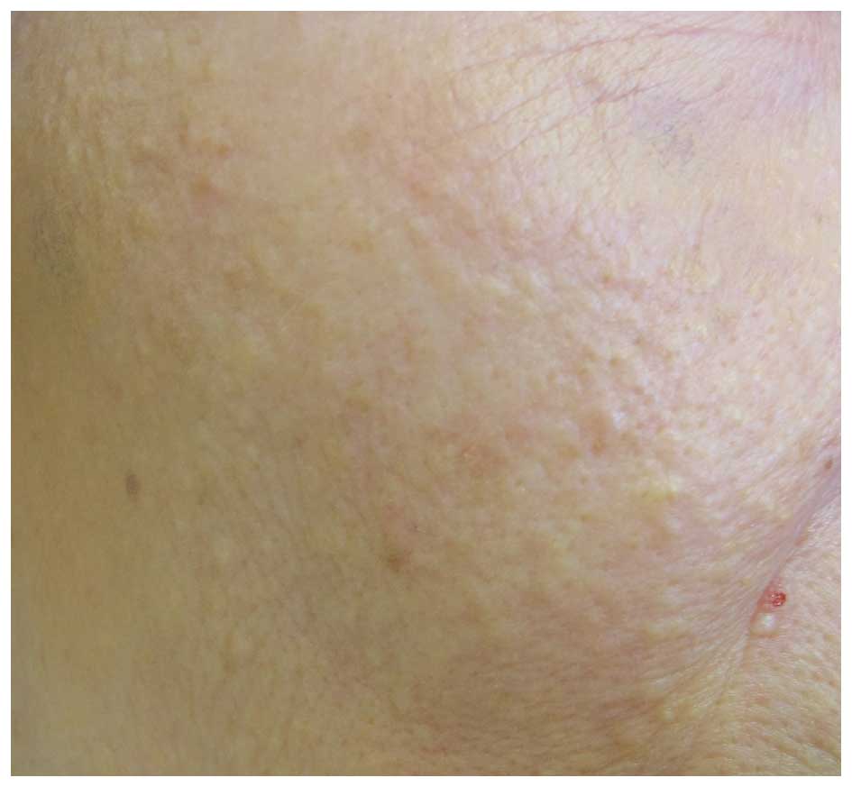
A 74-year-old woman presented to the Department of Dermatology, Meiwa Hospital (Nishinomiya, Japan) on May 2015 with multiple white papules on the face, which had persisted for 10 years (Fig. 1). Milia were diagnosed by the clinical feature of facial white papules.

Figure 1.

Multiple whitish papules on the face of the patient were observed clinically.

The skin biopsy was obtained from a white papule under local anesthesia. The specimens were fixed with formalin, embedded in paraffin, and cut into sections of 5-7-µm thickness. Selected sections were stained with hematoxylin and eosin. Analysis of a skin biopsy resulted in the diagnosis of primary milia, with hematoxylin and eosin staining indicating the presence of dilated follicular cysts. The epithelial lining of the cyst walls consisted of two sections: The more superficial and the deeper sections. Keratohyalin granules were detected in the more superficial section of the epithelial lining, but were not detected in the deeper section. Hair germ structures were identified in the deeper section of the epithelial lining.

On immunohistochemical examination for keratin and filaggrin expression, the labeled streptavidin-biotin (LSAB) method was used (Dako North America, Inc., Carpinteria, CA, USA). The following mouse anti-human keratin antibodies were used in the present study: 34βB4 (anti-K1; dilution 1:50), LP5K (anti-K7; dilution 1:10), LP3K (anti-K8; dilution 1:50), HP1 (anti-K10; dilution 1:50), LL002 (anti-K14; dilution 1:200), LHK15 (anti-K15; dilution 1:40), LL025 (anti-K16; dilution 1:20), E3 (anti-K17; dilution 1:25), 5D3 (anti-K18; dilution 1:20), b170 (anti-K19; dilution 1:100), Ks 20.8 (anti-K20; dilution 1:25), and 15C10 (anti-filaggrin; dilution 1:50), all from Novocastra Laboratories Ltd. (New castle upon Tyne, UK). The LSAB method was applied 
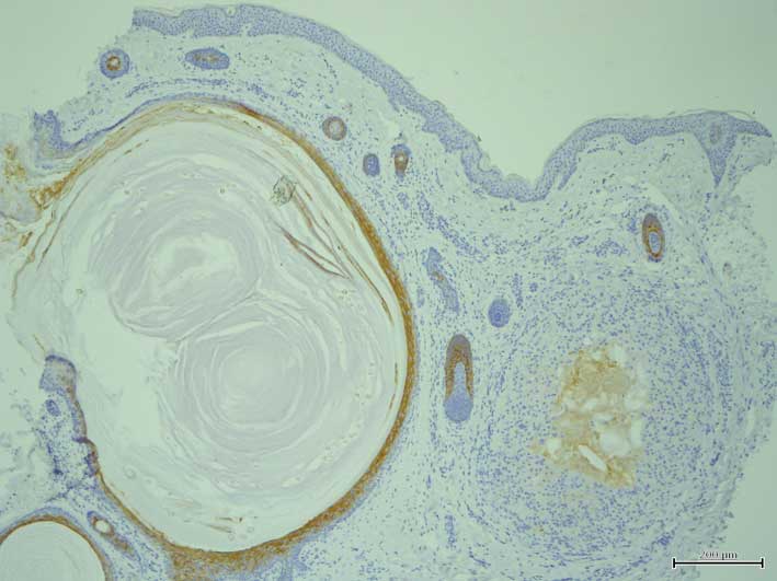
according to the manufacturer's protocol. The levels of keratin expression were assessed using anti-keratin antibodies against K1, 
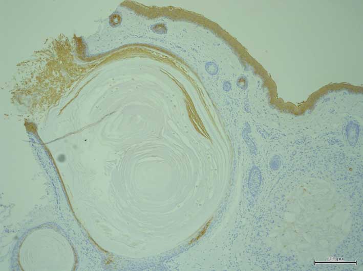
K7, K8, K10, K14, K15, K16, K17, K18, K19, K20 and filaggrin. K1 (Fig. 2) and K10 expression was detected in the suprabasal layers of the more superficial section of the cyst walls and their expression was attenuated in the deeper 
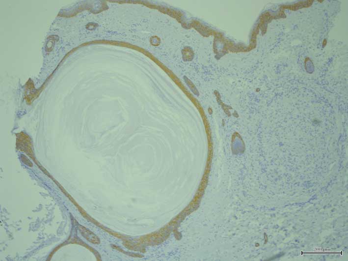
sections. By contrast, K14 expression was observed in all layers of 
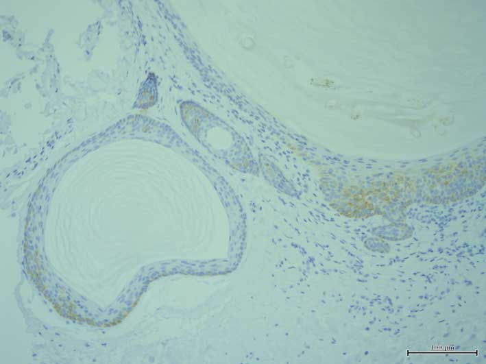
the cyst walls (Fig. 3). Notably, K15 was expressed in the outermost layer of the deeper area of the cyst walls and hair germ structures (Fig. 4), while K17 was expressed in all layers of the cyst walls (Fig. 5). The expression of other 
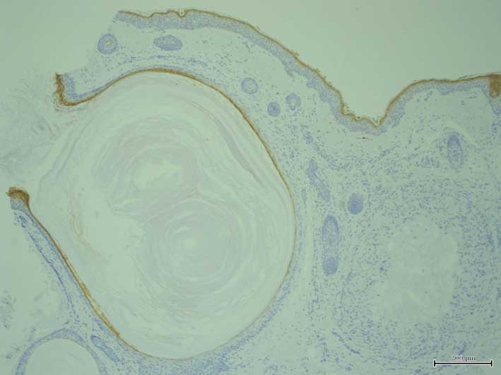
keratins was not detected in the cyst walls. Filaggrin was expressed in the superficial layer of the more superficial sections of the cyst walls (Fig. 6). The milia were treated with adapalene. However, no remarkable change was observed and the treatment was discontinued.

Figure 2.

K1 was expression was detected in the suprabasal layers of the more superficial section of the cyst walls. However, K1 expression was attenuated in the deeper section of the cyst walls (original magnification, ×4).

Figure 3.

K14 was expressed in all layers of the cyst walls (original magnification, ×4).

Figure 4.

K15 was expressed in the outermost layer of the deeper section of the cyst walls and hair germ structure (original magnification, ×20).

Figure 5.

K17 was expressed in all layers of the cyst walls (original magnification, ×4).

Figure 6.

Filaggrin was expressed in the superficial layer of the more superficial section of the cyst walls (original magnification, ×4).

Discussion

Milia are derived from the lowest portion of the infundibulum of vellus hairs at the level of the sebaceous duct (1). The current study demonstrated that K1 and K10 were expressed in the more superficial sections of the cyst walls, and that filaggrin expression was detected in the more superficial part of the cyst walls, while K15 and K17 were expressed in the deeper sections of the cyst walls. K14 expression was detected in all cyst wall layers. In addition, it was observed that K16 was not expressed in milia. This is different from the results of a previous study (3) and may be explained by the diversified heterogeneity of milia differentiation. It is possible that the present case may not be a conventional example of milia and that individual differences between various cases of milia may exist.

Regarding the previously described pattern of keratin expression in normal hair follicles (5), the current study revealed that keratin expression in the more superficial section of the cyst wall is the same as that of the infundibulum, and that its expression in the deeper section is the same as that of the hair bulge of the outer root sheath. These results suggested that keratin and filaggrin expression in milia correspond to the more superficial section of the infundibulum and the outermost layer of the hair bulge, respectively. Furthermore, K15, a stem pluripotent cell marker, is expressed in the outermost layer in normal hair follicles (5). K15 was present in the outermost layer of the deeper section of cyst walls and hair germ structures in the present study. In trichoepithelioma, K15 expression is observed (6). A previous study demonstrated that milia were the result of cystic alteration of the trichoepitheliomatous bulge proliferation (4).

In conclusion, the present study reported a case of milia with an immunohistological evaluation of keratin and filaggrin expression. According to the pattern of keratin and filaggrin expression observed in the current study, milia may originate from the outermost cells of the hair bulge of the outer root sheath. Further studies concerning keratin and filaggrin expression in milia are necessary to confirm this hypothesis.

References

1 

Berk DR and Bayliss SJ: Milia: A review and classification. J Am Acad Dermatol. 59:1050–1063. 2008. View Article : Google Scholar : PubMed/NCBI

2 

Epstein W and Kligman AM: The pathogenesis of milia and benign tumors of the skin. J Invest Dermatol. 26:1–11. 1956. View Article : Google Scholar : PubMed/NCBI

3 

Broekaert D, Leigh IM, Lane EB, Van Muijen GN, Ramaekers FC, De Bersaques J and Coucke P: An immunohistochemical and histochemical study of cytokeratin, involucrin and transglutaminase in seborrhoeic keratosis. Arch Dermatol Res. 285:482–490. 1993. View Article : Google Scholar : PubMed/NCBI

4 

van der Putte SC: The pathogenesis of familial multiple cylindromas, trichoepitheliomas, milia, and spiradenomas. Am J Dermatopathol. 17:271–280. 1995. View Article : Google Scholar : PubMed/NCBI

5 

Kurokawa I, Takahashi K, Moll I and Moll R: Expression of keratins in cutaneous epithelial tumors and related disorders-distribution and clinical significance. Exp Dermatol. 20:217–228. 2011. View Article : Google Scholar : PubMed/NCBI

6 

Kanitakis J, Bourchany D, Faure M and Claudy A: Expression of the hair stem cell-specific keratin 15 in pilar tumors of the skin. Eur J Dermatol. 9:363–365. 1999.PubMed/NCBI

Related Articles

  • Abstract
  • View
  • Download
  • Twitter
Copy and paste a formatted citation
Spandidos Publications style
Kurokawa I, Kakuno A and Tsubura A: Milia may originate from the outermost layers of the hair bulge of the outer root sheath: A case report. Oncol Lett 12: 5190-5192, 2016.
APA
Kurokawa, I., Kakuno, A., & Tsubura, A. (2016). Milia may originate from the outermost layers of the hair bulge of the outer root sheath: A case report. Oncology Letters, 12, 5190-5192. https://doi.org/10.3892/ol.2016.5335
MLA
Kurokawa, I., Kakuno, A., Tsubura, A."Milia may originate from the outermost layers of the hair bulge of the outer root sheath: A case report". Oncology Letters 12.6 (2016): 5190-5192.
Chicago
Kurokawa, I., Kakuno, A., Tsubura, A."Milia may originate from the outermost layers of the hair bulge of the outer root sheath: A case report". Oncology Letters 12, no. 6 (2016): 5190-5192. https://doi.org/10.3892/ol.2016.5335
Copy and paste a formatted citation
x
Spandidos Publications style
Kurokawa I, Kakuno A and Tsubura A: Milia may originate from the outermost layers of the hair bulge of the outer root sheath: A case report. Oncol Lett 12: 5190-5192, 2016.
APA
Kurokawa, I., Kakuno, A., & Tsubura, A. (2016). Milia may originate from the outermost layers of the hair bulge of the outer root sheath: A case report. Oncology Letters, 12, 5190-5192. https://doi.org/10.3892/ol.2016.5335
MLA
Kurokawa, I., Kakuno, A., Tsubura, A."Milia may originate from the outermost layers of the hair bulge of the outer root sheath: A case report". Oncology Letters 12.6 (2016): 5190-5192.
Chicago
Kurokawa, I., Kakuno, A., Tsubura, A."Milia may originate from the outermost layers of the hair bulge of the outer root sheath: A case report". Oncology Letters 12, no. 6 (2016): 5190-5192. https://doi.org/10.3892/ol.2016.5335
Follow us
  • Twitter
  • LinkedIn
  • Facebook
About
  • Spandidos Publications
  • Careers
  • Cookie Policy
  • Privacy Policy
How can we help?
  • Help
  • Live Chat
  • Contact
  • Email to our Support Team