Introduction
Osteosarcoma is the most common primary bone
malignancy. It is derived from primitive bone-forming mesenchymal
cells and frequently arises in the metaphyses of long bones
(1,2).
The introduction of chemotherapy significantly improves the outcome
for patients, and 5-year event-free survival for localized
osteosarcoma increases from <20 to 60–65% (2,3). However,
the overall 5-year survival rate for osteosarcoma remains unchanged
and has exhibited no marked improvement over recent decades
(2).
Cisplatin is one of the most widely used and
effective chemotherapy drugs for the treatment of various solid
tumors, including those of the breast, brain, lung and testis
(4–7).
It is an alkylating agent and works by causing DNA lesions via the
formation of intrastrand and interstrand crosslinks (8). Cisplatin has been widely used in the
treatment of osteosarcoma (9).
However, chemoresistance and pulmonary metastasis frequently lead
to treatment failure, and the underlying mechanisms remain to be
fully elucidated.
The epithelial-mesenchymal transition (EMT) is a
biological process by which epithelial cells lose their polarity,
disassemble the cell-cell adhesion and become mesenchymal-like
cells (10). The process of EMT is
accompanied by a reduction in the cell-cell adhesion molecule
E-cadherin, and upregulation of more plastic mesenchymal proteins,
including vimentin, N-cadherin and smooth muscle actin (11,12). A
number of transcription factors (TFs), including Snail/Slug and
zinc finger E-box binding homeobox (Zeb)1/2, are involved in this
process. EMT allows the tumor cells to gain elevated migratory
properties and increased invasiveness, which is a critical step in
the process of metastasis leading to cancer spreading and treatment
failure (13,14). Chemotherapy has been reported to
induce EMT in tumor cells. A previous study by the present authors
revealed that osteosarcoma cells expressed a number of
EMT-associated genes, which implied that EMT also has a role in
mesenchymal-derived sarcoma (15).
However, whether cisplatin induces EMT in osteosarcoma remains to
be elucidated. In the present study, the shape of U2OS cells tended
to be diamond and they exhibited an epithelial phenotype compared
with other osteosarcoma cell lines. Therefore, this cell line was
selected to investigate the process of EMT in osteosarcoma.
In the present study, it was observed that cisplatin
exposure promoted mesenchymal characteristics in osteosarcoma and
the underlying mechanisms involved upregulation of Snail. These
data may provide scientific information for targeted therapy of
osteosarcoma.
Materials and methods
Cell culture
The human osteosarcoma cell line U2OS was obtained
from the China Center for Type Culture Collection (Wuhan, China).
U2OS cells were cultured in Dulbecco's modified Eagle's medium
(DMEM; Thermo Fisher Scientific, Inc., Waltham, MA, USA) containing
10% (v/v) fetal bovine serum (FBS; Thermo Fisher Scientific, Inc.)
and 1% (v/v) antibiotics (105 U/ml penicillin,
105 µg/ml streptomycin; GE Healthcare Life Sciences,
Logan, UT, USA). Cells were propagated in a humidified environment
at 37°C with 5% CO2 and 100% humidity. Cell viability
was determined using trypan blue staining (Thermo Fisher
Scientific, Inc.). Medium was replaced every three days.
RNA interference (RNAi)
Small interfering RNA (siRNA) gene expression
knockdown studies were performed according to the manufacturer's
protocol. Each 27mer RNAi duplex was transfected into cells using
Lipofectamine™ 2000 transfection reagent (Thermo Fisher Scientific,
Inc.) according to the manufacturer's protocol. siRNA was
synthesized (Guangzhou RiboBio Co., Ltd., Guangzhou, China) using
the following sequences: Snail, 5′-CCACAGAAAUGGCCAUGGGAAGGCCUC-3′;
and negative control, 5′-UCACAAGGGAGAGAAAGAGAGGAAGGA-3′.
Cell cytotoxicity assay
The cells were seeded into 96-well culture plates
and cultured at 37°C for 24 h to attach. Subsequently, various
doses (0, 2, 4, 6, 8, 10, 12, 14 or 16 µmol/l) of cisplatin
(Sellack Chemicals, Houston, TX, USA) were used to treat cells as
indicated and cultured at 37°C for 24 h. The cells in each well
containing 100 µl medium were incubated with 10 µl cell counting
kit-8 reagent (Beyotime Institute of Biotechnology, Haimen, China)
at 37°C for 2 h. The optical density of each well was subsequently
measured at 450 nm using a microplate reader (Thermo Fisher
Scientific, Inc.).
Reverse transcription-quantitative
polymerase chain reaction
Total RNA was isolated by the RNeasy Plus Mini kit
(Qiagen China Co., Ltd, Shanghai, China). The concentration and
purity of RNA was determined by an ND-1000 spectrophotometer
(NanoDrop Technologies; Thermo Fisher Scientific, Inc.). Reverse
transcription was performed using the TaqMan Reverse Transcription
Reagents (Applied Biosystems; Thermo Fisher Scientific, Inc.).
RT-qPCR was subsequently performed using an ABI 7900 HT Fast
Real-Time PCR system (Applied Biosystems; Thermo Fisher Scientific,
Inc.) in the presence of SYBR-Green PCR Master Mix (Applied
Biosystems; Thermo Fisher Scientific, Inc.). The gene-specific
primers used are listed in Table I.
Target sequences were amplified at 95°C for 10 min, followed by 40
cycles at 95°C for 15 sec and 60°C for 1 min. β-actin was used as
an endogenous normalization control. All assays were performed in
triplicate. The fold change in mRNA expression was determined
according to the method of 2ΔΔCq (16).
 | Table I.Primer sequences used for
quantitative polymerase chain reaction. |
Table I.
Primer sequences used for
quantitative polymerase chain reaction.
| Gene | Primer
sequence |
|---|
| Actin |
|
|
Forward |
5′-CACCCAGCACAATGAAGATCAAGAT-3′ |
|
Reverse |
5′-CCAGTTTTTAAATCCTGAGTCAAGC-3′ |
| Snail |
|
|
Forward |
5′-TTACCTTCCAGCAGCCCTACGA-3′ |
|
Rerverse |
5′-GAGCCTTTCCCACTGTCCTCAT-3′ |
| Slug |
|
|
Forward |
5′-TCCTGGTCAAGAAGCATTTCA-3′ |
|
Reverse |
5′-CGCCCCAAAGATGAGGAGTAT-3′ |
| Zeb1 |
|
|
Forward |
5′-GCAGTCTGGGTGTAATCGTAAAT-3′ |
|
Reverse |
5′-TTGCCGTATCTGTGGTCGTG-3′ |
| Zeb2 |
|
|
Forward |
5′-TCCCTTCTGCGACATAAATACG-3′ |
|
Reverse |
5′-TGTGATTCATGTGCTGCGAGTA-3′ |
Immunocytofluorescence staining
The cells were seeded on square coverslips in
six-well plates for 24 h to allow them to attach. Subsequently, the
cells were fixed, permeated and blocked using the Immunol Fluorence
Staining kit (Beyotime Institute of Biotechnology). The cells were
then incubated with anti-E-cadherin antibody (diluted at 1:100;
701134; Thermo Fisher Scientific, Inc.), anti-N-cadherin antibody
(diluted at 1:200; PA5-19486; Thermo Fisher Scientific, Inc.) and
anti-vimentin antibody (diluted at 1:200; PA5-27231; Thermo Fisher
Scientific, Inc.) overnight at 4°C. Secondary antibody (diluted at
1:200; ab150077; Abcam, Cambridge, MA, USA) was applied for 1 h at
room temperature. The cells were counterstained with DAPI and
washed with PBS following each step of the staining procedure.
Coverslips were mounted using Anti-fade Fluorescence Mounting
Medium (Beyotime Institute of Biotechnology). The long and short
axes of cells were measured using the Zeiss LSM Image Examiner
software (version 4.2.0.121; Carl Zeiss AG, Oberkochen, Germany),
and the long/short axis ratio was determined by counting 100 cells
per experiment.
Western blotting
Cell lysates were extracted using
radioimmunoprecipitation assay lysis buffer containing protease
inhibitor cocktail (Sigma-Aldrich; EMD Millipore, Billerica, MA,
USA). Protein concentrations were determined using the
bicinchoninic acid method (Sigma-Aldrich; EMD Millipore). Cell
lysates containing 40 µg protein were loaded and separated on 10%
SDS-PAGE gels and subsequently transferred to polyvinylidene
fluoride membranes (Thermo Fisher Scientific, Inc.). The membranes
were blocked in Tris Buffered Saline with 5% (w/v) skimmed milk and
0.05% Tween 20 (Thermo Fisher Scientific, Inc.) for 1 h at 37°C.
Primary antibodies were incubated overnight at 4°C. The primary
antibodies and mouse monoclonal anti-β-actin were purchased from
Abcam (anti-Snail antibody; ab180714; diluted at 1:1,000; anti-Slug
antibody; ab27568; diluted at 1:1,000; anti-N-cadherin antibody;
PA5-19486; diluted at 1:1,000; anti-β-actin antibody; ab8226;
diluted at 1:2,000) The pH2AX antibody (MBS837487; diluted at
1:2,000) was purchased from MyBioSource, Inc. (San Diego, CA, USA).
The membranes were washed and incubated with secondary antibody
(ab6721; Abcam) at 1:5,000 dilution for 2 h at room temperature.
The membranes were washed again and developed using enhanced
chemiluminescence substrate (Sigma-Aldrich; EMD Millipore).
Quantitative analysis was performed using QuantiOne imaging
software (Bio-Rad Laboratories, Inc., Hercules, CA, USA).
Wound healing assay
A total of 5×10⁵ cells were seeded into
6-well plates and cultured overnight at 37°C to attach. When
adherent cells reached ~90% confluence, a scratch was made using a
200-µl pipette tip. The cells were washed three times and further
incubated at 37°C for 24 h. The migration was observed and recorded
under a phase contrast microscope (Nikon Eclipse TE2000-U; Nikon
Corporation, Tokyo, Japan).
Transwell assay
Matrigel-coated Transwell invasion assay plates
(Corning Inc., Corning, NY, USA) were used for this assay. Cells
were placed in the upper chamber (1×10⁵ cells/well) in
DMEM medium with 0.1% FBS. The lower chambers were filled with DMEM
medium with 10% FBS. Following culturing for 24 h at 37°C, the
inserts were removed and the inner side was wiped with cotton
swabs. The filters were stained with Harris's hematoxylin solution
(Sigma-Aldrich; EMD Millipore) for 20 min and peeled off following
washing three times. The migrated cells were counted by a light
microscope (Nikon Eclipse TE2000-U).
Animals and transplantation assay
For the in vivo assay, male NOD/SCID mice
(n=14; 6-week-old; 18–23 g; SPF) were purchased from and maintained
maintained (humidity, 50–60%; temperature, 18–22°C; light cycle,
10–14 h a day) at the Wuhan University Center for Animal Experiment
(Wuhan, China). The care and use of animals was reviewed and
approved by the Institutional Animal Care and Use Committee
(approval number, 2011006). A total of 5×10⁶ cells were
subcutaneously injected into 2 mice, and the xenografts were
obtained following two weeks of growth, and the 2 mice were
sacrificed by CO2. The tumor xenografts were divided
into small pieces of ~5 mm3 and transplanted
subcutaneously. The mice were divided into a cisplatin treated
group (peritoneal injection of 5 mg/kg cisplatin once a week; n=6)
and the control group (receiving the same amount of saline once a
week; n=6). Following 4 weeks of rearing, the mice were sacrificed
by CO2. Tumor samples and lung tissues were obtained and
usde for subsequent immunohistochemistry experiments.
Immunohistochemistry
Tissues were fixed in 10% neutral-buffered formalin,
processed and embedded in paraffin. Tissue sections were
deparaffinized and rehydrated in an ethanol series. Sections were
blocked for nonspecific binding with 1% normal serum (Thermo Fisher
Scientific, Inc.) and incubated with the primary anti-Snail
(ab53519; diluted at 1:500; Abcam) and anti-N cadherin (PA5-19486;
diluted at 1:300; Thermo Fisher Scientific, Inc.) antibodies
overnight at 4°C. Subsequently, immunostaining was developed using
3,3′-diaminobenzidine (Vector Laboratories, Inc., Burlingame, CA,
USA) followed by hematoxylin counterstaining (Sigma-Aldrich; EMD
Millipore). Immunostaining was visualized using a fluorescence
microscope (Eclipse 80i Fluorescence Microscope; Nikon
Corporation).
Statistics analysis
Each sample was analyzed in triplicate, and
experiments were repeated at least two times. The mean, standard
error and P-values base on the two-sample two-tailed t-test
were calculated with Excel 2013 software (Microsoft Corporation,
Redmond, WA, USA). P<0.05 was considered to indicate a
statistically significant difference.
Results
Cisplatin treatment promotes
mesenchymal-like properties in osteosarcoma
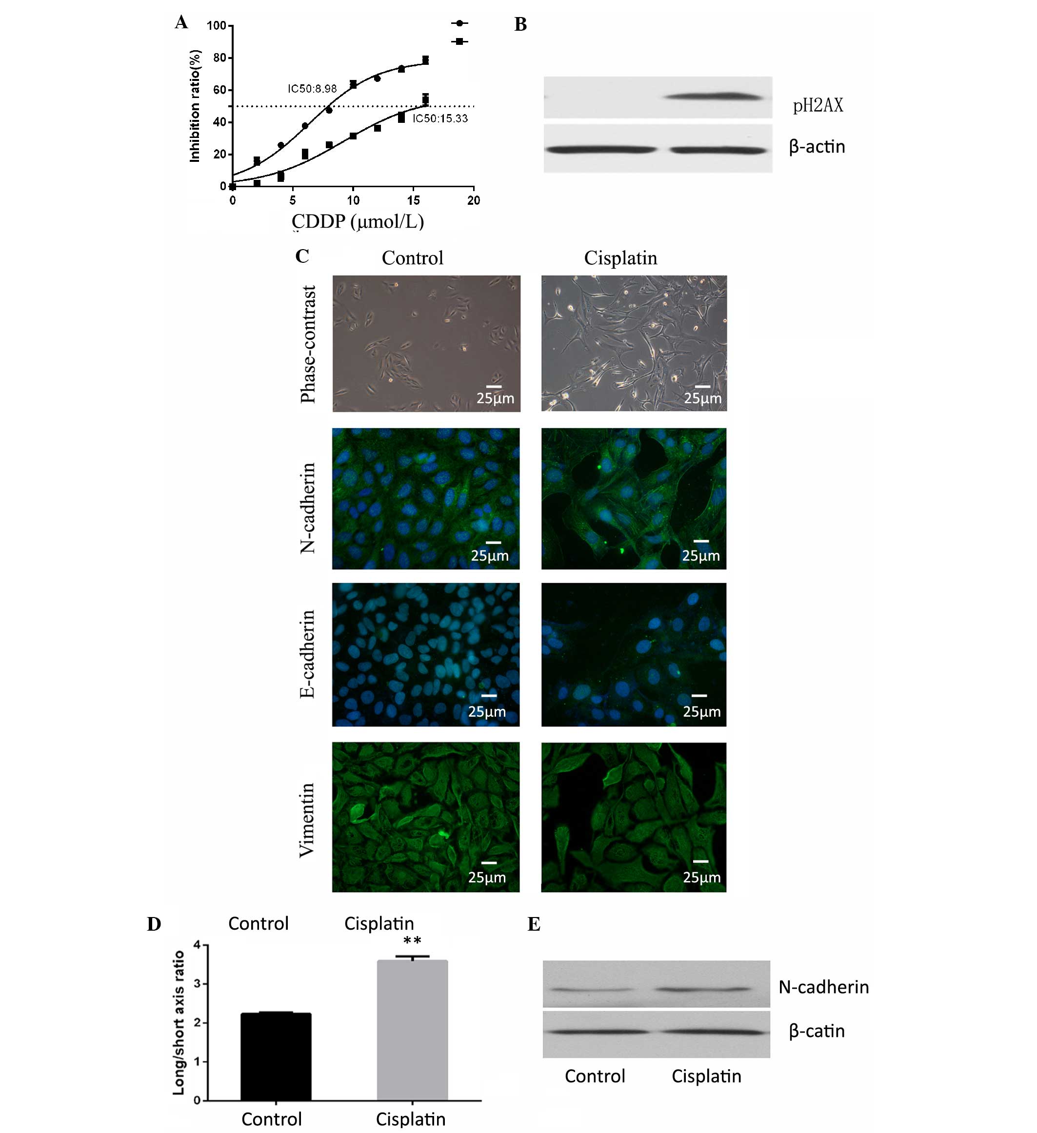
Cells were treated with 5 µM of cisplatin for 24 h
and maintained in normal conditions for 5 days to recover from the
chemotherapeutic stress. The surviving cells were observed to
exhibit increased resistance to cisplatin (Fig. 1A). The DNA damage marker pH2AX was
investigated by western blotting to observe the effect of cisplatin
on the cells, and the drug was confirmed to be effective (Fig. 1B). The present study observed cell
morphology changes in these cisplatin-resistant cells, in which
they appeared to possess a more spindle-like shape. Although there
were no differences between the two groups in terms of E-cadherin
and vimentin expression, N-cadherin expression was observed to be
significantly increased in the cisplatin treated group compared
with the control cells (Fig. 1C). EMT
is frequently accompanied by an alteration from a rounded to
spindle shape in terms of cell morphology (17). Therefore, the present study assessed
the long/short axis ratio in various cells. The cells treated with
cisplatin were observed to have an average ratio of 3.591±0.119,
which was increased compared with the cells of the control group
(average ratio, 2.232±0.041; P=0.001; Fig. 1D). In addition, the expression of
epithelial and mesenchymal markers was examined by western blot
analysis. The epithelial markers, E-cadherin and cytokeratin, were
not detectable in either the cisplatin of the control group.
Mesenchymal-marker N-cadherin was highly expressed in the cisplatin
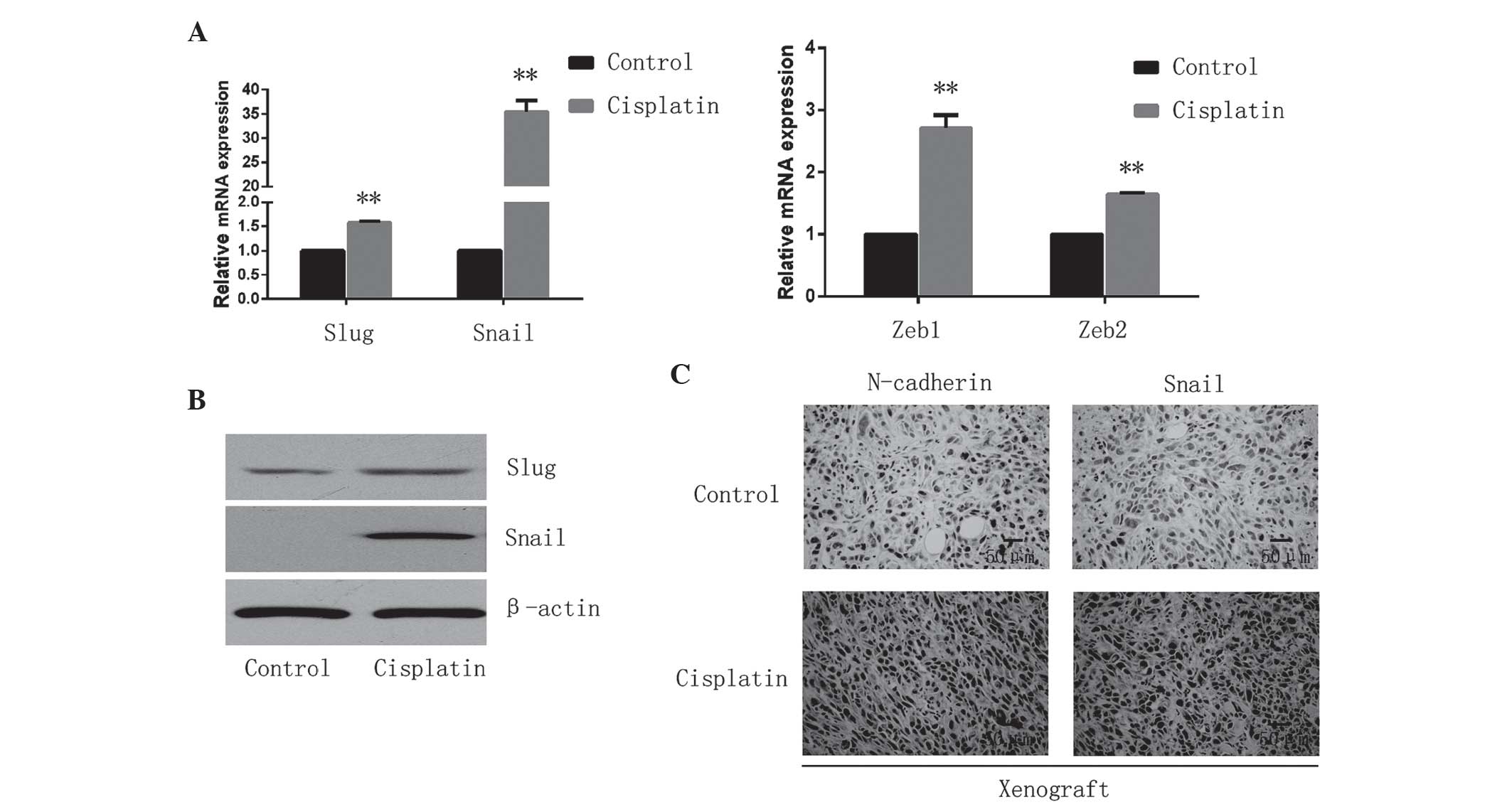
group (Fig. 1E). EMT-inducing TFs,
including Snail/Slug and Zeb1/2, suppress epithelial marker
expression and induce the expression of mesenchymal markers,
facilitating qPCR analysis that led to the observation that these
four EMT-TFs were significantly upregulated (Snail, P<0.001;
Slug, P<0.001; Zeb1, P=0.0011; Zeb2, P<0.001) in the
cisplatin treated cells (Fig. 2A),
among which the expression of the Snail gene exhibited the most
marked increase with levels of relative mRNA expression of
35.44±2.35. Furthermore, western blotting confirmed the upregulated
expression of Snail and Slug in the cisplatin treated cells
(Fig. 2B). In addition, the in
vivo xenograft assay confirmed that the expression of
N-cadherin and Snail was increased following cisplatin exposure
(Fig. 2C).
Cisplatin treated cells are prone to
migration and invasion
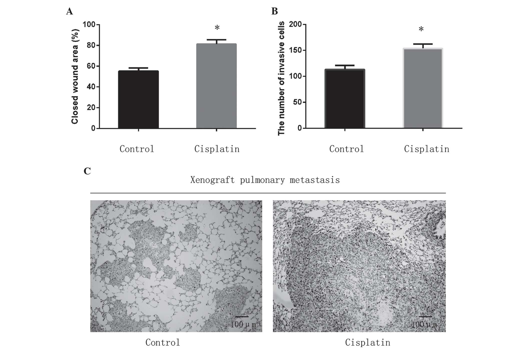
Subsequently, the present study investigated the
migratory and invasive capacity following cisplatin treatment. The
cisplatin treated cells exhibited significantly increased cell
migration compared with the control group (P=0.001; Fig. 3A). The invasive potential through the
Matrigel of the cisplatin treated group was also enhanced, with an
average fold increase of 1.31±0.05 (P=0.002; Fig. 3B). To investigate the in vivo
metastatic capacity, the present study examined pulmonary lesions
in both groups. It was observed that cisplatin exposure promoted
pulmonary metastasis and induced more severe lung destruction,
although the primary tumor was inhibited (Fig. 3C).
Knockdown of Snail increases cisplatin
sensitivity and reverses cisplatin-induced EMT
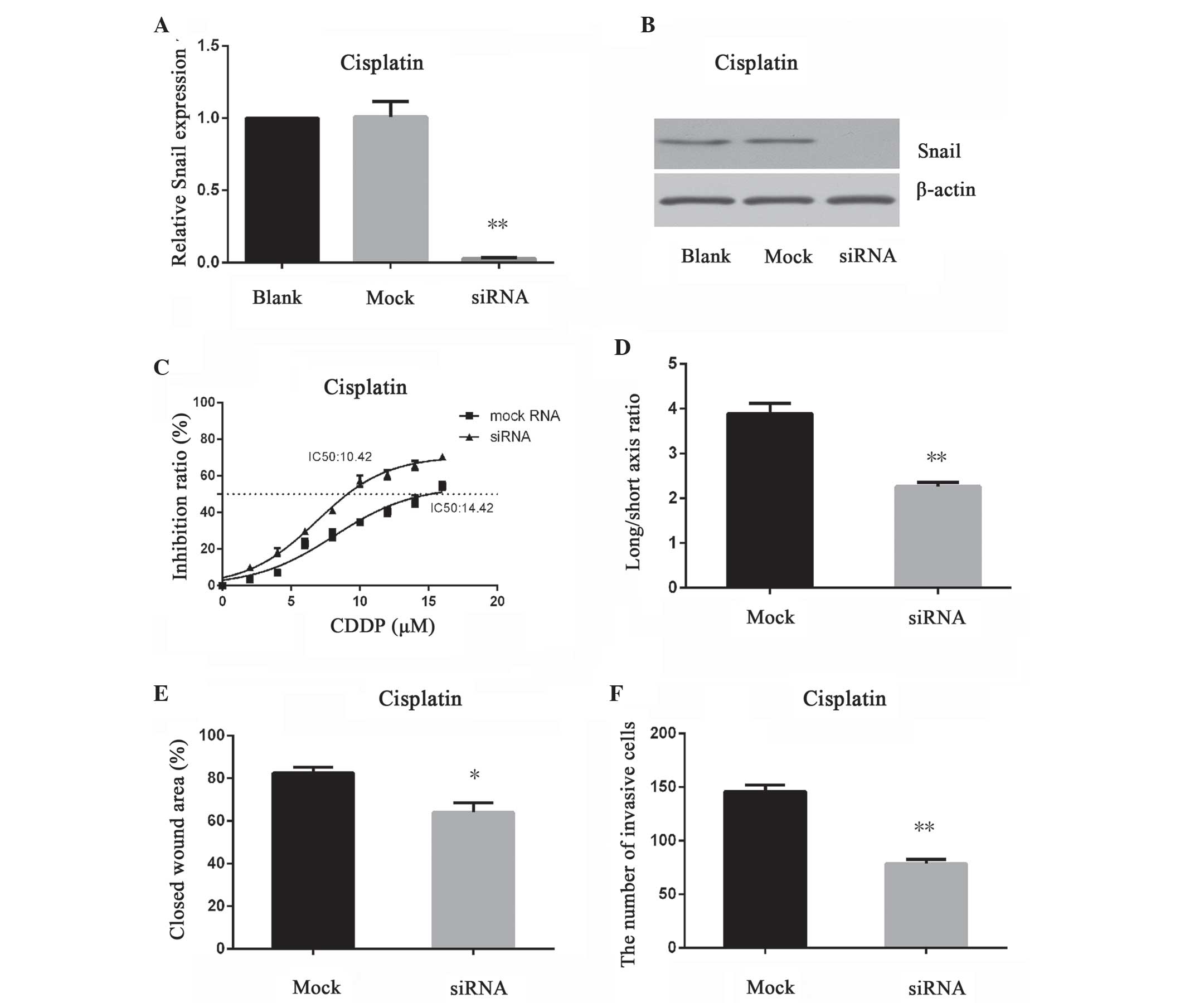
The present study applied RNAi techniques to
knockdown the expression of Snail. The efficiency of RNAi was
confirmed by qPCR and western blotting (blank vs. mock group,
P=0.88; blank vs. siRNA group, P<0.001; Fig. 4A and B). The IC50 for
cisplatin was decreased when Snail was inhibited, which indicated
that the sensitivity of osteosarcoma cells to cisplatin was
enhanced (Fig. 4C). When the Snail
knockdown cells were exposed to cisplatin, they exhibited a less
spindle-like shape, with a decreased long/short axis ratio compared
with mock cells (P<0.001; Fig.
4D). Furthermore, Snail inhibition blocked cisplatin-induced
cell migration (P<0.001) and invasion (P<0.001) in
vitro (Fig. 4E and F).
Discussion
The introduction of chemotherapy has improved
five-year survival rates of osteosarcoma (1). However, recurrence and metastasis lead
to poor prognosis and are frequently associated with
chemoresistance (18). EMT, a common
biological process, has been reported to be associated with tumor
invasiveness and migration in breast, skin and lung cancer
(19–21). A previous study by the present authors
reported that the concept of EMT involvement in invasiveness and
migration also applies to osteosarcoma (15). In the present study, osteosarcoma
cells were treated with a sublethal dose of cisplatin, and any
surviving cells presented with enhanced mesenchymal-like
characteristics. These cells were observed to be more resistant to
cisplatin treatment, as shown by cell cytotoxicity assay. In
addition, the cells demonstrated an increased expression of
mesenchymal markers and an increased long/short axis ratio compared
with control cells, which indicated a mesenchymal phenotype.
Furthermore, the cells exhibited increased expression of
EMT-inducing TFs, including Snail, Slug and Zeb1/2, which are
critical in the process of EMT. The cells were also more likely to
invade and migrate in vitro. In addition, xenografts treated
with cisplatin demonstrated increased levels of EMT-TFs compared
with those injected with saline. As the treatment time was short,
it may be considered that cells with high expression of EMT-TFs,
particularly Snail, demonstrated resistance to cisplatin and
survived drug treatment.
In addition, to elucidate the underlying mechanisms
involved in cisplatin-induced EMT, cells were transfected with
siRNA targeting Snail, which was observed to have the most marked
increase following cisplatin exposure in the present study.
Following transfection, it was observed that the expression of
Snail in the cisplatin group was decreased, while the mock cells
remained unchanged. The results of the present study confirmed that
the siRNA was successfully constructed and transfected into cells.
Snail is a zinc-finger transcriptional repressor, which is critical
to numerous biological processes, particularly in EMT (22,23). A
number of studies have demonstrated that Snail is able to suppress
the expression of epithelial genes, primarily E-cadherin, and
activate the expression of mesenchymal proteins, including
N-cadherin and fibronectin (17,24,25). In
the present study, cells of the cisplatin group demonstrated
increased expression of N-cadherin and Snail, and exhibited a
mesenchymal phenotype. When Snail was silenced, the cells reverted
to an epithelial-like phenotype. As osteosarcoma is a
mesenchymal-derived tumor, the expression of E-cadherin, an
epithelial gene, was low in both groups.
Snail participates in the process of EMT; in
addition, recent studies have proven that Snail is involved in
chemoresistance to numerous chemotherapeutic reagents (24). Hsu et al (26) reported that the expression of Snail
determined the resistance to cisplatin in head and neck squamous
cell carcinoma and non-small cell lung carcinoma cells and Zhang
et al (27) discovered that
Snail conferred resistance to 5-fluorouracil in breast cancer
cells. Similarly, in the present study, it was observed that
cisplatin induced the expression of Snail in osteosarcoma cells.
When Snail was suppressed, the cells became more sensitive to
cisplatin. These observations appeared to indicate that Snail was
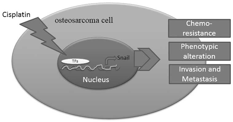
involved in resistance to cisplatin in osteosarcoma. The schematic
diagram in Fig. 5 shows that
cisplatin promoted the binding of TFs with Snail promoter and
induced its expression. Snail subsequently induced EMT leading to
resistance to chemotherapy, phenotypic alteration and an increased
capability of invasion and metastasis (Fig. 5).
Kudo-Saito et al (28) observed that knockdown of Snail halted
tumor metastasis in melanoma. Accordingly, it was observed that the
knockdown of Snail may suppress the process of EMT and inhibited
the invasion and metastasis of osteosarcoma cells. Cancer stem
cells are subpopulations in tumors that possess self-renewing
capabilities (29). For example,
cells with a cluster of differentiation
(CD)44high/CD24low phenotype were regarded as
possessing stem cell traits in breast cancer (30). Mani et al (31) reported that
CD44high/CD24low cells demonstrated a
decrease in E-cadherin expression and elevations in Snail and
vimentin expression. Furthermore, in human mammary epithelial
cells, Snail-induced EMT cells increased the capability of
mammosphere-forming by 30-fold compared with the control group
(32). The pluripotent capability of
stem cells was maintained in part by homeobox protein Nanog,
octamer-binding transcription factor 4 (Oct4) and sex-determining
region Y-box 2 (Sox2) (32,33). In a similar study of lung cancer, Wang
et al (34) used A549/CDDP, a
cisplatin resistance cell line, and observed that A549/CDDP cells
exhibited increased expression levels of Nanog, Oct4 and Sox2.
A549/CDDP cells also demonstrated increased capacities of migration
and invasiveness and a higher expression of Snail. The knockdown of
Snail caused a decline in migration and invasiveness in A549/CDDP
cells, and the phosphoinositide 3-kinase (PI3K)/Akt signaling
pathway was reported to be involved in this process (35). As well as PI3K/Akt, Snail was also
able to activate the mitogen-activated protein kinase survival
signaling pathways, and be activated by nuclear factor (NF)-κB
(35,36). Therapies with nitric oxide, the
proteasome inhibitor NPI-0052 targeting NF-κB and Snail were proven
to be effective in prostate cancer and B-non-Hodgkin's lymphoma
(37).
In conclusion, the EMT concept may be applied to
osteosarcoma, which is a mesenchymal-derived tumor. Targeting Snail
and other EMT-TFs may assist with preventing metastasis of
osteosarcoma patients. Chemical inhibitors targeting Snail and
other EMT-TFs have been reported to be effective in cell lines and
animal experiments (38–41). As well as U2OS, future studies must
investigate other cell lines in osteosarcoma to increase
credibility. Additional studies and clinical trials are required to
evaluate the efficacy of targeting EMT-TFs in the prevention of
cancer recurrence, metastasis and chemoresistance.
Acknowledgments
The present study was supported by grants from the
National Natural Science Foundation of China (grant no., 81341078)
and the Science and Technology Foundation of Wuhan (grant no.,
2014062801011264).
References
|
1
|
Ottaviani G and Jaffe N: The epidemiology
of osteosarcoma. Cancer Treat Res. 152:3–13. 2009. View Article : Google Scholar : PubMed/NCBI
|
|
2
|
Mirabello L, Troisi RJ and Savage SA:
Osteosarcoma incidence and survival rates from 1973 to 2004: Data
from the Surveillance, Epidemiology, and End Results Program.
Cancer. 115:1531–1543. 2009. View Article : Google Scholar : PubMed/NCBI
|
|
3
|
Clark JC, Dass CR and Choong PF: A review
of clinical and molecular prognostic factors in osteosarcoma. J
Cancer Res Clin Oncol. 134:281–297. 2008. View Article : Google Scholar : PubMed/NCBI
|
|
4
|
Chen Y, Han F, Cao LH, Li C, Wang JW, Li
Q, Zheng W, Guo ZX, Li AH and Zhou JH: Dose-response relationship
in cisplatin-treated breast cancer xenografts monitored with
dynamic contrast-enhanced ultrasound. BMC Cancer. 15:1362015.
View Article : Google Scholar : PubMed/NCBI
|
|
5
|
Shahmabadi H Ebrahimi, Movahedi F,
Esfahani M Koohi Moftakhari, Alavi SE, Eslamifar A, Anaraki G
Mohammadi and Akbarzadeh A: Efficacy of Cisplatin-loaded polybutyl
cyanoacrylate nanoparticles on the glioblastoma. Tumour Biol.
35:4799–4806. 2014. View Article : Google Scholar : PubMed/NCBI
|
|
6
|
Pirker R: Adjuvant chemotherapy in
patients with completely resected non-small cell lung cancer.
Transl Lung Cancer Res. 3:305–310. 2014.PubMed/NCBI
|
|
7
|
Hjelle LV, Gundersen PO, Oldenburg J,
Brydøy M, Tandstad T, Wilsgaard T, Fosså SD, Bremnes RM and Haugnes
HS: Long-term platinum retention after platinum-based chemotherapy
in testicular cancer survivors: A 20-year follow-up study.
Anticancer Res. 35:1619–1625. 2015.PubMed/NCBI
|
|
8
|
Woźniak K and Błasiak J: Recognition and
repair of DNA-cisplatin adducts. Acta Biochim Pol. 49:583–596.
2002.PubMed/NCBI
|
|
9
|
Haddox CL, Han G, Anijar L, Binitie O,
Letson GD, Bui MM and Reed DR: Osteosarcoma in pediatric patients
and young adults: A single institution retrospective review of
presentation, therapy and outcome. Sarcoma. 2014:4025092014.
View Article : Google Scholar : PubMed/NCBI
|
|
10
|
Jeanes A, Gottardi CJ and Yap AS:
Cadherins and cancer: How does cadherin dysfunction promote tumor
progression? Oncogene. 27:6920–6929. 2008. View Article : Google Scholar : PubMed/NCBI
|
|
11
|
Han SP and Yap AS: The cytoskeleton and
classical cadherin adhesions. Subcell Biochem. 60:111–135. 2012.
View Article : Google Scholar : PubMed/NCBI
|
|
12
|
Azmi AS: Unveiling the role of nuclear
transport in epithelial-to-mesenchymal transition. Curr Cancer Drug
Targets. 13:906–914. 2013. View Article : Google Scholar : PubMed/NCBI
|
|
13
|
Patel IS, Madan P, Getsios S, Bertrand MA
and MacCalman CD: Cadherin switching in ovarian cancer progression.
Int J Cancer. 106:172–177. 2003. View Article : Google Scholar : PubMed/NCBI
|
|
14
|
Lim J and Thiery JP:
Epithelial-mesenchymal transitions: Insights from development.
Development. 139:3471–3486. 2012. View Article : Google Scholar : PubMed/NCBI
|
|
15
|
Yu L, Liu S, Guo W, Zhang C, Zhang B, Yan
H and Wu Z: hTERT promoter activity identifies osteosarcoma cells
with increased EMT characteristics. Oncol Lett. 7:239–244.
2014.PubMed/NCBI
|
|
16
|
Livak and Schmittgen, . Analysis of
relative gene expression data using real-time quantitative PCR and
the 2-ΔΔCt method. Methods. 25:402–408. 2001. View Article : Google Scholar : PubMed/NCBI
|
|
17
|
Wang Y, Shi J, Chai K, Ying X and Zhou BP:
The Role of Snail in EMT and Tumorigenesis. Curr Cancer Drug
Targets. 13:963–972. 2013. View Article : Google Scholar : PubMed/NCBI
|
|
18
|
Bruheim S, Xi Y, Ju J and Fodstad O: Gene
expression profiles classify human osteosarcoma xenografts
according to sensitivity to doxorubicin, cisplatin, and ifosfamide.
Clin Cancer Res. 15:7161–7169. 2009. View Article : Google Scholar : PubMed/NCBI
|
|
19
|
Zhou F, Liu H, Zhang X, Shen Y, Zheng D,
Zhang A, Lai Y and Li H: Proline-rich protein 11 regulates
epithelial-to-mesenchymal transition to promote breast cancer cell
invasion. Int J Clin Exp Pathol. 7:8692–8699. 2014.PubMed/NCBI
|
|
20
|
Chen CH, Lin DS, Cheng CW, Lin CJ, Lo YK,
Yen CC, Lee AY and Hsiao CD: Cdc6 cooperates with c-Myc to promote
genome instability and epithelial to mesenchymal transition EMT in
zebrafish. Oncotarget. 5:6300–6311. 2014. View Article : Google Scholar : PubMed/NCBI
|
|
21
|
Grelet S, Andries V, Polette M, Gilles C,
Staes K, Martin AP, Kileztky C, Terryn C, Dalstein V, Cheng CW, et
al: The human NANOS3 gene contributes to lung tumour invasion by
inducing epithelial-mesenchymal transition. J Pathol. 237:25–37.
2015. View Article : Google Scholar : PubMed/NCBI
|
|
22
|
Barrallo-Gimeno A and Nieto MA: The Snail
genes as inducers of cell movement and survival: Implications in
development and cancer. Development. 132:3151–3161. 2005.
View Article : Google Scholar : PubMed/NCBI
|
|
23
|
Peinado H, Olmeda D and Cano A: Snail, Zeb
and bHLH factors in tumour progression: An alliance against the
epithelial phenotype? Nat Rev Cancer. 7:415–428. 2007. View Article : Google Scholar : PubMed/NCBI
|
|
24
|
Kim YS, Yi BR, Kim NH and Choi KC: Role of
the epithelial-mesenchymal transition and its effects on embryonic
stem cells. Exp Mol Med. 46:e1082014. View Article : Google Scholar : PubMed/NCBI
|
|
25
|
Kaufhold S and Bonavida B: Central role of
Snail1 in the regulation of EMT and resistance in cancer: A target
for therapeutic intervention. J Exp Clin Cancer Res. 33:622014.
View Article : Google Scholar : PubMed/NCBI
|
|
26
|
Hsu DS, Lan HY, Huang CH, Tai SK, Chang
SY, Tsai TL, Chang CC, Tzeng CH, Wu KJ, Kao JY and Yang MH:
Regulation of excision repair cross-complementation group 1 by
Snail contributes to cisplatin resistance in head and neck cancer.
Clin Cancer Res. 16:4561–4571. 2010. View Article : Google Scholar : PubMed/NCBI
|
|
27
|
Zhang W, Feng M, Zheng G, Chen Y, Wang X,
Pen B, Yin J, Yu Y and He Z: Chemoresistance to 5-fluorouracil
induces epithelial-mesenchymal transition via up-regulation of
Snail in MCF7 human breast cancer cells. Biochem Biophys Res
Commun. 417:679–685. 2012. View Article : Google Scholar : PubMed/NCBI
|
|
28
|
Kudo-Saito C, Shirako H, Takeuchi T and
Kawakami Y: Cancer metastasis is accelerated through
immunosuppression during Snail-induced EMT of cancer cells. Cancer
Cell. 15:195–206. 2009. View Article : Google Scholar : PubMed/NCBI
|
|
29
|
Gupta PB, Chaffer CL and Weinberg RA:
Cancer stem cells: Mirage or reality? Nat Med. 15:1010–1012. 2009.
View Article : Google Scholar : PubMed/NCBI
|
|
30
|
Al-Hajj M, Wicha MS, Benito-Hernandez A,
Morrison SJ and Clarke MF: Prospective identification of
tumorigenic breast cancer cells. Proc Natl Acad Sci USA.
100:3983–3988. 2003. View Article : Google Scholar : PubMed/NCBI
|
|
31
|
Mani SA, Guo W, Liao MJ, Eaton EN, Ayyanan
A, Zhou AY, Brooks M, Reinhard F, Zhang CC, Shipitsin M, et al: The
epithelial-mesenchymal transition generates cells with properties
of stem cells. Cell. 133:704–715. 2008. View Article : Google Scholar : PubMed/NCBI
|
|
32
|
Takahashi K and Yamanaka S: Induction of
pluripotent stem cells from mouse embryonic and adult fibroblast
cultures by defined factors. Cell. 126:663–676. 2006. View Article : Google Scholar : PubMed/NCBI
|
|
33
|
Moon JH, Yun W, Kim J, Hyeon S, Kang PJ,
Park G, Kim A, Oh S, Whang KY, Kim DW, et al: Reprogramming of
mouse fibroblasts into induced pluripotent stem cells with Nanog.
Biochem Biophys Res Commun. 431:444–449. 2013. View Article : Google Scholar : PubMed/NCBI
|
|
34
|
Wang H, Zhang G, Zhang H, Zhang F, Zhou B,
Ning F, Wang HS, Cai SH and Du J: Acquisition of
epithelial-mesenchymal transition phenotype and cancer stem
cell-like properties in cisplatin-resistant lung cancer cells
through AKT/β-catenin/Snail signaling pathway. Eur J Pharmacol.
723:156–166. 2014. View Article : Google Scholar : PubMed/NCBI
|
|
35
|
Vega S, Morales AV, Ocaña OH, Valdés F,
Fabregat I and Nieto MA: Snail blocks the cell cycle and confers
resistance to cell death. Genes Dev. 18:1131–1143. 2004. View Article : Google Scholar : PubMed/NCBI
|
|
36
|
Vega MI, Baritaki S, Huerta-Yepez S,
Martinez-Paniagua MA and Bonavida B: A potential mechanism of
rituximab-induced inhibition of tumor growth through its
sensitization to tumor necrosis factor-related apoptosis-inducing
ligand-expressing host cytotoxic cells. Leuk Lymphoma. 52:108–121.
2011. View Article : Google Scholar : PubMed/NCBI
|
|
37
|
Baritaki S, Yeung K, Palladino M, Berenson
J and Bonavida B: Pivotal roles of snail inhibition and RKIP
induction by the proteasome inhibitor NPI-0052 in tumor cell
chemoimmunosensitization. Cancer Res. 69:8376–8385. 2009.
View Article : Google Scholar : PubMed/NCBI
|
|
38
|
Shah P, Gau Y and Sabnis G: Histone
deacetylase inhibitor entinostat reverses epithelial to mesenchymal
transition of breast cancer cells by reversing the repression of
E-cadherin. Breast Cancer Res Treat. 143:99–111. 2014. View Article : Google Scholar : PubMed/NCBI
|
|
39
|
Miller CR, Oliver KE and Farley JH: MEK1/2
inhibitors in the treatment of gynecologic malignancies. Gynecol
Oncol. 133:128–137. 2014. View Article : Google Scholar : PubMed/NCBI
|
|
40
|
McCubrey JA, Steelman LS, Chappell WH,
Abrams SL, Franklin RA, Montalto G, Cervello M, Libra M, Candido S,
Malaponte G, et al: Ras/Raf/MEK/ERK and PI3K/PTEN/Akt/mTOR cascade
inhibitors: How mutations can result in therapy resistance and how
to overcome resistance. Oncotarget. 3:1068–1111. 2012. View Article : Google Scholar : PubMed/NCBI
|
|
41
|
Bruheim S, Xi Y, Ju J and Fodstad O: Gene
expression profiles classify human osteosarcoma xenografts
according to sensitivity to doxorubicin, cisplatin, and ifosfamide.
Clin Cancer Res. 15:7161–7169. 2009. View Article : Google Scholar : PubMed/NCBI
|