Introduction
Hepatocellular carcinoma (HCC) is one of the leading
causes of cancer-associated mortality worldwide (1). The primary treatments for liver cancer
include surgery, radiation and chemotherapy, however, the success
rate of these treatments is limited by toxicity and a high rate of
relapse (2). Therefore, the
identification of efficient novel treatment methods with low side
effect profiles is of high priority in HCC research.
Antimicrobial peptides (AMPs), which are isolated
from a range of organisms, including insects, fish, amphibians and
mammals (3,4), are typically cationic and amphipathic
molecules (5). AMPs exhibit a wide
range of antibacterial activities through direct electrostatic
bonding to the anionic surfaces of bacteria, leading to the
disruption of their plasma membrane (6). Furthermore, recent studies have reported
that AMPs exhibit anti-tumor activity (7–9). This may
be because cancer cells contain increased levels of anionic plasma
membrane phospholipids compared with normal cells, allowing AMPs to
bind and kill cancer cells with increased efficacy (10).
Bovine lactoferricin (LfcinB) is a
lactoferrin-derived cationic AMP, which exhibits chemotherapeutic
properties in a variety of cancer cells, while demonstrating low
toxicity to healthy cells (11). The
classical AMP LfcinB is composed of 25 amino acids
(FKCRRWQWRMKKLGAPSITCVRRAF) and is limited in its use as a
chemotherapeutic due to its high toxicity, poor efficacy and high
synthesis cost (7). Therefore, the
development of novel AMPs comprised of fewer amino acids and with
decreased toxicity is essential for applications in cancer
treatment.
In the present study, the novel peptide LfcinB-P13
(KCRRWLKRMKKLG), an analog of LfcinB, was designed and synthesized
as an amphipathic AMP of 13 amino acids in length. The effect of
LfcinB-P13 on the human HCC cell line SMMC7721 was studied. The
results demonstrated that the synthetic LfinB-P13-induced apoptosis
in SMMC7721 cells was caspase-dependent and reactive oxygen species
(ROS)-mediated.
Materials and methods
Reagents and experimental animals
LfcinB-P13 (KCRRWLKRMKKLG) was synthesized by GL
Biochem, Ltd. (Shanghai, China) using stepwise solid-phase peptide
synthesis. Sixty male Nude mice (age, 6–8 weeks old; weight, 18–20
g) were obtained from Yangzhou University (Yangzhou, China) and
housed in a rodent facility at 22±1°C with a 12 h light/dark cycle.
The mice were provided with continuous standard rodent feed and
water for acclimatization. All procedures involving animals and
their care were conducted in accordance with protocols approved by
the Ethics Committee of Anhui Medical University (Hefei,
China).
Cell culture
Wild-type liver (L02) and HCC (SMMC7721) human cell
lines were obtained from the American Type Culture Collection
(Manassas, VA, USA) and maintained in RMPI-1640 medium supplemented
with 10% fetal calf serum (FBS) (both GibcThermo Fisher Scientific,
Inc., Waltham, MA, USA), 100 U/ml penicillin and 100 U/ml
streptomycin (both Beyotime Institute of Biotechnology, Haimen,
China). Cells were cultured at 37°C in a humidified incubator with
5% CO2.
Analysis of cell viability
To evaluate the in vitro cytotoxic effect of
LfcinB-P13 and LfcinB (GL Biochem, Ltd.) on SMMC7721 and L02 cells,
an MTT assay was performed as previously described (8). Cells were harvested and seeded into
96-well plates at a density of 5×10³ cells/well in
medium containing 10% FBS and incubated at 37°C overnight.
Increasing concentrations (0, 20, 40, 60, 80 and 100 µg/ml) of
LfcinB-P13 or LfcinB were added to the cells, which were
subsequently incubated for a further 12, 24 or 48 h. Cells were
incubated with 5 µg/ml MTT solution for 4 h and the purple formazan
crystals formed were solubilized using dimethyl sulfoxide (Beyotime
Institute of Biotechnology). The absorbance at 490 nm measured
using a microplate reader (Bio-Rad Laboratories, Inc., Hercules,
CA, USA). Cell viability was calculated using the following
formula: Cell viability (%) = (ODA / ODB) ×
100. ODA and ODB refer to the absorbance at
490 nm of the experimental and control groups, respectively.
Flow cytometric detection of apoptotic
cells
The apoptotic effect of LfcinB-P13 on SMMC7721 cells
was detected using an Annexin V-FITC (AV-FITC) and propidium iodide
(PI) Apoptosis Detection kit (eBioscience, Inc., San Diego, CA,
USA). SMMC7721 cells were incubated at 37°C with multiple
concentrations of LfcinB-P13 (range, 0–60 µg/ml) for 24 h. Treated
cells were harvested via trypsin digestion, washed twice with PBS
and subsequently used in the AV/PI assay, which was performed
according the manufacturer's protocol. Assayed cells were analyzed
using a flow cytometer (BD Biosciences, San Jose, CA, USA). AV-FITC
signals were measured at 635 nm for excitation and 660 nm for
emission, and PI signals were measured at 535 nm for excitation and
615 nm for emission. Cell population fractions in four quadrants
were analyzed by performing quadrant statistics using FlowJo
(version 7.6; Tree Star, Inc., Ashland, OR, USA) software. Early
and late apoptotic cells were observed in the lower and upper right
quadrants, respectively. The percentage of apoptotic cells was
calculated using the following formula: Apoptotic rate (%) =
(number of apoptotic cells / total number of cells observed) ×
100.
ROS assay
ROS generation was assessed using
2′,7′-dichlorodihydrofluorescein diacetate (DCFH-DA), as previously
described (9). SMMC7721 cells were
treated with LfcinB-P13 at a number of concentrations (0, 20, 40
and 60 µg/ml) and incubated at 37°C for 24 h. A total of 30 min
prior to the end of the experiment, 50 µM DCFH-DA was added to the
cell culture medium. Cells were harvested via trypsin digestion and
ROS production was detected by measuring the fluorescent intensity
at 240 nm for excitation and 530 nm for emission using a flow
cytometer.
Caspase-3 and −9 activity assay
Activation of caspase-3 and −9 was measured using a
Caspase-3 or Caspase-9 Colorimetric Assay kit (BioVision, Inc.,
Milpitas, CA, USA), according to the manufacturer's protocol.
SMMC7721 cells were treated with a number of LfcinB-P13
concentrations (range, 0–60 µg/ml) for 24 h at 37°C. The treated
cells were harvested prior to the preparation of cell lysates using
Triton X-100 (Sigma-Aldrich; Merck Millipore, Darmstatd, Germany)
as previously described (12). The
reaction buffer containing the fluorogenic peptide substrates
Ac-DEVD-AMC (caspase-3) or Ac-LEHD-AMC (caspase-9) was added to the
cell lysates, which were incubated at 37°C in the dark for 2 h.
Caspase-3 and −9 activation in SMMC7721 cells was measured using
the Infinite® 200 PRO (Tecan Group, Ltd., Männedorf,
Switzerland) microplate reader at 390 nm for excitation and 500 nm
for emission. In addition, groups were pretreated with 5 µM
antioxidant, N-acetyl-cysteine (NAC), prior to treatment with
LfcinB-P13.
SMMC7721-xenograft mouse model and in
vivo LfcinB-P13 administration
SMMC7721 cells were harvested in the logarithmic
phase of growth and diluted with normal saline. The cell suspension
(0.1 ml; 5×10⁷ cells) was transplanted subcutaneously
into the right axilla of each nude mouse. When tumors reached
between 100 and 300 mm3 in size, mice were randomly
divided into the following four groups: A control group
administered with normal salinand three LfcinB-P13-treated groups
(10, 30 and 50 mg/kg body weight). Each group contained six mice.
LfcinB-P13 was dissolved in normal saline and filtered through a
0.22 µm filter (EMD Millipore, Billerica, MA, USA) prior to
administration by intra-tumoral injection (50 µl) three times a
week. Following three weeks of treatment, mice were sacrificed by
cervical dislocation 24 h following the final administration and
the tumor weights of each group were measured. During the
treatment, the tumor volume (TV) of each mouse was measured every
three days. TV was calculated using the following formula: TV = ½ ×
a × b2. In this formula a and b represent the length and
width of each tumor, respectively. The rate of inhibition (%) was
calculated as follows: [(A-B) / A] × 100. A and B represent the
average tumor weight of the control and LfcinB-P13-treated groups,
respectively.
Statistical analysis
Values are presented as the mean ± standard
deviation of three independent experiments. A one-way analysis of
variance was performed to statistically identify differences
between the control and treatment groups, followed by Dunnett's
test. GraphPad Prism software (version 5; GraphPad Software, Inc.,
La Jolla, CA, USA) was used for statistical analyses. P<0.05 was
considered to indicate a statistically significant difference.
Results
LfcinB-P13 reduces cell viability in
SMMC7721 cells
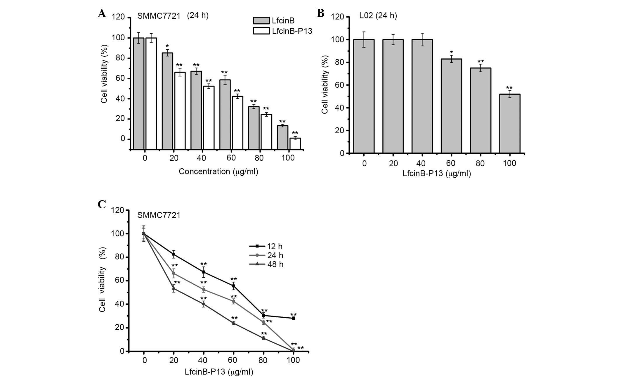
To determine the cytotoxicity of LfcinB-P13 in
vitro, the MTT assay was performed. The results of the present
study indicated that, following treatment with a number of
LfcinB-P13/LfcinB concentrations for 24 h, SMMC7721 cell viability
was significantly inhibited in a dose-dependent manner at all doses
(P=0.032 vs. the control group; Fig.
1A). Furthermore, LfcinB-P13 notably decreased SMMC7721 cell
viability compared with Lfcin-B (Fig.
1A). Treatment of L02 cells, a wild-type human liver cell line,
with LfcinB-P13 for 24 h indicated that LfcinB-P13 exhibits a
significant cytotoxic effect in L02 cells at 60, 80 and 100 µg/ml
compared with the untreated cells (P=0.032, 0.003 and 0.001,
respectively; Fig. 1B). Specifically,
following 48 h treatment with 80 µg/ml LfcinB-P13, the cell
viabilities of SMMC7721 and L02 cells following 48 h were reduced
to 24.5 and 75.1%, respectively, suggesting that healthy liver
cells are less susceptible to LfcinB-P13 toxicity compared with
cancerous cells (Fig. 1B and C). In
addition, the inhibition of SMMC7721 cell viability by LfcinB-P13
was dose- and time-dependent (Fig.
1C). The results of the present study demonstrate that
LfcinB-P13 has an inhibitory effect on SMMC7721 cell viability
in vitro.
LfcinB-P13 induces apoptosis in
SMMC7721 cells
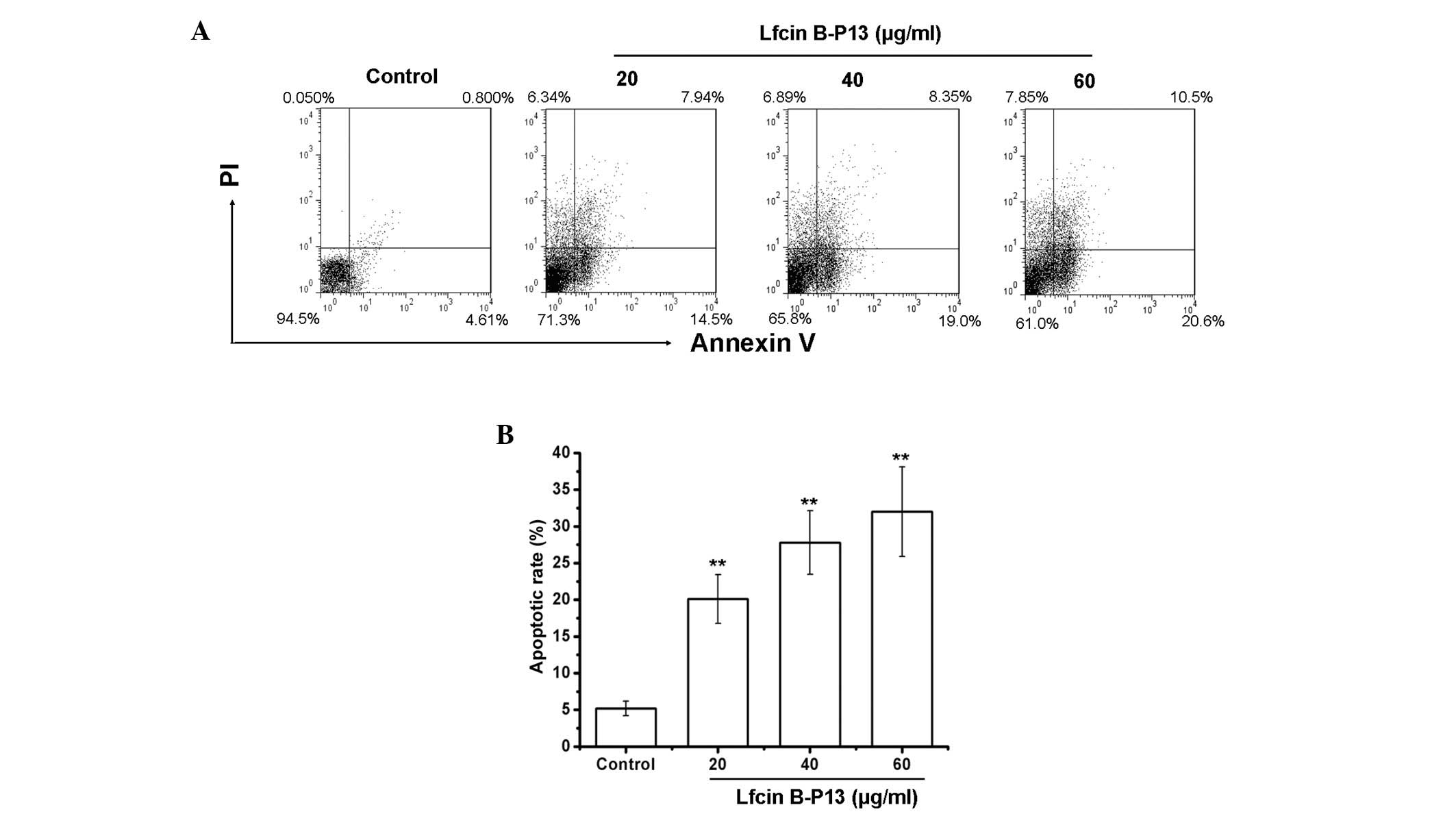
To determine whether treatment with LfcinB-P13
inhibits the viability of SMMC7721 cells by inducing apoptosis, the
percentage of apoptotic cells was measured using the AV/PI assay.
Positive staining with AV is associated with the loss of membrane
polarity and PI staining is associated with the loss of membrane
integrity, which are markers of apoptosis. Therefore, dual staining
with AV and PI was used to discriminate between apoptotic and
non-apoptotic cells. As demonstrated in Fig. 2A, treatment with LfcinB-P13 induced
apoptosis in SMMC7721 cells in a dose-dependent manner. Apoptosis
rates were 20.1, 27.8 and 32.1% following treatment with 20, 40 and
60 µg/ml LfcinB-P13, respectively, demonstrating significant
upregulation of apoptosis compared with the control group
(P=0.0053; Fig. 2B). These results
indicate that LfcinB-P13 reduces SMMC7721 cell viability through
inducing apoptosis.
LfcinB-P13 increases ROS generation in
SMMC7721 cells
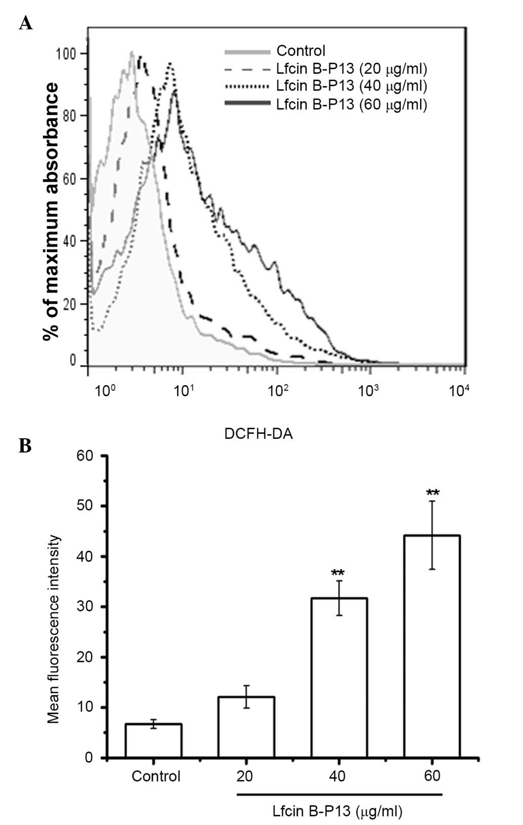
Intracellular ROS generation is one mechanism of
mediating apoptosis. The results of the present study indicated
that ROS levels in SMMC7721 cells were significantly increased
following treatment with LfcinB-P13 (P<0.01; Fig. 3A and B). The mean fluorescence
intensity in the 60 µg/ml LfcinB-P13-treated cells was increased by
6.58 fold compared with the control group. These results indicate
that LfcinB-P13 treatment induces apoptosis in SMMC7721 cells by
upregulating ROS generation.
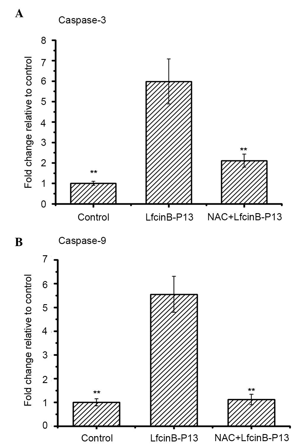
LfcinB-P13 activates caspase-3 and −9
in SMMC7721 cells
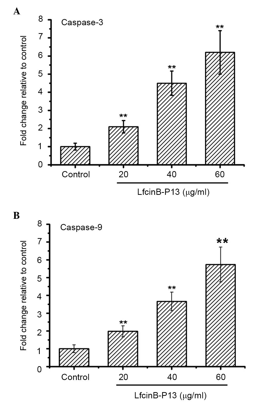
Caspase-3 and −9, markers of apoptotic cells, are
activated during the early stages of apoptosis. In the present
study caspase-3 and −9 were significantly upregulated in the
LfcinB-P13-treated cells at all doses compared with the control
group (P<0.01; Fig. 4A and B).
Treatment with 60 µg/ml LfcinB-P13 increased the activities of
caspase-3 and −9 by 6.2 and 5.7 fold compared with the control
group. These results suggest that caspase-3 and −9 activation is an
underlying mechanism of LfcinB-P13-induced apoptosis.
LfcinB-P13-induced activation of
caspase-3 and −9 in SMMC7721 cells is ROS-mediated
To examine whether LfcinB-P13-induced caspase
activity was dependent on ROS, SMMC7721 cells were treated with
LfcinB-P13 in the absence and presence of the antioxidant NAC, an
ROS scavenger. Fig. 5A and B
demonstrate that combined treatment with NAC and LfcinB-P13
significantly decreased activation of caspase-3 and −9 compared
with treatment with LfcinB-P13 alone (P<0.01). The results of
the present study indicate that upregulated ROS generation is
required for LfcinB-P13-mediated caspase-3 and −9 activation.
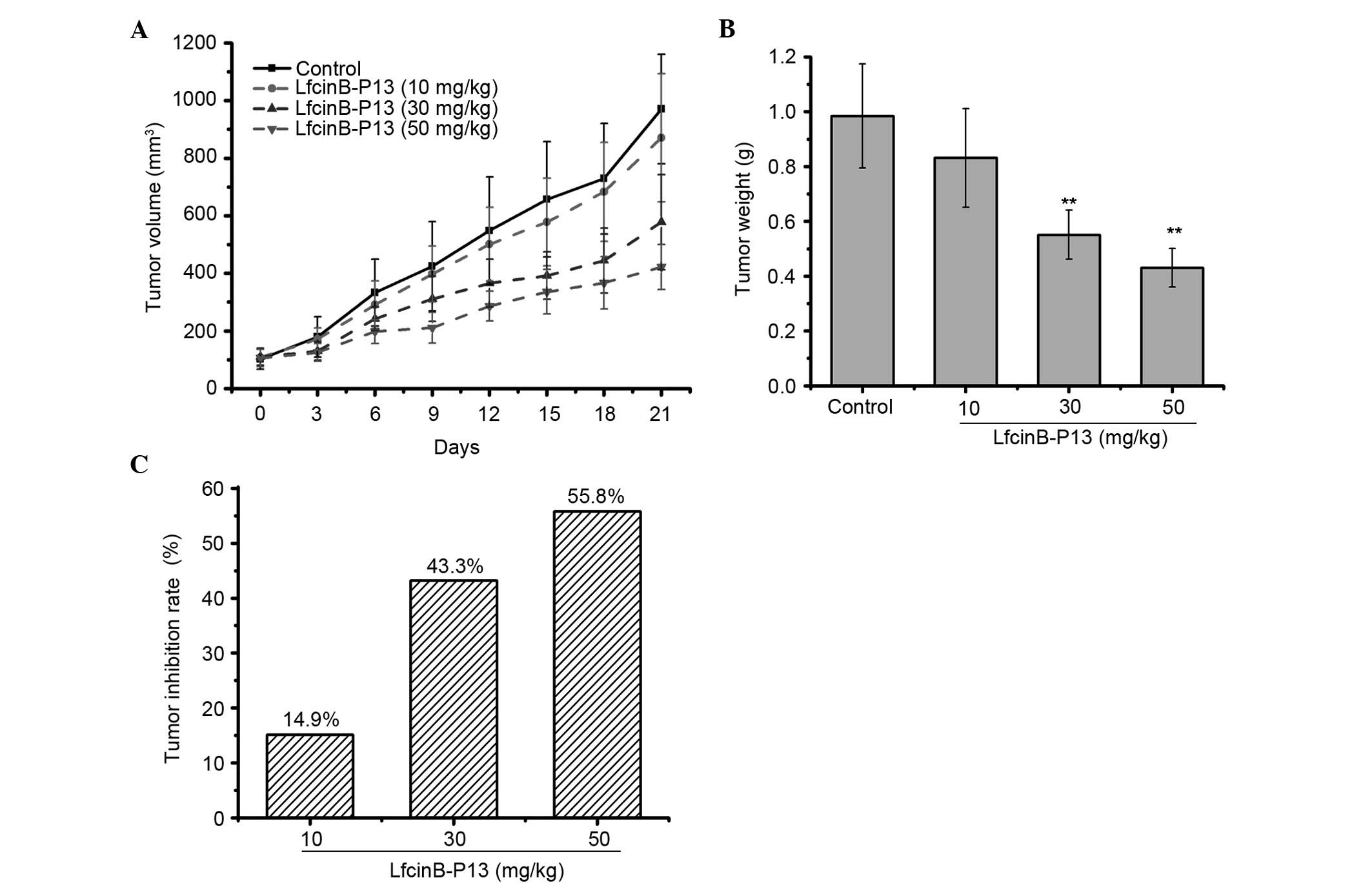
LfcinB-P13 suppresses tumor growth in
SMMC7721-xenografted nude mice
To determine whether LfcinB-P13 prevents SMMC7721
cell growth in vivo, a SMMC7721-xenografted nude mouse model
was established. These results indicate that LfcinB-P13 inhibits
SMMC7721 cell growth, in terms of tumor volume and weight, and
tumor inhibition rate, in a dose-dependent manner (Fig. 6A-C). Treatment with 30 and 50 mg/kg
LfcinB-P13 significantly decreased tumor weight compared with the
control group (P=0.006 and 0.005, respectively; Fig. 6B). Tumor inhibition rates were 14.9,
43.3 and 55.8% following treatment with 10, 30 and 50 mg/kg
LfcinB-P13, respectively (Fig.
6C).
Discussion
HCC is one of the most common malignancies, is
highly metastatic and is the third-leading cause of
cancer-associated mortality worldwide (13,14). The
majority of traditional chemotherapeutic agents exhibit an
extensive profile of side effects, therefore the development of
novel drugs with high efficiency and low toxicity is warranted.
LfcinB, a lactoferrin-derived AMP, is composed of 25 amino acids
and has been demonstrated to possess broad-spectrum antimicrobial
activity, in addition to anti-tumorigenic effects (15–17). In
the present study, the peptide LfcinB-P13, an analog of LfcinB, was
designed and synthesized as an amphipathic AMP of 13 amino acids in
length, and the effect of this synthetic compound on SMMC7721 cells
was investigated.
The cytotoxic effect of LfcinB-P13 on SMMC7721 and
L02 cells was initially investigated. The results of this indicated
that LfcinB-P13 significantly inhibits the proliferation of
SMMC7721 cells in vitro, and that the cells exhibited
increased sensitivity to LfcinB-P13 compared with Lfcin-B. The
half-maximal inhibitory concentration (IC50) of
LfcinB-P13 in SMMC7721 cells was 41.8 µg/ml at 24 h, while the
equivalent IC50 of LfcinB-P13 in L02 cells was >100
µg/ml, demonstrating increased cytotoxicity in the HCC cell line.
The results of this suggest that LfcinB-P13 exhibits
cancer-selective activity. The underlying mechanism of LfcinB-P13
action in SMMC7721 cells was subsequently investigated.
Apoptosis is a tightly regulated process of
programmed cell death, leading to the elimination of undesirable
and defective cells. Induction of apoptosis is a mechanism involved
in the action of AMPs (18–20). Dual staining with AV and PI
demonstrated that rates of apoptosis were significantly increased
in cells treated with LfcinB-P13 compared with the control group,
suggesting that induction of apoptosis is a mechanism by which
LfcinB-P13 prevents the growth of SMMC7721 cells in
vitro.
ROS, typically regarded as toxic products of
cellular metabolism, are able to function as signaling molecules
that regulate a number of physiological processes (21). ROS are important in the induction of
apoptosis in physiological and pathological conditions. Previous
studies have reported that moderate levels of ROS promote cell
proliferation and survival (22),
whereas excessive levels of ROS induce cell death and lead to
ischemia-reperfusion-induced cellular injury, resulting in lipid
peroxidation, DNA damage and apoptosis (23,24). In
the present study, LfcinB-P13 significantly elevated ROS levels in
SMMC7721 cells compared with the control group, which may be the
underlying mechanism of LfcinB-P13-induced apoptosis.
Caspase family members serve important roles in the
regulation of apoptosis. Caspase stimulation is a marker of
apoptotic induction in response to death-inducing signals, which
originate from cell surface receptors, mitochondria or the
endoplasmic reticulum (25).
Caspase-3 and −9 are activated in the early stages of apoptosis,
and are therefore frequently used as markers of apoptotic cells
(26). Caspase-9 is the primary
initiator caspase in the intrinsic pathway of apoptosis (27). Following cleavage and activation of
its pro-form, caspase-9 activates caspase-3, which leads to
activation of the apoptotic caspase cascade. In the present study,
caspase-3 and −9 were significantly upregulated following treatment
with LfcinB-P13 in SMMC7721 cells compared with the control group.
Furthermore, it was demonstrated that chelation of ROS by NAC
inhibits LfcinB-P13-mediated caspase-3 and −9 activation,
suggesting that LfcinB-P13-induced ROS generation is associated
with the activation of caspase-3 and −9.
In conclusion, the results of the present study
indicate that LfcinB-P13 suppresses the proliferation of SMMC7721
cells in vitro and in vivo by increasing
intracellular ROS generation, which leads to the subsequent
activation of caspase-3 and −9, followed by apoptosis. These
results suggest that LfcinB-P13 is a novel candidate therapeutic
agent for the treatment of liver cancer.
Acknowledgements
The present study was supported by the Program for
the Science and Technology Development of the Anhui Province (grant
no. 11010402163) and the National Natural Science Foundation of
China (grant no. 81272398).
References
|
1
|
Heindryckx F and Gerwins P: Targeting the
tumor stroma in hepatocellular carcinoma. World J Hepatol.
7:165–176. 2015. View Article : Google Scholar : PubMed/NCBI
|
|
2
|
Wang M, Wang H, Tang Y, Kang D, Gao Y, Ke
M, Dou J, Xi T and Zhou C: Effective inhibition of a
Strongylocentrotus nudus eggs polysaccharide against hepatocellular
carcinoma is mediated via immunoregulation in vivo. Immunol Lett.
141:74–82. 2011. View Article : Google Scholar : PubMed/NCBI
|
|
3
|
Wang G, Mishra B, Lau K, Lushnikova T,
Golla R and Wang X: Antimicrobial peptides in 2014. Pharmaceuticals
(Basel). 8:123–150. 2015. View Article : Google Scholar : PubMed/NCBI
|
|
4
|
Cheung RC, Ng TB and Wong JH: Marine
Peptides: Bioactivities and Applications. Mar Drugs. 13:4006–4043.
2015. View Article : Google Scholar : PubMed/NCBI
|
|
5
|
Tossi A and Sandri L: Molecular diversity
in gene-encoded, cationic antimicrobial polypeptides. Curr Pharm
Des. 8:743–761. 2002. View Article : Google Scholar : PubMed/NCBI
|
|
6
|
Sun Y, Dong W, Sun L, Ma L and Shang D:
Insights into the membrane interaction mechanism and antibacterial
properties of chensinin-1b. Biomaterials. 37:299–311. 2015.
View Article : Google Scholar : PubMed/NCBI
|
|
7
|
Zhang T, Chan CF, Lan R, Li H, Mak NK,
Wong WK and Wong KL: Porphyrin-based ytterbium complexes targeting
anionic phospholipid membranes as selective biomarkers for cancer
cell imaging. Chem Commun (Camb). 49:7252–7254. 2013. View Article : Google Scholar : PubMed/NCBI
|
|
8
|
Hilchie AL, Vale R, Zemlak TS and Hoskin
DW: Generation of a hematologic malignancy-selective membranolytic
peptide from the antimicrobial core (RRWQWR) of bovine
lactoferricin. Exp Mol Pathol. 95:192–198. 2013. View Article : Google Scholar : PubMed/NCBI
|
|
9
|
Sheng M, Zhao Y, Zhang A, Wang L and Zhang
G: The effect of LfcinB9 on human ovarian cancer cell SK-OV-3 is
mediated by inducing apoptosis. J Pept Sci. 20:803–810. 2014.
View Article : Google Scholar : PubMed/NCBI
|
|
10
|
Wang H, Ke M, Tian Y, Wang J, Li B, Wang
Y, Dou J and Zhou C: BF-30 selectively inhibits melanoma cell
proliferation via cytoplasmic membrane permeabilization and
DNA-binding in vitro and in B16F10-bearing mice. Eur J Pharmacol.
707:1–10. 2013. View Article : Google Scholar : PubMed/NCBI
|
|
11
|
Pieme CA, Guru SK, Ambassa P, Kumar S,
Ngameni B, Ngogang JY, Bhushan S and Saxena AK: Induction of
mitochondrial dependent apoptosis and cell cycle arrest in human
promyelocytic leukemia HL-60 cells by an extract from Dorstenia
psilurus: A spice from Cameroon. BMC Complement Altern Med.
13:2232013. View Article : Google Scholar : PubMed/NCBI
|
|
12
|
Rouhollahi E, Moghadamtousi S Zorofchian,
Paydar M, Fadaeinasab M, Zahedifard M, Hajrezaie M, Hamdi OA Ahmed,
Looi CY, Abdulla MA, Awang K and Mohamed Z: Inhibitory effect of
Curcuma purpurascens BI. rhizome on HT-29 colon cancer cells
through mitochondrial-dependent apoptosis pathway. BMC Complement
Altern Med. 15:152015. View Article : Google Scholar : PubMed/NCBI
|
|
13
|
Dai L, Lei N, Liu M and Zhang JY:
Autoantibodies to tumor-associated antigens as biomarkers in human
hepatocellular carcinoma (HCC). Exp Hematol Oncol. 2:152013.
View Article : Google Scholar : PubMed/NCBI
|
|
14
|
Yang SF, Chang CW, Wei RJ, Shiue YL, Wang
SN and Yeh YT: Involvement of DNA damage response pathways in
hepatocellular carcinoma. Biomed Res Int. 2014:1538672014.
View Article : Google Scholar : PubMed/NCBI
|
|
15
|
Enrique M, Manzanares P, Yuste M, Martínez
M, Vallés S and Marcos JF: Selectivity and antimicrobial action of
bovine lactoferrin derived peptides against wine lactic acid
bacteria. Food Microbiol. 26:340–346. 2009. View Article : Google Scholar : PubMed/NCBI
|
|
16
|
Furlong SJ, Mader JS and Hoskin DW: Bovine
lactoferricin induces caspase-independent apoptosis in human
B-lymphoma cells and extends the survival of immune-deficient mice
bearing B-lymphoma xenografts. Exp Mol Pathol. 88:371–375. 2010.
View Article : Google Scholar : PubMed/NCBI
|
|
17
|
Wang S, Tu J, Zhou C, Li J, Huang L, Tao L
and Zhao L: The effect of Lfcin-B on non-small cell lung cancer
H460 cells is mediated by inhibiting VEGF expression and inducing
apoptosis. Arch Pharm Res. 38:261–271. 2015. View Article : Google Scholar : PubMed/NCBI
|
|
18
|
Park HJ, Choi SY, Hong SM, Hwang SG and
Park DK: The ethyl acetate extract of Phellinus linteus grown on
germinated brown rice induces G0/G1 cell cycle arrest and apoptosis
in human colon carcinoma HT29 cells. Phytother Res. 24:1019–1026.
2010.PubMed/NCBI
|
|
19
|
Tsai TL, Li AC, Chen YC, Liao YS and Lin
TH: Antimicrobial peptide m2163 or m2386 identified from
Lactobacillus casei ATCC 334 can trigger apoptosis in the human
colorectal cancer cell line SW480. Tumour Biol. 36:3775–3789. 2015.
View Article : Google Scholar : PubMed/NCBI
|
|
20
|
Pan WR, Chen YL, Hsu HC and Chen WJ:
Antimicrobial peptide GW-H1-induced apoptosis of human gastric
cancer AGS cell line is enhanced by suppression of autophagy. Mol
Cell Biochem. 400:77–86. 2015. View Article : Google Scholar : PubMed/NCBI
|
|
21
|
Kamogashira T, Fujimoto C and Yamasoba T:
Reactive oxygen species, apoptosis, and mitochondrial dysfunction
in hearing loss. Biomed Res Int. 2015:6172072015. View Article : Google Scholar : PubMed/NCBI
|
|
22
|
Trachootham D, Lu W, Ogasawara MA, Nilsa
RD and Huang P: Redox regulation of cell survival. Antioxid Redox
Sign. 10:1343–1374. 2008. View Article : Google Scholar
|
|
23
|
Marchi S, Giorgi C, Suski JM, Agnoletto C,
Bononi A, Bonora M, De Marchi E, Missiroli S, Patergnani S, Poletti
F, et al: Mitochondria-ros crosstalk in the control of cell death
and aging. J Signal Transduct. 2012:3296352012. View Article : Google Scholar : PubMed/NCBI
|
|
24
|
Riedl SJ and Salvesen GS: The apoptosome:
Signalling platform of cell death. Nat Rev Mol Cell Bio. 8:405–413.
2007. View
Article : Google Scholar
|
|
25
|
Fiandalo MV and Kyprianou N: Caspase
control: Protagonists of cancer cell apoptosis. Exp Oncol.
34:165–175. 2012.PubMed/NCBI
|
|
26
|
Cakir E, Yilmaz A, Demirag F, Oguztuzun S,
Sahin S, Yazici UE and Aydin M: Prognostic significance of
micropapillary pattern in lung adenocarcinoma and expression of
apoptosis-related markers: Caspase-3, bcl-2 and p53. APMIS.
119:574–580. 2011. View Article : Google Scholar : PubMed/NCBI
|
|
27
|
Würstle ML, Laussmann MA and Rehm M: The
central role of initiator caspase-9 in apoptosis signal
transduction and the regulation of its activation and activity on
the apoptosome. Exp Cell Res. 318:1213–1220. 2012. View Article : Google Scholar : PubMed/NCBI
|