Introduction
Despite recent advances in surgery, radiotherapy and
chemotherapy for the treatment of various types of cancer,
morbidity remains at a high level (1)
and the five-year survival rate for oral cancer has only moderately
improved (2,3). Therefore, novel therapeutic strategies
are required. As the majority of types of oral cancer are oral
squamous cell carcinoma (OSCC), one feature of which is progressive
local invasion (4,5), it is necessary to elucidate its
underlying invasion mechanisms in order to improve currently
available treatments for OSCC. The processes involved in tumor
invasion include cell migration, interaction between the tumor and
stroma at the invasive front and the involvement of growth factors
and external stimuli that affect the invading cells (6–10). It is
important to understand the signaling mechanisms underlying the
regulation of cell migration and invasive growth, in order to
facilitate the identification of novel therapeutic targets
(11–13).
Signal transduction via receptor tyrosine kinases
(RTKs) is stimulated by the respective extracellular ligands, which
regulate critical cellular processes, including cell proliferation
and cell migration (14). Therefore,
genetic changes and abnormalities in RTKs often lead to a malignant
transformation (14). A notable
example of extracellular growth factors activating RTKs is the
epidermal growth factor (EGF) family, the members of which function
via the EGF receptor (EGFR) (15).
Although EGFR is expressed in the normal oral epithelium as well as
in the majority of OSCC cells (15,16), it is
also a therapeutic target for the treatment of oral cancer
(12,13). Previous studies using various cell
types have demonstrated that the downstream signaling pathways of
numerous RTKs are involved in the regulation of cell motility
(7,17).
Certain mitogen-activated protein (MAP) kinases,
including extracellular-regulated kinase (ERK), Jun kinase and
tumor protein (p)38, are able to affect various cell functions,
including migration (18).
Phosphatidylinositol-3 kinase (PI3K) controls cell motility through
the activation of protein kinase B (Akt) and other targets
(19,20); however, cell-dependent differences in
these regulatory mechanisms exist (18,21–23).
In head and neck cancer, the signaling pathways
involved in RTK-mediated migration have yet to be elucidated and
understood. EGFR stimulation induces cell migration through the
activation of matrix metalloproteinases (MMPs) (24), signal transducer and activator of
transcription 3 (STAT3) (25,26) or the MEK/ERK and PI3K signaling
pathways (9), and may be associated
with an epithelial-mesenchymal transition (EMT)-like phenotype
(27). Furthermore, cross-talk
between EGFR and G-protein-coupled receptors contributes to cell
migration (28,29).
Our previous study reported that cetuximab, an
EGFR-specific monoclonal antibody, inhibits migration of the SAS
OSCC cell line, but not of the HSC4 OSCC cell line; however, the
proliferation of HSC4 cells was observed to be sensitive to
cetuximab (30). These results
suggested that EGFR signaling may induce cell migration in a cell
type-dependent manner. In the present study, the underlying
mechanisms of EGFR signal transduction involved in the migration of
the SAS and HSC4 OSCC cell lines were investigated and
compared.
Materials and methods
Cell culture and reagents
The HSC4 and SAS OSCC cell lines were purchased from
RIKEN Bioresource Center (Ibaraki, Japan). Cells were cultured in
Dulbecco's modified Eagle's medium (DMEM) supplemented with 10%
(v/v) fetal bovine serum (FBS) at 37°C in a humidified atmosphere
of 5% CO2. DMEM and FBS were purchased from Gibco
(Thermo Fisher Scientific, Inc., Waltham, MA, USA). The antibodies
used consisted of anti-AKT (rabbit monoclonal; cat. no. 4681;
dilution, 1:1,000; Cell Signaling Technology, Inc., Danvers, MA,
USA), anti-phospho-AKT (rabbit polyclonal; cat. no. 9271; dilution;
1:1,000, Cell Signaling Technology, Inc.), anti-ERK (rabbit
polyclonal; cat. no. sc-93; dilution, 1:1,000; Santa Cruz
Biotechnology, Inc. Santa Cruz, CA, USA), anti-phospho-ERK (mouse
monoclonal; cat. no. sc-7383; dilution, 1:1,000; Santa Cruz, Inc.),
anti-profilin-1 (rabbit polyclonal; cat. no. 3237; dilution,
1:1,000; Cell Signaling Technology, Inc.), anti-cofilin (rabbit
polyclonal; cat. no. sc-33729; dilution, 1:1,000; Santa Cruz
Biotechnology, Inc.), anti-phospho-cofilin (rabbit polyclonal; cat.
no. sc-12912; dilution, 1:1,000; Santa Cruz Biotechnology) and
anti-α-tubulin (mouse monoclonal; cat. no. T6074; dilution,
1:1,000; Sigma-Aldrich; Merck Millipore, Darmstadt, Germany). The
secondary antibodies used consisted of ECL™ anti-mouse IgG,
horseradish peroxidase (HRP)-linked (dilution, 1:10,000; cat. no.
NA931V; GE Healthcare Japan, Tokyo, Japan) and ECL™ anti-rabbit
IgG, HRP linked (dilution 1, 10,000; cat. no. NA934V; GE Healthcare
Japan). Cetuximab (Erbitux®) was purchased from Merck
Serono (Tokyo, Japan). EGFR inhibitor AG1478, Akt inhibitor MK2206
and MEK inhibitor PD98059 were from Calbiochem (Merck Millipore,
Darmstadt, Germany). Acti-stain™ 488 (Cytoskeleton, Inc., Denver,
CO, USA) was used for actin filament staining.
Scratch wound healing assay
Cell migration was evaluated using a scratch wound
healing assay, as previously described (30), with certain modifications. Briefly,
OSCC HSC4 and SAS cells were seeded at 1–2×10⁴ in
12-well plates, cultured for 24 h, and the semiconfluent cells were
treated with 10 µg/ml mitomycin C for 4 h at 37°C to block
proliferation and were subsequently wounded with a sterile 200 µl
pipette tip in order to generate a cell-free gap. The cells were
then washed with PBS and an image was captured using inverted
phase-contrast microscopy (IX71; Olympus Corporation, Tokyo, Japan)
and a digital CCD camera (DP72-SET; Olympus Corporation) to record
the wound width at 0 h. One group of HSC4 or SAS cells was then
cultured in DMEM supplemented with 10% FBS as a control. The HSC4
and SAS cells were treated with reagents including cetuximab (10
and 20 µg/ml), AG1478 (10 and 20 µg/ml), FBS (10%), EGF (10, 20 and
50 ng/ml), MK2206 (1, 5 and 10 µM) or PD98059 (1, 5 and 10 µM).
Following incubations for 12–20 h at 37°C, images of the cells were
captured using inverted phase-contrast microscopy (IX71, Olympus,
Tokyo, Japan) in order to evaluate the rate of migration.
Western blotting
The OSCC HSC4 and SAS cells were washed with PBS and
then lysed with radioimmunoprecipitation assay buffer, consisting
of 150 mM NaCl, 10 mM Tris-HCl, pH 8.0, 1% (v/v) Nonidate P-40,
0.5% (w/v) deoxycholic acid, 0.1% (w/v) SDS, 5 mM EDTA, 1X Halt™
protease inhibitor cocktail (Thermo Fisher Scientific, Inc.) and 1X
Halt™ protein phosphatase inhibitor (Thermo Fisher Scientific,
Inc). The protein concentration of the lysates was determined using
a BCA™ Protein Assay kit (Thermo Fisher Scientific, Inc.) and equal
amounts of the proteins (10–50 µg) were subjected to SDS-PAGE analysis.
The separated proteins were electrophoretically transferred onto
polyvinylidene fluoride membranes (Clear Trans SP; Wako Pure
Chemical Industries, Ltd., Osaka, Japan). Non-specific binding was
blocked by incubation in 5% (w/v) bovine serum albumin (BSA;
Sigma-Aldrich; Merck Millipore) in TBS/Tween-20 (TBS-T) for 1 h at
room temperature. The membranes were probed with primary antibodies
including anti-ERK, anti-phospho-ERK, anti-Akt, anti-phospho-Akt,
anti-profilin, anti-cofilin, anti-phospho-cofilin and
anti-α-tubulin in TBS-T overnight at 4°C and then incubated with
horseradish peroxidase-conjugated secondary antibodies for 1 h at
room temperature. Antibody-antigen complexes were detected using
the Enhanced Chemiluminescence Plus Western Blotting Detection
Reagent (GE Healthcare Japan, Tokyo, Japan).
Staining of actin fiber
Cultured HSC4 and SAS cells were fixed in 3.5% (w/v)
paraformaldehyde for 10 min at room temperature, permeabilized in
0.2% (v/v) Triton X-100 for 5 min at room temperature, and blocked
in 2% (w/v) BSA for 30 min at room temperature. Fixed cells were
incubated in 100 nM of Acti-stain™ 488 phalloidin in the dark for
30 min at room temperature. Phalloidin staining was observed under
fluorescent microscopy (Olympus, Tokyo, Japan).
Statistical analysis
All data are presented as the mean ± standard error
and an unpaired student's t-test was used to determine the
significant differences between the groups. P<0.05 was
considered to indicate a statistically significant difference.
Results
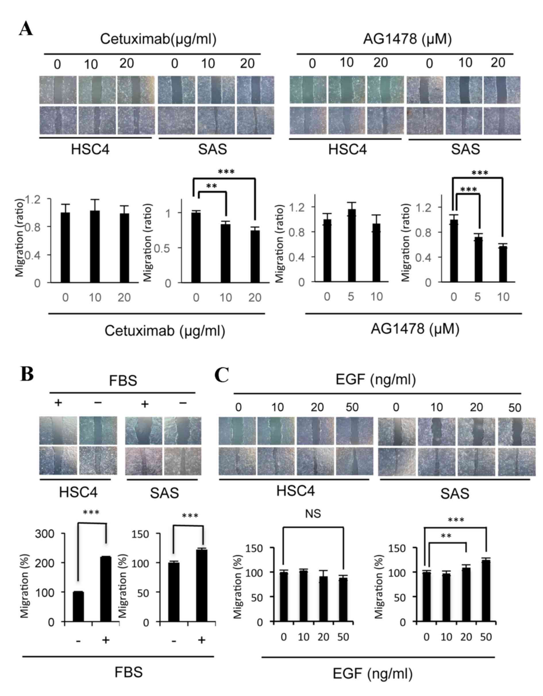
The EGF-EGFR signaling pathway is
associated with the migration of SAS cells, but not the migration
of HSC4 cells
Changes in the EGFR signaling pathway induced by
cetuximab have an important role in the migration of SAS cells, but
not in the migration of HSC4 cells (30). In the present study, the effects of
the AG1478 EGFR inhibitor on cell migration were examined and
compared with those of cetuximab (Fig.
1A). The migration of SAS cells was significantly inhibited by
AG1478 (10 µM P=0.00036, 20 µM P=0.00000055) as well as cetuximab
(10 µM P=0.0027, 20 µM P=0.000045) treatment, whereas the migration
of HSC4 cells was not affected by these inhibitors (Fig. 1A). As the migration of HSC4 and SAS
cells was significantly stimulated (HSC4 P=2.8×10⁻¹⁸;
SAS P=0.00012) by the addition of FBS in culture medium (Fig. 1B), the effects of EGF, an EGFR ligand
present in serum, on migration were examined. EGF was observed to
significantly stimulate the migration of SAS cells (20 ng/ml,
P=0.0022, 50 ng/ml, P=0.000023), but not of the HSC4 cells
(Fig. 1C). These results suggest that
the EGF-EGFR signaling pathway induces the migration of SAS cells,
whereas the signaling pathways induced by various other factors
present in serum may contribute to the migration of HSC4 cells.
The EGFR-Akt signaling pathway is
associated with the migration of SAS cells
The ability of distinct RTKs to stimulate the
migration of certain cell lines (14)
indicates that various signal transduction pathways may be involved
in the motility of HSC4 and SAS cells. In order to investigate the
downstream signaling factors involved in OSCC cell motility, the
effects of specific Akt and MEK inhibitors on the migration of
these cells were evaluated. The Akt inhibitor MK2206 significantly
suppressed the migration of HSC4 (1 µM, P=0.0097; 5 µM, P=0.000018;
10 µM, P=0.000023) and SAS cells (5 µM, P=0.047; 10 µM, P=0.0056;
Fig. 2A). In addition, EGF was
observed to induce Akt phosphorylation in SAS cells, but not in
HSC4 cells (Fig. 2C). These results
indicate that the Akt signaling pathway is required for the
migration of these cell lines, and that EGF induces Akt
phosphorylation in SAS cells, but not in HSC4 cells. The MEK
inhibitor PD98059 was identified to significantly inhibit the
migration of SAS cells (5 µM, P=0.000039; 10 µM,
P=8.9×10⁻⁹), but not of HSC4 cells (Fig. 2B). The phosphorylation of ERK was
highly and moderately induced by EGF in HSC4 and SAS cells,
respectively (Fig. 2C). These data
indicate that EGF is able to activate the ERK signaling pathway in
certain OSCC cell lines; however, the ERK signaling pathway is only
involved in the migration of SAS cells.
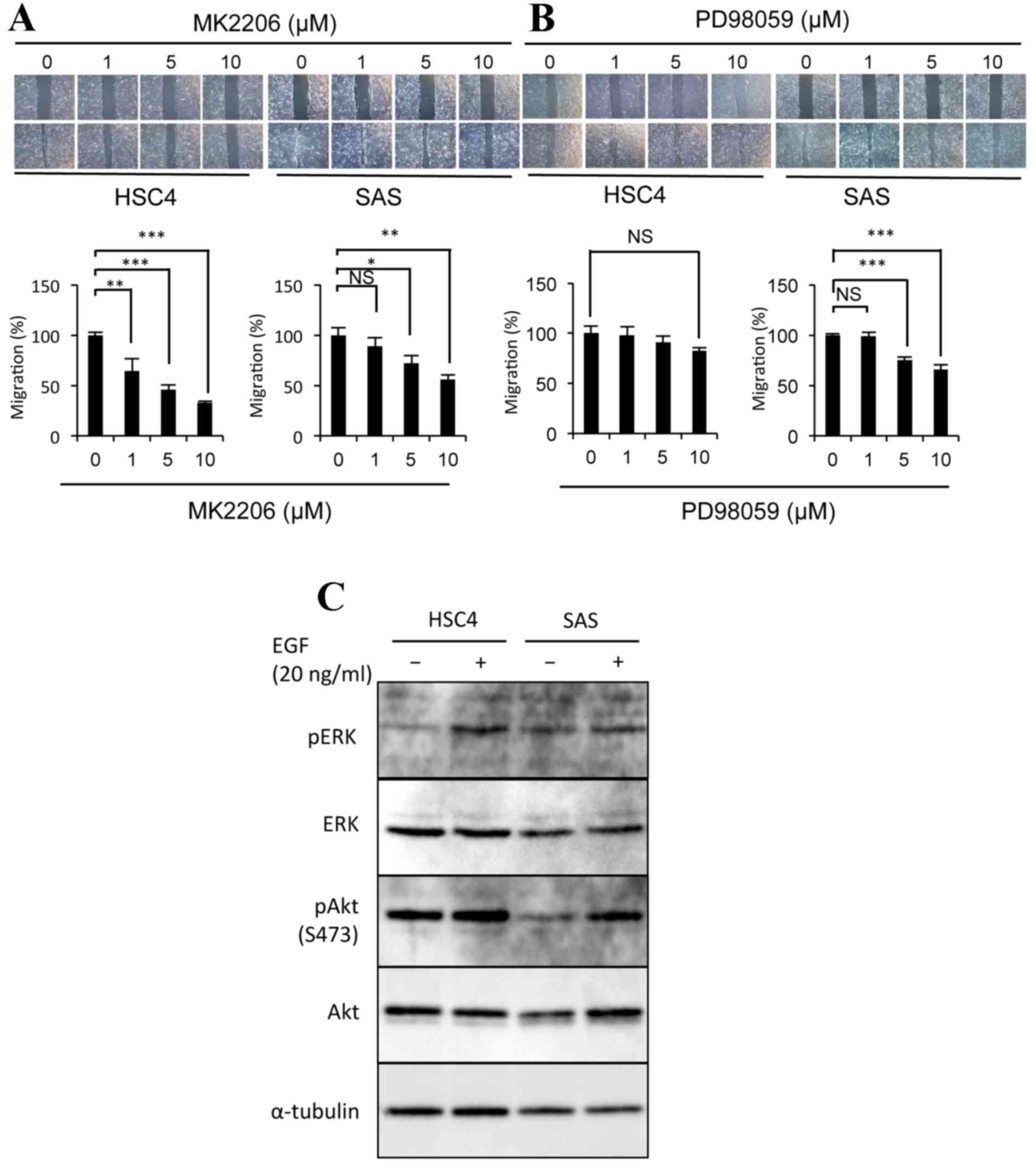 | Figure 2.The EGF-EGFR signaling pathway
induces the migration of SAS cells via Akt phosphorylation. The
effects of (A) MK2206 or (B) PD98059 treatment on the migration of
HSC4 and SAS cells. Phase-contrast micrographs and graphs are as
described in Fig. 1. (C)
Representative western blot demonstrating that EGF promotes ERK
phosphorylation in HSC4 cells, and Akt phosphorylation in SAS
cells. Protein extracts from each cell sample were probed with
anti-ERK, anti-phospho-ERK, anti-Akt and anti-phospho-Akt, and
anti-α-tubulin as the loading control. Error bars, SEM; n=3;
*P<0.05, **P<0.01, ***P<0.001; EGF, epidermal growth
factor; EGFR, EGF receptor; Akt, protein kinase B; ERK,
extracellular-regulated kinase; p, phosphorylated. |
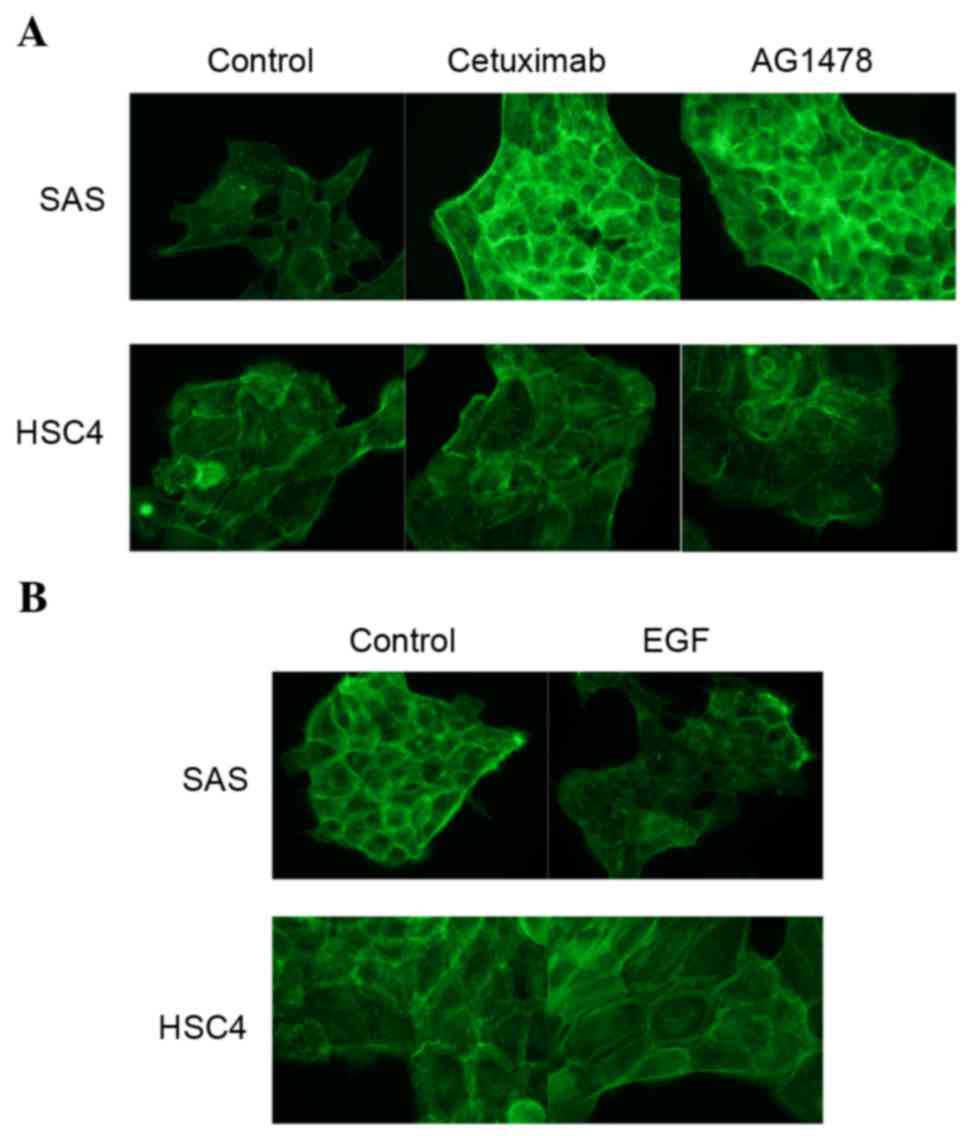
EGFR inhibitors promote, and EGF
suppresses, the accumulation of actin filaments in SAS cells
As the remodeling of the actin network is considered
to be involved in cell migration (31), the effects of cetuximab or AG1478
treatment on actin filament levels were examined using phalloidin
staining. The degree of staining observed was markedly increased
following the treatment of SAS cells with cetuximab (P=0.000020)
and AG1478 (P=0.00036), as compared with untreated cells (Fig. 3A). By contrast, the EGFR inhibitors
cetuximab (P=0.73) and AG1478 (P=0.89) were not observed to affect
the degree of phalloidin staining in HSC4 cells (Fig. 3A). EGFR is induced by EGF binding, it
was then examined whether EGF affects actin organization in HSC4
and SAS cells. EGF treatment reduced the degree of palloidin
staining in SAS cells (P=0.00014), but not in HSC4 cells (P=0.94;
Fig. 3B). These results indicate that
the EGF-EGFR signaling pathway regulates actin filament turnover,
and that it is necessary for cell motility. Thus, we examined
effect of EGFR signaling on profiling and cofilin levels, because
these proteins play key roles in actin polymerization and
depolymerization, respectively. However, following treatment of the
HSC4 and SAS cells with EGFR inhibitors, profiling-1 expression
levels were not observed to be altered and cofilin-1
phosphorylation increased in both cells (data not shown).
Discussion
The directed migration of tumor cells into the
surrounding tissues and blood vessels promotes the tissue invasion
of metastatic cancer cells (32).
This directed cell migration is often regulated by responses to
extracellular stimuli (33–35). Notably, signal transduction via EGFR
has an important role in cell motility in various types of cancer
(36,37). In the present study, EGF-EGFR
signaling was identified to be required for the migration of SAS
OSCC cells, but not for HSC4 OSCC cells. In our previous study, it
was demonstrated that EGFR activity is essential for the
proliferation of HSC4 cells, but not SAS cells (30). The Akt inhibitor MK2206 and ERK
inhibitor PD98059, which did not affect HSC4 cell migration, were
observed to inhibit SAS cell migration. EGF stimulated the
phosphorylation of ERK in HSC4 cells and Akt in SAS cells. These
results indicate that the EGF-EGFR signaling pathway induces
fundamental phenomena, including cell proliferation and migration,
in a cell type-dependent manner. Furthermore, it is possible that
the migration of SAS cells is regulated by EGF-EGFR signal
transduction via Akt, and that the proliferation of HSC4 cells is
regulated by EGF-EGFR signal transduction via the MEK-ERK signaling
pathway.
The migration of HSC4 cells was promoted by the
addition of FBS; however, the addition of EGF did not affect their
migration, indicating that HSC4 cell migration requires certain
extracellular stimuli present in serum other than EGF. At present,
~60 RTKs have been identified and their activities have been
demonstrated to affect various physiological functions, including
cell migration (38). In addition to
EGF, previous studies have revealed that the following RTKs are
also involved in regulating the migration of various cancer cells:
growth arrest-specific 6 (GAS6) receptor (AXL); hepatocyte growth
factor receptor (MET); fibroblast growth factor; and discoidin
domain receptor family, member 1 (39–43).
Therefore, further examination of how these candidate RTKs are
involved in the regulation of OSCC cell migration is required.
The cytoskeleton, which is composed of actin
filaments, is a highly organized network that enables cellular
motion (44). In the present study,
it was demonstrated that the degree of phalloidin staining in SAS
cells treated with EGFR inhibitors was markedly increased, as
compared with the control cells. Untreated control cells exhibited
a normal arrangement of actin filaments near the outer layer of the
cell periphery. Although this staining pattern was fundamentally
unchanged following treatment of the control cells with EGFR
inhibitors, intense overall cortical staining was observed. By
contrast, the degree of phalloidin staining of the actin filaments
was reduced in SAS cells by the addition of EGF; however, the
staining pattern was not altered. It was hypothesized that the
accumulation of actin filaments may produce irregular tension in
the cytoskeletal system that is able to disrupt the orchestrated
generation of force and interfere with directional cell migration.
Therefore, it was concluded that the effects of EGFR inhibitors on
the motility of SAS cells are possibly due to the accumulation of
actin filaments in the cytoskeleton.
Previous studies have indicated that profilin and
cofilin have key roles in regulating the assembly of actin
filaments beneath the plasma membrane, in order to promote cell
motility and other actin-associated processes (45,46).
Profilin-1 is an actin monomer-binding protein considered to be an
essential factor in actin polymerization (47,48). In
addition, the depolymerization of actin filaments is regulated by
cofilin, the phosphorylated form of which cannot sever actin
filaments and therefore shows negative regulation (49–51).
Therefore, the effects of EGFR inhibitors on the expression levels
of profilin-1 and phosphorylated cofilin-1 were examined. However,
our unpublished western blot analysis data suggested that
profiling-1 and cofilin-1 may not necessarily have direct roles in
the actin dynamics regulated by EGFR signaling. It was hypothesized
that various other actin-binding proteins involved in the
reorganization of the actin cytoskeleton are regulated downstream
of the EGFR signaling pathway, and that EGFR inhibition promotes
the overexpression of these proteins, resulting in the accumulation
of actin filaments and cell dysmotility.
The proliferation of SAS cells, which possess a stem
cell-like potency (52), was
resistant to cetuximab in monolayer culture conditions, despite the
phosphorylation of EGFR; however, the growth of SAS cell
aggregates, in which EGFR levels were increased, was sensitive to
cetuximab (30). In the present
study, it was demonstrated that SAS cell migration is also
sensitive to the EGFR inhibitors cetuximab and AG1478. Therefore,
EGFR may be a candidate therapeutic target for the prevention of
cancer stem cell dissociation from the primary tumor, migration
into the surrounding tissues during the early stage and
colonization at distant sites in OSCC metastasis.
Acknowledgements
This manuscript has been edited for English language
by Textcheck English consultants. Funding for the present study was
provided by Osaka University (grant no. 1508000001) and Osaka
Dental University (grant no. 217006).
Glossary
Abbreviations
Abbreviations:
|
EGF
|
epidermal growth factor
|
|
EGFR
|
EGF receptor
|
|
OSCC
|
oral squamous cell carcinoma
|
|
DMEM
|
Dulbecco's modified Eagle's medium
|
|
FBS
|
fetal bovine serum
|
|
RTK
|
receptor tyrosine kinase
|
|
ERK
|
extracellular-regulated kinase
|
|
PBS
|
phosphate-buffered saline
|
References
|
1
|
Jemal A, Bray F, Center MM, Ferlay J, Ward
E and Forman D: Global cancer statistics. CA Cancer J Clin.
61:69–90. 2011. View Article : Google Scholar : PubMed/NCBI
|
|
2
|
Cohen EE, Lingen MW and Vokes EE: The
expanding role of systemic therapy in head and neck cancer. J Clin
Oncol. 22:1743–1752. 2004. View Article : Google Scholar : PubMed/NCBI
|
|
3
|
Siegel RL, Miller KD and Jemal A: Cancer
statistics, 2015. CA Cancer J Clin. 65:5–29. 2015. View Article : Google Scholar : PubMed/NCBI
|
|
4
|
Kramer R, Shen X and Zhou H: Tumor cell
invasion and survival in head and neck cancer. Cancer Metastasis
Rev. 24:35–45. 2005. View Article : Google Scholar : PubMed/NCBI
|
|
5
|
Ziober AF, Falls EM and Ziober BL: The
extracellular matrix in oral squamous cell carcinoma: Friend or
foe? Head Neck. 28:740–749. 2006. View Article : Google Scholar : PubMed/NCBI
|
|
6
|
De Hert MJ and de Jong RJ Baatenburg: HGF
and c-MET as potential orchestrators of invasive growth in head and
neck squamous cell carcinoma. Front Biosci. 13:2516–2526. 2008.
View Article : Google Scholar : PubMed/NCBI
|
|
7
|
Kalyankrishna S and Grandis JR: Epidermal
growth factor receptor biology in head and neck cancer. J Clin
Oncol. 24:2666–2672. 2006. View Article : Google Scholar : PubMed/NCBI
|
|
8
|
Rorth P: Collective cell migration. Annu
Rev Cell Dev Biol. 25:407–429. 2009. View Article : Google Scholar : PubMed/NCBI
|
|
9
|
Neiva KG, Zhang Z, Miyazawa M, Warner KA,
Karl E and Nör JE: Cross talk initiated by endothelial cells
enhances migration and inhibits anoikis of squamous cell carcinoma
cells through STAT3/Akt/ERK signaling. Neoplasia. 11:583–593. 2009.
View Article : Google Scholar : PubMed/NCBI
|
|
10
|
Friedl P and Wolf K: Plasticity of cell
migration: A multiscale tuning model. J Cell Biol. 188:11–19. 2010.
View Article : Google Scholar : PubMed/NCBI
|
|
11
|
Seiwert TY, Jagadeeswaran R, Faoro L,
Janamanchi V, Nallasura V, El Dinali M, Yala S, Kanteti R, Cohen
EE, Lingen MW, et al: The MET receptor tyrosine kinase is a
potential novel therapeutic target for head and neck squamous cell
carcinoma. Cancer Res. 69:3021–3031. 2009. View Article : Google Scholar : PubMed/NCBI
|
|
12
|
Bonner JA, Harari PM, Giralt J, Cohen RB,
Jones CU, Sur RK, Raben D, Baselga J, Spencer SA, Zhu J, et al:
Radiotherapy plus cetuximab for locoregionally advanced head and
neck cancer: 5-year survival data from a phase 3 randomised trial
and relation between cetuximab-induced rash and survival. Lancet
Oncol. 11:21–28. 2010. View Article : Google Scholar : PubMed/NCBI
|
|
13
|
Fung C and Grandis JR: Emerging drugs to
treat squamous cell carcinomas of the head and neck. Expert Opin
Emerg Drugs. 15:355–373. 2010. View Article : Google Scholar : PubMed/NCBI
|
|
14
|
Lemmon MA and Schlessinger J: Cell
signaling by receptor tyrosine kinases. Cell. 141:1117–1134. 2010.
View Article : Google Scholar : PubMed/NCBI
|
|
15
|
Jorissen RN, Walker F, Pouliot N, Garrett
TP, Ward CW and Burgess AW: Epidermal growth factor receptor:
Mechanisms of activation and signalling. Exp Cell Res. 284:31–53.
2003. View Article : Google Scholar : PubMed/NCBI
|
|
16
|
Laimer K, Spizzo G, Gastl G, Obrist P,
Brunhuber T, Fong D, Barbieri V, Jank S, Doppler W, Rasse M and
Norer B: High EGFR expression predicts poor prognosis in patients
with squamous cell carcinoma of the oral cavity and oropharynx: A
TMA-based immunohistochemical analysis. Oral Oncol. 43:193–198.
2007. View Article : Google Scholar : PubMed/NCBI
|
|
17
|
Citri A and Yarden Y: EGF-ERBB signalling:
Towards the systems level. Nat Rev Mol Cell Biol. 7:505–516. 2006.
View Article : Google Scholar : PubMed/NCBI
|
|
18
|
Huang C, Jacobson K and Schaller MD: MAP
kinase and cell migration. J Cell Sci. 117:4619–4628. 2004.
View Article : Google Scholar : PubMed/NCBI
|
|
19
|
Bunney TD and Katan M: Phosphoinositide
signalling in cancer: Beyond PI3K and PTEN. Nat Rev Cancer.
10:342–352. 2010. View
Article : Google Scholar : PubMed/NCBI
|
|
20
|
Kölsch V, Charest PG and Firtel RA: The
regulation of cell motility and chemotaxis by phospholipid
signaling. J Cell Sci. 121:551–559. 2008. View Article : Google Scholar : PubMed/NCBI
|
|
21
|
Irie HY, Pearline RV, Grueneberg D, Hsia
M, Ravichandran P, Kothari N, Natesan S and Brugge JS: Distinct
roles of Akt1 and Akt2 in regulating cell migration and
epithelial-mesenchymal transition. J Cell Biol. 171:1023–1034.
2005. View Article : Google Scholar : PubMed/NCBI
|
|
22
|
Loesch M and Chen G: The p38 MAPK stress
pathway as a tumor suppressor or more? Front Biosci. 13:3581–3593.
2008. View Article : Google Scholar : PubMed/NCBI
|
|
23
|
Ye M, Hu D, Tu L, Zhou X, Lu F, Wen B, Wu
W, Lin Y, Zhou Z and Qu J: Involvement of PI3K/Akt signaling
pathway in hepatocyte growth factor-induced migration of uveal
melanoma cells. Invest Ophthalmol Vis Sci. 49:497–504. 2008.
View Article : Google Scholar : PubMed/NCBI
|
|
24
|
Zuo JH, Zhu W, Li MY, Yi H, Zeng GQ, Wan
XX, He QY, Li JH, Qu JQ, Chen Y and Xiao ZQ: Activation of EGFR
promotes squamous carcinoma SCC10A cell migration and invasion via
inducing EMT-like phenotype change and MMP-9-mediated degradation
of E-cadherin. J Cell Biochem. 112:2508–2517. 2011. View Article : Google Scholar : PubMed/NCBI
|
|
25
|
Wheeler SE, Suzuki S, Thomas SM, Sen M,
LeemanNeill RJ, Chiosea SI, Kuan CT, Bigner DD, Gooding WE, Lai SY
and Grandis JR: Epidermal growth factor variant III mediates head
and neck cancer cell invasion via STAT3 activation. Oncogene.
29:5135–5145. 2009. View Article : Google Scholar
|
|
26
|
Lin WL, Lin YS, Shi GY, Chang CF and Wu
HL: Lewisy promotes migration of oral cancer cells by glycosylation
of epidermal growth factor receptor. PLoS One. 10:e01201622015.
View Article : Google Scholar : PubMed/NCBI
|
|
27
|
Holz C, Niehr F, Boyko M, Hristozova T,
Distel L, Budach V and Tinhofer I:
Epithelial-mesenchymal-transition induced by EGFR activation
interferes with cell migration and response to irradiation and
cetuximab in head and neck cancer cells. Radiother Oncol.
101:158–164. 2011. View Article : Google Scholar : PubMed/NCBI
|
|
28
|
Thomas SM, Bhola NE, Zhang Q, Contrucci
SC, Wentzel AL, Freilino ML, Gooding WE, Siegfried JM, Chan DC and
Grandis JR: Cross-talk between G protein-coupled receptor and
epidermal growth factor receptor signaling pathways contributes to
growth and invasion of head and neck squamous cell carcinoma.
Cancer Res. 66:11831–11839. 2006. View Article : Google Scholar : PubMed/NCBI
|
|
29
|
Liebmann C: EGF receptor activation by
GPCRs: An universal pathway reveals different versions. Mol Cell
Endocrinol. 331:222–231. 2011. View Article : Google Scholar : PubMed/NCBI
|
|
30
|
Ohnishi Y, Yasui H, Kakudo K and Nozaki M:
Cetuximab-resistant oral squamous cell carcinoma cells become
sensitive in anchorage-independent culture conditions through the
activation of the EGFR/AKT pathway. Int J Oncol. 47:2165–2172.
2015.PubMed/NCBI
|
|
31
|
Pollard TD and Borisy GG: Cellular
motility driven by assembly and disassembly of actin filaments.
Cell. 112:453–465. 2003. View Article : Google Scholar : PubMed/NCBI
|
|
32
|
Wyckoff JB, Jones JG, Condeelis JS and
Segall JE: A critical step in metastasis: In vivo analysis of
intravasation at the primary tumor. Cancer Res. 60:2504–2511.
2000.PubMed/NCBI
|
|
33
|
Seppa H, Grotendorst G, Seppä S,
Schiffmann E and Martin GR: Platelet derived growth factor in
chemotactic for fibroblasts. J Cell Biol. 92:584–588. 1982.
View Article : Google Scholar : PubMed/NCBI
|
|
34
|
Yip SC, ElSibai M, Coniglio SJ, Mouneimme
G, Eddy RJ, Drees BE, Neilsen PO, Goswami S, Symons M, Condeelis JS
and Backer JM: The distinct roles of Ras and Rac in PI
3-kinase-dependent protrusion during EGF-stimulated cell migration.
J Cell Sci. 120:3138–3146. 2007. View Article : Google Scholar : PubMed/NCBI
|
|
35
|
Barrientos J, Stojadinoric O, Golinko MS,
Brem H and Tomic-Canic M: Growth factors and cytokines in wound
healing. Wound Repair Regen. 16:585–601. 2008. View Article : Google Scholar : PubMed/NCBI
|
|
36
|
Yue P, Zhang X, Paladino D, Sengupta B,
Ahmad S, Holloway RW, Ingersoll SB and Turkson J: Hyperactive EGFR
receptor, Jaks and Stat3 signaling promote enhanced colony-forming
ability, motility and migration of cisplatin-resistant ovarian
cancer cells. Oncogene. 31:2309–2322. 2012. View Article : Google Scholar : PubMed/NCBI
|
|
37
|
Liu SV, Subramaniam D, Cyriac GC,
AbdoulKhalek FJ and Giaccone G: Emberging protein kinase inhibitors
for non-small cell lung cancer. Expert Opin Emerg Drugs. 19:51–65.
2014. View Article : Google Scholar : PubMed/NCBI
|
|
38
|
Potratz J, Tillmanns A, Berning P,
Korsching E, Schaefer C, Lechtape B, Schleithoff C, Unland R,
Schäfer KL, Müller-Tidow C, et al: Receptor tyrosine kinase gene
expression profiles of Ewing sarcomas reveal ROR1 as a potential
therapeutic target in metastatic disease. Mol Oncol. 10:677–692.
2016. View Article : Google Scholar : PubMed/NCBI
|
|
39
|
Birchmeier C, Birchmeier W, Gherardi E and
Woude GF Vande: Met, metastasis, motility and more. Nat Rev Mol
Cell Biol. 4:915–925. 2003. View
Article : Google Scholar : PubMed/NCBI
|
|
40
|
Gherardi E, Birchmeier W, Birchmeier C and
Woude G Vande: Targeting MET in cancer: Rationale and progress. Nat
Rev Cancer. 12:89–103. 2012. View Article : Google Scholar : PubMed/NCBI
|
|
41
|
Penzes K, Baumann C, Szabadkai I, Orfi L,
Kéri G, Ullrich A and Torka R: Combined inhibition of AXL, Lyn and
p130Cas kinase block migration of triple negative breast cancer
cells. Cell Biol Ther. 15:1571–1582. 2014.
|
|
42
|
Sie M, den Dunne WF, Lourens HJ,
Meeuwsen-de Boer TG, Scherpen FJ, Zomerman WW, Kampen KR, Horing EW
and de Bant ES: Growth-factor-driven rescue to receptor tyrosine
kinase (RTK) inhibitors through Akt and Erk phosphorylation in
pediatric low grade astrocytoma and ependymoma. PLoS One.
10:e01225552015. View Article : Google Scholar : PubMed/NCBI
|
|
43
|
Favreau AJ, Vary CP, Brooks PC and
Sathyanarayana P: Cryptic collagen IV promotes cell migration and
adhesion in myeloid leukemia. Cancer Med. 3:265–272. 2014.
View Article : Google Scholar : PubMed/NCBI
|
|
44
|
Small JV and Resch GP: The comings and
goings of actin: Coupling protrusion and retraction in cell
motility. Curr Opin Cell Biol. 17:517–523. 2005. View Article : Google Scholar : PubMed/NCBI
|
|
45
|
Kang F, Purich DL and Southwick FS:
Profilin promotes barbed-end actin filament assembly without
lowering the critical concentration. J Biol Chem. 274:36963–36972.
1999. View Article : Google Scholar : PubMed/NCBI
|
|
46
|
Konakahara S, Ohashi K, Mizuno K, Itoh K
and Tsuji T: CD29 integrin-β and LIMK1/cofilin-mediated actin
reorganization regulates the migration of haematopoietic progenitor
cells underneath bone marrow stromal cells. Genes Cells. 9:345–358.
2004. View Article : Google Scholar : PubMed/NCBI
|
|
47
|
Yun SP, Ryu JM, Jang MW and Han HJ:
Interaction of profiling-1 and F-actin via a β-arrestin-1/JNK
signaling pathway involved in prostaglandin E(2)-induced human
mesenchymal stem cells migration and proliferation. J Cell Physiol.
226:559–571. 2011. View Article : Google Scholar : PubMed/NCBI
|
|
48
|
Pantalon D and Carlier MF: How profiling
promotes actin filament assembly in the presence of thymosin beta
4. Cell. 75:1007–1014. 1993. View Article : Google Scholar : PubMed/NCBI
|
|
49
|
Kobayashi M, Nishita M, Mishima T, Ohashi
K and Mizuno K: MAPKAPK-2 mediated LIM-kinase activation is
critical for VEGF-induced actin remodeling and cell migration. EMBO
J. 25:713–726. 2006. View Article : Google Scholar : PubMed/NCBI
|
|
50
|
McGough A, Pope B, Chlu W and Weeds A:
Cofilin changes the twist of F-actin: Implications for actin
filament dynamics and cellular function. J Cell Biol. 138:771–781.
1997. View Article : Google Scholar : PubMed/NCBI
|
|
51
|
Yamaguchi H and Condeelis J: Regulation of
the actin cytoskeleton in cancer cell migration and invasion.
Biochim Biophys Acta. 1773:642–652. 2007. View Article : Google Scholar : PubMed/NCBI
|
|
52
|
Ohnishi Y, Minamino Y, Kakudo K and Nozaki
M: Resistance of oral squamous cell carcinoma cells to cexuximab is
associated with EGFR insensitivity and enhanced stem cell-like
potency. Oncol Rep. 32:780–786. 2014.PubMed/NCBI
|