Introduction
Pancreatic neuroendocrine tumors (PNETs) are a
heterogeneous group of neoplasms with varying clinical findings,
and are considered as more indolent tumors with higher long-term
survival rates than tumors of the exocrine pancreas (1–3). PNETs
account for 1–2% of all pancreatic neoplasms and 7.0% of all
neuroendocrine tumors (4). PNETs in
the United States currently have an estimated annual incidence
ranging between 2 and 5 cases per million individuals, but this
appears to be increasing (5). PNETs
can be classified as either functional or non-functional. The
majority of PNETs (60–90%) are non-functional (6).
It has been suggested that a complete surgical
resection of a PNET is the only potentially curative treatment;
this is similar to the suggestion for pancreatic adenocarcinoma.
However, unlike pancreatic adenocarcinoma, surgical resection of
the tumor and metastases can be performed where the entire tumor
and metastatic disease can be removed, or surgery can be performed
for palliation in patients with PNETs (6). The choice of surgical procedure,
including enucleation, middle pancreatectomy, distal pancreatectomy
and splenectomy, spleen-preserving distal pancreatectomy,
pancreaticoduodenectomy or total pancreatectomy, depends on the
tumor size and location (7).
Subsequent to an R0 resection, the 5-year survival rate has been
recorded at 86.4%, but this rate decreases significantly following
an incomplete resection (8).
Although a considerable amount of research has been
undertaken, our understanding of the natural history and predictors
of survival for PNETs remains incomplete. Several studies reported
that the main factors predicting survival after the resection of
PNETs were the tumor grade, the presenting symptoms, the size of
the lesion, lymph node involvement and the presence of metastases
(4,9–14). To
evaluate the experience of a single institution with regard to
these uncommon tumors, a retrospective review of 104 consecutive
resections of PNETs was conducted in the present study in order to
analyze clinical characteristics, to assess long-term survival
following surgical treatment, and to discuss the prognosis of
affected patients.
Patients and methods
Patients
A total of 104 patients who underwent surgical
resection for PNETs between September 2002 and September 2013 at
the First Affiliated Hospital, Zhejiang University School of
Medicine (Hangzhou, Zhejiang, China) were retrospectively
reviewed.
Methods
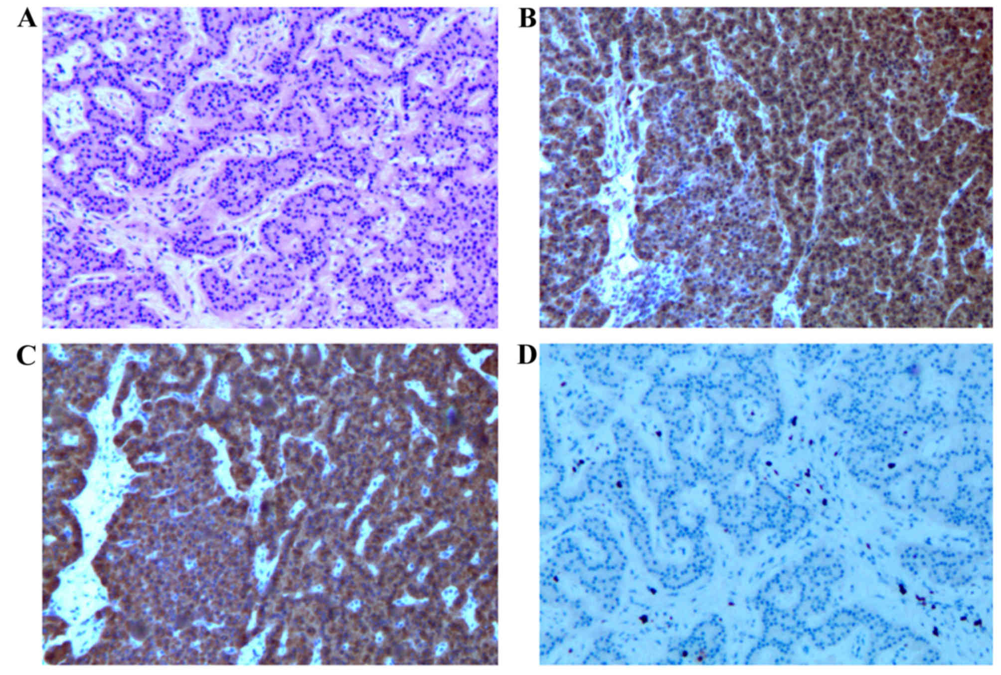
The diagnosis of PNET was made based on standard
histological criteria (Fig. 1).
Hematoxylin and eosin (HE) staining and immunohistochemical
staining for chromogranin A (dilution, 1:2,000; Dako, Carpinteria,
CA, USA), synaptophysin (dilution, 1:75; Dako) and Ki-67 (dilution,
1:50; Thermo Scientific Lab Vision) were performed. Tissue samples
were fixed in 10% formaldehyde overnight, dehydrated and embedded
in paraffin. Sections (5 mm) were stained with HE and also used for
immunohistochemistry.
The following characteristics were collected for
each patient: Age, gender, presenting symptoms, location of primary
tumor, type of surgery, complications, pathological features,
including tumor size, mitotic count and Ki-67 index,
Tumor-Node-Metastasis (TNM) stage (15), adjuvant therapy, the patient's disease
status (13) at the last hospital
visit and the most recent follow-up information.
For the determination of disease stage, the PNETs
were classified into the localized, regional and distant groups. A
localized PNET was defined as an invasive tumor that was completely
confined to the organ of origin. A regional PNET was defined as a
tumor with extension further than the limits of the organ of origin
directly into the surrounding organs or tissue, or the involved
regional lymph nodes, or a tumor that fulfilled each of the
aforementioned criteria. Furthermore, a distant PNET was defined as
a tumor that had spread to regions of the body that were remote
from the primary tumor (16).
The present study was approved by the Institutional
Review Boards of the First Affiliated Hospital, Zhejiang University
School of Medicine, and informed consent was obtained from the
patient for the publication of this report and any accompanying
images.
Statistical analysis
Results are presented as median (range) and all
statistical analyses were performed using SPSS 16.0 (SPSS, Inc.,
Chicago, IL, USA) for Windows. Overall survival (OS) was defined as
the time between the initial diagnosis and mortality from any cause
or to the time of the last known contact. Disease-free survival
(DFS) was defined as the time between surgery and PNET recurrence.
Those individuals who did not exhibit evidence of local recurrence
or metastasis at the last follow-up and those patients who
succumbed from causes that were unrelated to PNETs were censored in
the analysis of DFS rate. OS and DFS rate analyses were performed
by Kaplan-Meier methodology with log-rank testing. Cox proportional
hazard models were used to estimate hazard ratios for OS and DFS
rates, and to determine independent risk factors. All tests were
two-sided, with P<0.05 considered to indicate a statistically
significant difference.
Results
Clinicopathological characteristics of
PNETs
Among the 104 Chinese patients with PNETs, 49
(47.1%) were men and 55 (52.9%) were women. The median age at
presentation was 52 years (range, 19–76 years). Of the 104 PNETs,
30 (28.8%) were found incidentally during a health examination. The
most common presentation of the symptomatic PNETs was abdominal
pain in 31 (29.8%) patients, followed by hypoglycemia in 28
(26.9%), abdominal discomfort in 9 (8.7%), jaundice in 4 (3.8%) and
diarrhea in 2 (1.9%). The median size of the PNETs was 3 cm (range,
0.8–19.0 cm). The PNETs were located in the head of the pancreas
(n=49; 47.1%), followed by the tail (n=38; 36.5%) and body (n=17;
16.3%) (Table I). A total of 99
patients underwent curative resections (R0 resection, 95.2%), in
which the distal pancreatectomy was the most common procedure
(51.0%), followed by pancreaticoduodenectomy (27.9%) and local
resection of the pancreas (16.3%). Palliative surgery (R1
resection, 4.8%) was performed for only 5 patients, where
symptomatic chemotherapy and somatostatin analog therapy were
simultaneously performed.
 | Table I.Clinical and pathological
characteristics for patients (n=104). |
Table I.
Clinical and pathological
characteristics for patients (n=104).
| Variable | Value |
|---|
| Age, years |
|
|
Median | 52 |
|
Range | 19–76 |
| Gender, n (%) |
|
| Male | 49 (47.1) |
|
Female | 55 (52.9) |
| Presentation, n
(%) |
|
| Abdominal
pain | 31 (29.8) |
|
Incidental finding | 30 (28.8) |
| Abdominal
discomfort | 9 (8.7) |
|
Jaundice | 4 (3.8) |
|
Hypoglycemia | 28 (26.9) |
|
Diarrhea | 2 (1.9) |
| Tumor size, cm |
|
|
Median | 3 |
|
Range | 0.8–19 |
| Location, n
(%) |
|
|
Head/uncinate | 49 (47.1) |
|
Body | 17 (16.3) |
|
Tail | 38 (36.5) |
| Surgical
approaches, n (%) |
|
| R0
resection |
|
DP | 53 (51.0) |
|
PD | 29 (27.9) |
|
LP | 17 (16.3) |
| R1
resection | 5
(4.8) |
| Stage, n (%) |
|
|
Local | 83 (79.8) |
|
Regional | 13 (12.5) |
|
Distant | 8
(7.7) |
| Grade, n (%) |
|
| 1 | 47 (45.2) |
| 2 | 50 (48.1) |
| 3 | 7
(6.7) |
| Distant metastasis,
n (%) |
|
| At
initial diagnosis | 8
(7.7) |
| During
follow-up | 18 (17.3) |
| Lymph node
metastasis, n (%) |
|
|
Negative | 84 (80.8) |
|
Positive | 20 (19.2) |
The majority of tumors were of low or moderate grade
(93.3%, grade 1 or 2) and local or regional stage (92.3%). In
total, 8 (7.7%) patients were classified as presenting with distant
metastasis at the initial diagnosis, while 17 (17.3%) patients
presented with distant metastasis during the follow-up. The most
frequent metastatic site was the liver. The patients received
treatments that included radiofrequency ablation (n=8),
transarterial chemoembolization (n=7), symptomatic chemotherapy
(n=4), with intravenous infusion of 100 mg/m2 etoposide
and 75 mg/m2 cisplatin daily for 1–3 days, repeated
every 21 days, reoperation (n=1) and somatostatin analog therapy
(n=2) with 20 mg intramuscular Sandostatin LAR every 4 weeks for 3
months. The pathology showed lymph node metastasis in 20 (19.2%)
patients. Only 20 tumors (19.2%) were functional, including 5
gastrinomas, 8 insulinomas, 4 glucagonomas and 3 cases of multiple
endocrine neoplasia-1.
Natural course and prognostic factors
of PNETs
The median duration of post-operative follow-up was
31 months. The 1, 3 and 5-year OS rates were 95, 85 and 73%,
respectively. Meanwhile, the 1, 3 and 5-year DFS rates were 86, 72
and 68%, respectively. The 5-year OS rates for grades 1 to 3 were
89.1, 63.6 and 43.8% (P=0.002), while the 5-year OS rates for
local, regional and distant disease stages were 90.4, 39.5 and
18.8% (P<0.001).
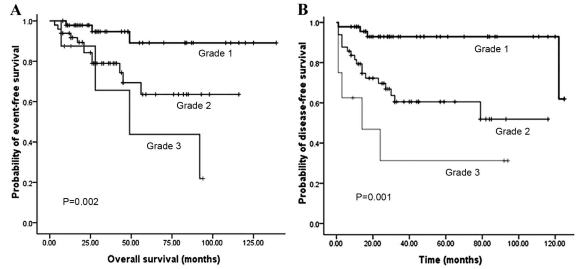
Fig. 2 illustrates
that the OS and DFS rates were significantly different with regard
to grade (grades 1, 2 and 3). Compared with grades 2 and 3, grade 1
PNET exhibited superior OS (P=0.002) and DFS (P<0.001) rates.
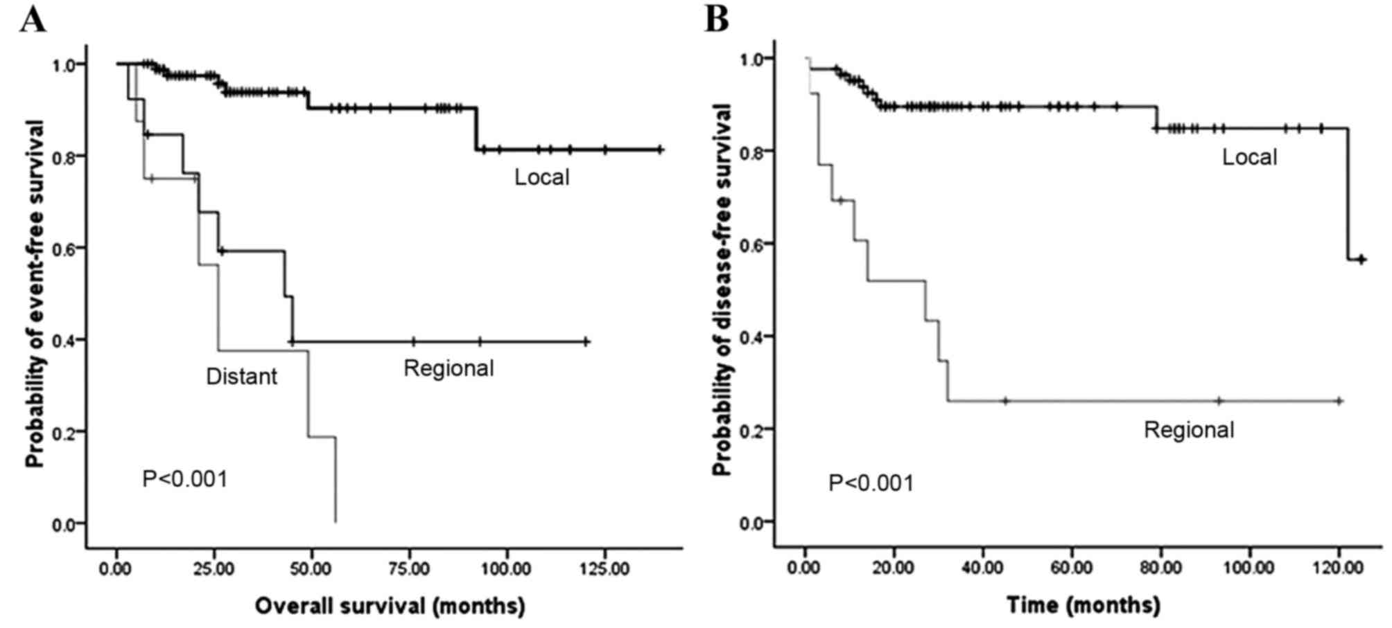
Fig. 3 illustrates that the OS and
DFS rates were also significantly different with regard to stage
(local, regional and distant disease) (all P<0.001). Compared
with the regional or distant stage, PNET at the local stage
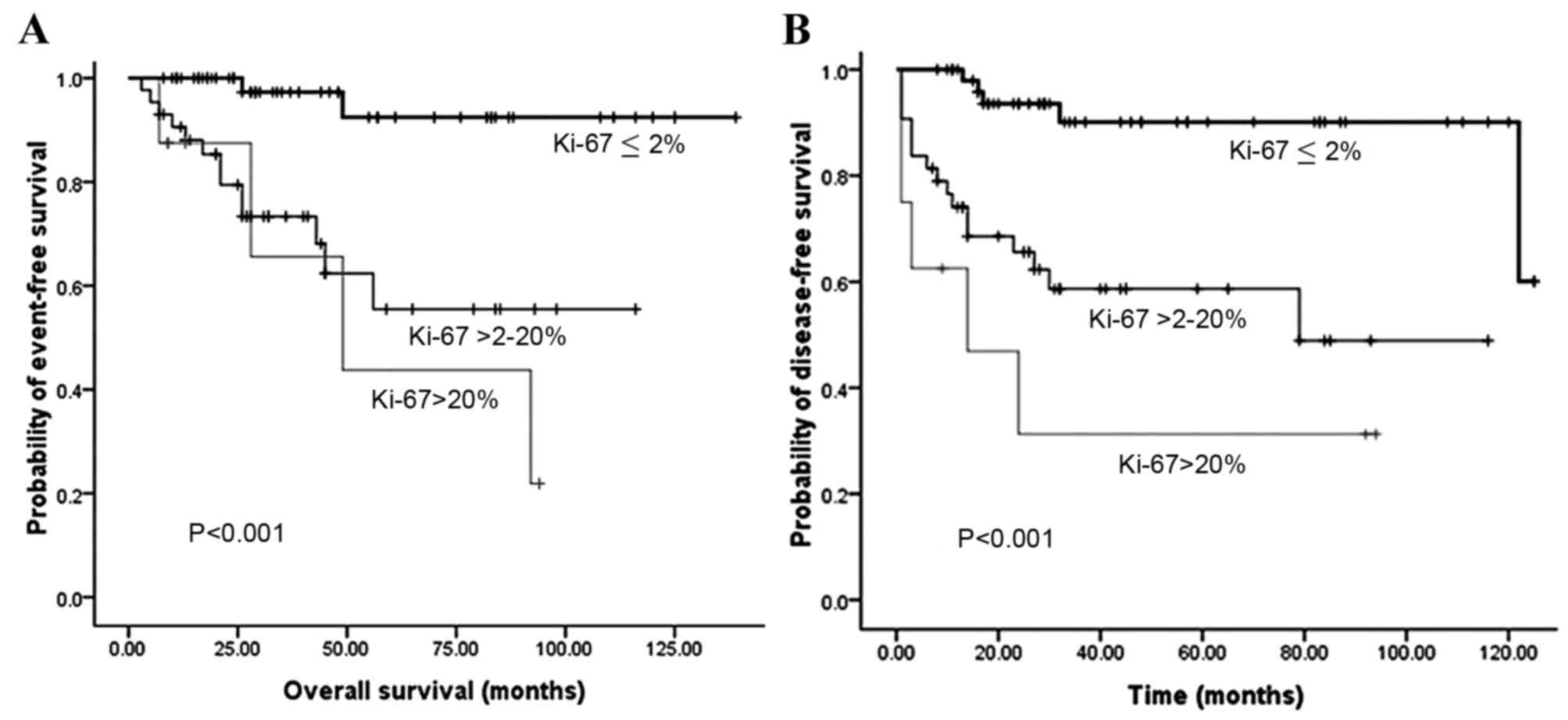
exhibited superior OS and DFS rates. Furthermore, patients with a
low Ki-67 index exhibited superior OS and DFS rates compared with
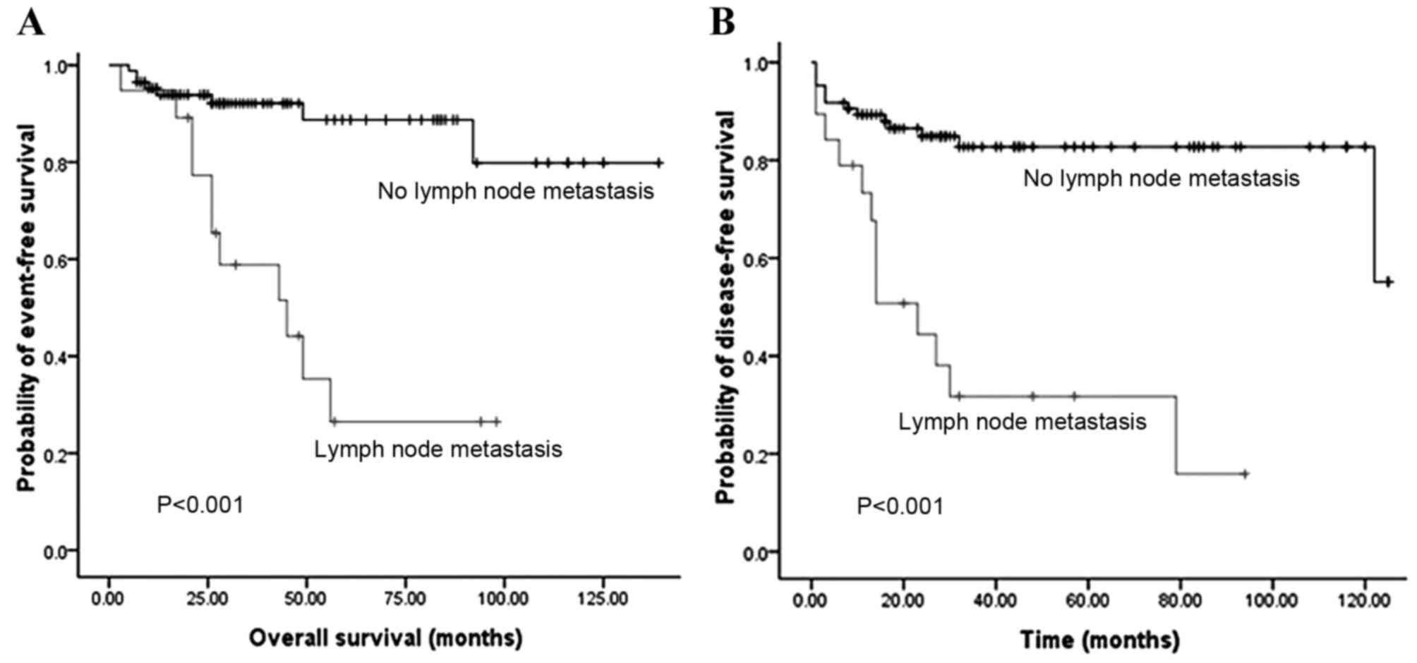
patients with a high Ki-67 index (P<0.001) (Fig. 4). The presence of lymph node
metastasis was predictive of inferior OS and DFS rates compared
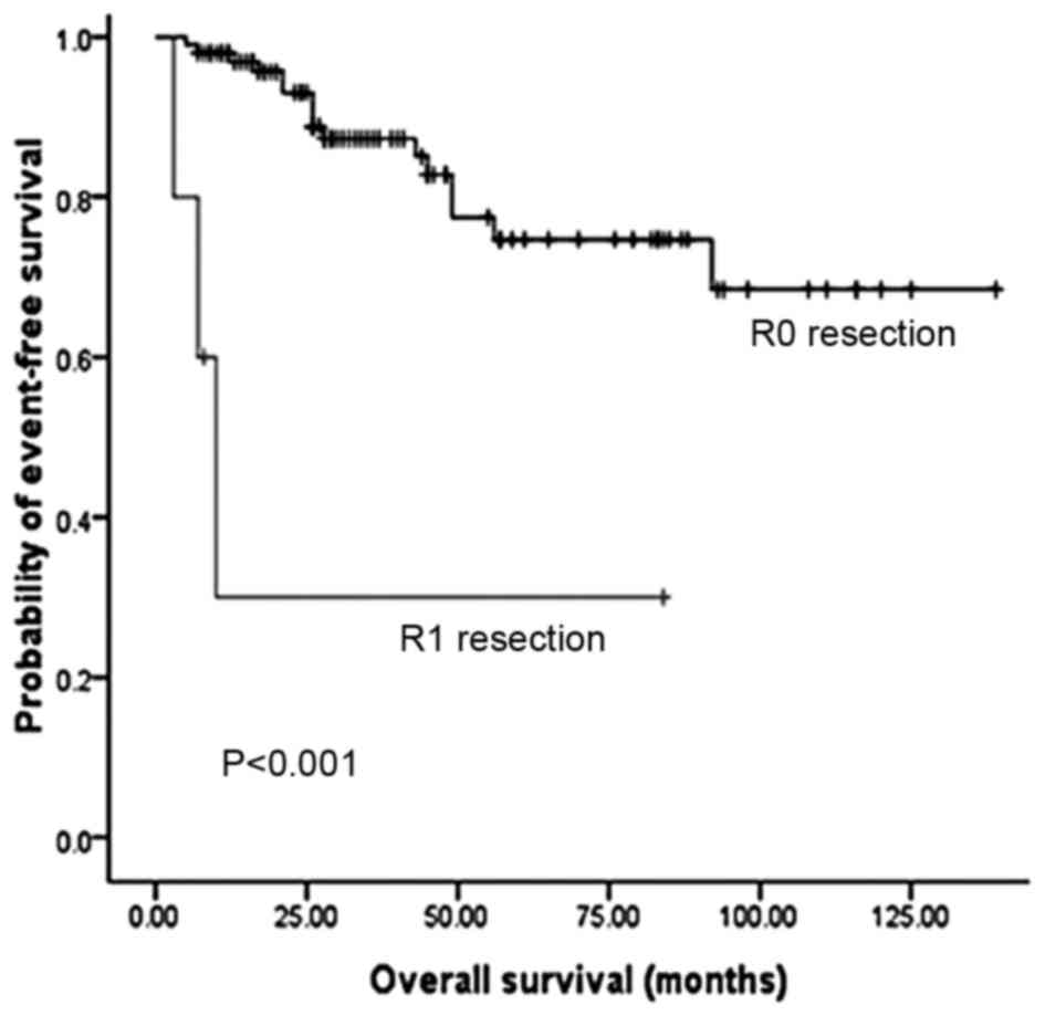
with the absence of lymph node metastasis (Fig. 5). In addition, the OS rate for
patients with PNETs was significantly decreased following R1
resection (P<0.001) (Fig. 6). In
addition, when the PNET patients were divided into 2 groups by age
using a cutoff value of 50 years, a significant difference in OS
rate (P=0.033) was observed, whereas no statistical difference was
observed in DFS rate (P=0.388). When the PNET patients were divided
into the 2 groups of incidental diagnosis and symptomatic
diagnosis, there was no statistical difference in OS or DFS rate.
Compared with symptomatic PNETs, the incidental detection of PNETs
was associated with longer OS (116 vs. 101 months; P=0.114) and DFS
(103 vs. 86 months; P=0.146) times. DFS rate (P=0.010) was
significantly different with regard to gender, whereas there was no
significantly difference in OS rate (P=0.055). When the PNETs were
divided into 3 groups according to primary tumor size, a
significant difference in OS rate (P=0.044) was observed, whereas
no statistical difference in DFS rate was observed, although it
appeared that patients with smaller PNETs experienced longer DFS
times than those with larger PNETs (P=0.054) (Table II).
| Table II.Univariate analyses of OS and
DFS. |
Table II.
Univariate analyses of OS and
DFS.
|
Characteristics | OS, months | 5-year survival
rate, % | P-value | DFS, months | 5-year DFS rate,
% | P-value |
|---|
| Age, years |
|
|
|
|
|
|
|
<50 | 121.0 | 81.0 |
0.033 |
96.0 | 75.1 |
0.388 |
|
≥50 | 90.0 | 67.7 |
|
89.0 | 70.9 |
|
| Gender |
|
|
|
|
|
|
|
Male |
88.0 | 64.3 |
0.055 |
78.0 | 60.5 |
0.010 |
|
Female | 119.0 | 81.5 |
| 103.0 | 83.4 |
|
| Incidental
diagnosis |
|
|
|
|
|
|
|
Yes | 116.0 | 91.8 |
0.114 | 103.0 | 87.4 |
0.146 |
| No
(symptomatic) | 101.0 | 68.2 |
|
86.0 | 67.2 |
|
| Tumor grade |
|
|
|
|
|
|
| Grade
1 | 127.0 | 89.1 |
0.002 | 116.0 | 92.9 | <0.001 |
| Grade
2 | 85.0 | 63.6 |
|
72.0 | 60.5 |
|
| Grade
3 | 58.0 | 43.8 |
|
36.0 | 31.3 |
|
| Stage |
|
|
|
|
|
|
|
Local | 124.0 | 90.4 | <0.001 | 111.0 | 89.5 | <0.001 |
|
Regional | 62.0 | 39.5 |
|
42.0 | 26.0 |
|
|
Distant | 30.0 | 18.8 |
| NA | NA |
|
| Lymph node
metastasis |
|
|
|
|
|
|
|
Yes | 50.0 | 26.5 | <0.001 |
37.0 | 31.7 | <0.001 |
| No | 121.0 | 88.7 |
| 104.0 | 82.7 |
|
| Size |
|
|
|
|
|
|
| ≤2
cm | 121.0 | 87.0 |
0.044 | 111.0 | 87.0 |
0.054 |
| >2
cm to 4 cm | 90.0 | 73.2 |
|
84.0 | 69.6 |
|
| >4
cm | 87.0 | 74.0 |
|
80.0 | 59.7 |
|
| Ki-67 index
(%) |
|
|
|
|
|
|
|
0–2 | 131.0 | 92.3 | <0.001 | 114.0 | 90.1 | <0.001 |
| >2
to 20 | 77.0 | 55.5 |
|
69.0 | 58.7 |
|
|
>20 | 58.0 | 43.8 |
|
36.0 | 31.3 |
|
| Resection
status |
|
|
|
|
|
|
| R0 | 109.0 | 74.7 | <0.001 | NA | NA | NA |
| R1 | 30.0 | 30.0 |
| NA | NA |
|
Multivariate OS rate variables of significance
remaining in the final model included stage (P=0.02), grade
(P=0.025), lymph node status (P<0.001), Ki-67 index (P=0.031)
and surgical margin status (P<0.001) (Table III). Tumor size (P=0.044) and age
(P=0.033) were those variables with a significant association with
OS rate on univariate analysis, but no significant multivariate
impact (P=0.254 for tumor size; P=0.229 for age). Meanwhile, upon
multivariate analysis, stage (P=0.001), grade (P=0.017), lymph
nodes status (P=0.02) and Ki-67 index (P=0.016) were prognostic
factors for DFS rate (Table IV).
| Table III.Cox regression analysis for overall
survival. |
Table III.
Cox regression analysis for overall
survival.
| Variable | HR | 95% CI | P-value |
|---|
| Age |
|
|
|
|
<50 |
|
|
|
|
≥50 | 2.17 | 0.61–7.69 |
0.229 |
| Tumor grade |
|
|
|
| G1 |
|
|
|
|
G2/G3 | 5.04 | 1.23–20.66 |
0.025 |
| Stage |
|
|
|
|
Local |
|
|
|
|
Regional/distant | 5.731 | 1.31–25.04 |
0.020 |
| Lymph node
metastasis |
|
|
|
| No |
|
|
|
|
Yes | 6.361 | 2.06–19.58 | <0.001 |
| Ki-67 index, % |
|
|
|
| ≤2 |
|
|
|
|
>2 | 4.13 | 1.14–15.02 |
0.031 |
| Resection
status |
|
|
|
| R0 |
|
|
|
| R1 | 20.16 | 3.37–120.72 | <0.001 |
| Table IV.Cox regression analysis for
disease-free survival. |
Table IV.
Cox regression analysis for
disease-free survival.
| Variable | HR | 95% CI | P-value |
|---|
| Gender |
|
|
|
|
Female |
|
|
|
|
Male | 2.11 | 0.83–5.40 | 0.119 |
| Tumor grade |
|
|
|
| G1 |
|
|
|
|
G2/G3 | 4.25 | 1.30–13.92 | 0.017 |
| Stage |
|
|
|
|
Local |
|
|
|
|
Regional | 5.71 | 2.14–15.21 | 0.001 |
| Lymph node
metastasis |
|
|
|
| No |
|
|
|
|
Yes | 3.85 | 1.23–11.98 | 0.020 |
| Ki-67 index, % |
|
|
|
| ≤2 |
|
|
|
|
>2 | 3.22 | 1.92–8.27 | 0.016 |
Discussion
PNETs, a group of endocrine tumors arising in the
pancreas, are rare and account for only 1–3% of all primary
pancreatic malignancies (4). Several
previous studies suggested that factors predicting survival
following the resection of PNETs included age, gender, functional
status of the tumor, pancreatic resection, lymph node involvement,
distant metastases, grade of the tumor, and TNM stage, while others
suggested that the presence or absence of cancer at the surgical
margin, tumor location, tumor size and incidental or symptomatic
tumors were the prognostic factors (6–14).
TNM stage and grade represent a simple and accurate
instrument for mortality risk and disease-free assessment, as they
accurately reflect the biology and natural history of the cancer. A
study by Strosberg et al showed that the 5-year
recurrence-free survival (RFS) rates for AJCC stages I, II and III
were 78, 53 and 33%, respectively (P=0.01 for stage I vs. stages
II/III), which suggested that the American Joint Cancer Committee
(AJCC) classifications for PNETs were prognostic for RFS (17). In addition, the use of the
International Union for Cancer Control/AJCC/World Health
Organization 2010 TNM staging results as an independent predictor
of survival upon multivariable analysis suggests that measuring the
extent of the cancer is relevant itself, as confirmed by a single
series investigation in 2011 (18).
Yang et al also indicated that the European Neuroendocrine
Tumor Society TNM staging system may be superior in clinical
practice when compared with the AJCC Staging Manual (seventh
edition) for use in PNETs. Tumor grade, radical resection and the
new AJCC system have all been confirmed as independent predictors
for PNETs (13). In the present
study, tumor stage was correlated with grade. The results showed
that OS rate in the local disease stage was significantly higher
compared with regional and distant disease stage (P<0.001),
which was similar to the results found by Kim et al
(16). Meanwhile, the present study
also found that tumor grade was a critically important prognostic
factor for OS rate (P=0.002). The present study documented that
higher grade PNETs exhibited greater recurrence than PNETs of a
lower grade (P<0.001). The most important factors to affect OS
and DFS rates were stage and grade, as observed previously in a
number of studies (2,6,9,13). Greater recurrence was predicted in
association with regional disease rather than local disease.
Certain studies, including the Surveillance,
Epidemiology, and End Results report (19), showed that upon multivariate analysis,
female gender was associated with a better prognosis, whereas in
other previous studies, this was not observed (20). Moreover, women also experienced
significantly longer DFS times compared with men. A study by
Casadei et al appeared to suggest that factors (young age,
female gender and the absence of comorbidities) represented a
strong predictor of survival (21).
In the present study, DFS rate (P=0.010) was found to be
significantly different with regard to gender, whereas there was no
significant difference in OS rate, although it appeared that women
had longer OS times (119 vs. 88.0 months, P=0.055). This result was
similar to that of a study by Rindi et al, which found
gender to be a prognostic factor for DFS in patients with
neuroendocrine tumors (22). Possible
explanations for these differences in tumor biology and survival
may be gender imbalances for risk factors such as smoking, alcohol
consumption, a different constitution or hormonal effects.
It is known that tumor size is an important
prognostic factor in PNETs (23). In
the present study, a smaller tumor size was associated with a
longer OS time. Although tumor size was not significantly
associated with DFS, patients with smaller PNETs experienced longer
DFS times. In addition, age was an important prognostic factor for
OS rate in the present study (P=0.033), as has been found in one
previous study (19), but in contrast
to other study results (6,13).
Another factor potentially associated with adverse
survival is lymph invasion (6). In
the current study, the presence of lymph node metastasis was
predictive of inferior OS and DFS rates compared with the absence
of lymph node metastasis. This was supported by the studies of
Scarpa et al (24) and Ito
et al (25), which also
identified lymph node metastasis as a relevant prognostic factor.
By contrast, Bahra et al reported that lymph node metastases
was not significant in determining the survival of non-functional
PNETs, and a multivariate analysis of nodal stage revealed no
significant differences with regard to the predicted cumulative
survival probability (P=0.81) (26).
As a marker of cellular proliferation, Ki-67 has
prognostic value in a number of malignancies, including PNETs
(26,27). Mitotic grade and Ki-67 grade have each
been used to distinguish between patients with a good prognosis and
those with a poor prognosis. The univariate analysis in the study
by Liu et al showed that mitotic count, Ki-67 index and
tumor necrosis were all significant prognostic factors for regional
well-differentiated PNETs (28).
Another study also demonstrated that the predictive power of Ki-67
was enhanced when combined with metastasis status and tumor size
(29). Cherenfant et al
reported that a cut-off value of >3% for Ki-67 was the best
mortality predictor (83%), with an area under the curve of 0.85.
This Ki-67 value was also predictive of distant metastasis
occurrence, with odds ratio and 95% confidence interval values of
9.22 and 1.55–54.55, respectively (P<0.015) (27). Scarpa et al observed no
difference in outcome between those individuals with a Ki-67 value
of ≤2% and those with a value of ≤5%. In this study, the
statistically significant cut-off value for Ki-67 was 5%. A Ki-67
index of 5–20% was associated with a 2-times higher risk of
mortality from disease compared with a Ki-67 of ≤5%. Patients with
a Ki-67 of >20% were 11 times more likely to succumb to disease
compared with patients with a Ki-67 of ≤5% (24). In the present study, patients with a
Ki-67 of >2% showed significantly decreased OS and DFS rates
upon multivariate analysis (P<0.05), which was similar to the
results by Bahra et al (26)
and La Rosa et al (30). As
these large variations exist in the Ki-67 index values in PNETs, a
requirement exists for a larger study or a meta-analysis in order
to identify the ideal cut-off point.
The risk of progression and mortality for
incidentally diagnosed PNETs appears to be lower than patients who
are symptomatic from their tumors at diagnosis. In the present
study, there was no statistical difference between the incidental
diagnosis and the symptomatic diagnosis with regard to OS (P=0.114)
and DFS (P=0.146) rates. Cheema et al reported an 86% 5-year
progression-free survival rate for incidentally diagnosed tumors
compared with a 59% 5-year progression-free survival rate for
symptomatic tumors (P=0.007). Upon multivariate analysis, the
strongest prognostic factor for progression was the incidental
detection of the tumors (31).
In conclusion, the results of the present study
suggested that stage, grade, Ki-67 index and lymph nodes
involvement are significant prognostic factors for OS and DFS rates
in PNETs. Furthermore, surgical margin status may also be an
independent predictor for the prognosis of PNETs. These findings
may serve as future useful clinical survival predictors,
particularly with regard to resected disease.
Acknowledgements
This study was funded by the Projects of the
Ministry of Public Health (grant no. 201002004).
Glossary
Abbreviations
Abbreviations:
|
PNETs
|
pancreatic neuroendocrine tumors
|
|
OS
|
overall survival
|
|
DFS
|
disease-free survival
|
References
|
1
|
Klimstra DS, Modlin IR, Coppola D, Lloyd
RV and Suster S: The pathologic classification of neuroendocrine
tumors: A review of nomenclature, grading, and staging systems.
Pancreas. 39:707–712. 2010. View Article : Google Scholar : PubMed/NCBI
|
|
2
|
Fischer L, Kleeff J, Esposito I, Hinz U,
Zimmermann A, Friess H and Büchler MW: Clinical outcome and
long-term survival in 118 consecutive patients with neuroendocrine
tumours of the pancreas. Br J Surg. 95:627–635. 2008. View Article : Google Scholar : PubMed/NCBI
|
|
3
|
Niederle MB, Hackl M, Kaserer K and
Niederle B: Gastroenteropancreatic neuroendocrine tumours: The
current incidence and staging based on the WHO and European
Neuroendocrine Tumour Society classification: An analysis based on
prospectively collected parameters. Endocr Relat Cancer.
17:909–918. 2010. View Article : Google Scholar : PubMed/NCBI
|
|
4
|
Bilimoria KY, Talamonti MS, Tomlinson JS,
Stewart AK, Winchester DP, Ko CY and Bentrem DJ: Prognostic score
predicting survival after resection of pancreatic neuroendocrine
tumors: Analysis of 3851 patients. Ann Surg. 247:490–500. 2008.
View Article : Google Scholar : PubMed/NCBI
|
|
5
|
Hauso O, Gustafsson BI, Kidd M, Waldum HL,
Drozdov I, Chan AK and Modlin IM: Neuroendocrine tumor
epidemiology: Contrasting Norway and North America. Cancer.
113:2655–2664. 2008. View Article : Google Scholar : PubMed/NCBI
|
|
6
|
Yang M, Zeng L, Zhang Y, Su AP, Yue PJ and
Tian BL: Surgical treatment and clinical outcome of nonfunctional
pancreatic neuroendocrine tumors: A 14-year experience from one
single center. Medicine (Baltimore). 93:e942014. View Article : Google Scholar : PubMed/NCBI
|
|
7
|
Centonze DC, Cinardi N and Giannone G:
Surgical resection for neuroendocrine tumors of the pancreas: A
fourteen years single institutional observation. Eur Rev Med
Pharmacol Sci. 18:(Suppl 2). 32–35. 2014.PubMed/NCBI
|
|
8
|
Wang SE, Su CH, Kuo YJ, Shyr YM, Li AF,
Chen TH, Wu CW and Lee CH: Comparison of functional and
nonfunctional neuroendocrine tumors in the pancreas and
peripancreatic region. Pancreas. 40:253–259. 2011. View Article : Google Scholar : PubMed/NCBI
|
|
9
|
Casadei R, Ricci C, Pezzilli R, Campana D,
Tomassetti P, Calculli L, Santini D, Antonacci N and Minni F: Value
of both WHO and TNM classification systems for patients with
pancreatic endocrine tumors: Results of a single-center series.
World J Surg. 33:2458–2463. 2009. View Article : Google Scholar : PubMed/NCBI
|
|
10
|
Casadei R, Ricci C, Rega D, D'Ambra M,
Pezzilli R, Campana D, Nori F, Minni F and Tomassetti P: Pancreatic
endocrine tumors less than 4 cm in diameter: Resect or enucleate? A
single-center experience. Pancreas. 39:825–828. 2010. View Article : Google Scholar : PubMed/NCBI
|
|
11
|
Franko J, Feng W, Yip L, Genovese E and
Moser AJ: Non-functional neuroendocrine carcinoma of the pancreas:
Incidence, tumor biology, and outcomes in 2,158 patients. J
Gastrointest Surg. 14:541–548. 2010. View Article : Google Scholar : PubMed/NCBI
|
|
12
|
Casadei R, Ricci C, Pezzilli R, Campana D,
Tomassetti P, Calculli L, Santini D, D'Ambra M and Minni F: Are
there prognostic factors related to recurrence in pancreatic
endocrine tumors? Pancreatology. 10:33–38. 2010. View Article : Google Scholar : PubMed/NCBI
|
|
13
|
Yang M, Zeng L, Zhang Y, Wang WG, Wang L,
Ke NW, Liu XB and Tian BL: TNM staging of pancreatic neuroendocrine
tumors: An observational analysis and comparison by both AJCC and
ENETS systems from 1 single institution. Medicine (Baltimore).
94:e6602015. View Article : Google Scholar : PubMed/NCBI
|
|
14
|
Partelli S, Inama M, Rinke A, Begum N,
Valente R, Fendrich V, Tamburrino D, Keck T, Caplin ME, Bartsch D,
et al: Long-term outcomes of surgical management of pancreatic
neuroendocrine tumors with synchronous liver metastases.
Neuroendocrinology. 102:68–76. 2015. View Article : Google Scholar : PubMed/NCBI
|
|
15
|
Rindi G, Klöppel G, Alhman H, Caplin M,
Couvelard A, de Herder WW, Erikssson B, Falchetti A, Falconi M,
Komminoth P, et al: TNM staging of foregut (neuro)endocrine tumors:
A consensus proposal including a grading system. Virchows Arch.
449:395–401. 2006. View Article : Google Scholar : PubMed/NCBI
|
|
16
|
Kim SJ, Kim JW, Oh DY, Han SW, Lee SH, Kim
DW, Im SA, Kim TY, Heo DS and Bang YJ: Clinical course of
neuroendocrine tumors with different origins (the pancreas,
gastrointestinal tract, and lung). Am J Clin Oncol. 35:549–556.
2012. View Article : Google Scholar : PubMed/NCBI
|
|
17
|
Strosberg JR, Cheema A, Weber JM, Ghayouri
M, Han G, Hodul PJ and Kvols LK: Relapse-free survival in patients
with nonmetastatic, surgically resected pancreatic neuroendocrine
tumors: An analysis of the AJCC and ENETS staging classifications.
Ann Surg. 256:321–325. 2012. View Article : Google Scholar : PubMed/NCBI
|
|
18
|
Strosberg JR, Cheema A, Weber J, Han G,
Coppola D and Kvols LK: Prognostic validity of a novel American
Joint Committee on Cancer Staging Classification for pancreatic
neuroendocrine tumors. J Clin Oncol. 29:3044–3049. 2011. View Article : Google Scholar : PubMed/NCBI
|
|
19
|
Yao JC, Hassan M, Phan A, Dagohoy C, Leary
C, Mares JE, Abdalla EK, Fleming JB, Vauthey JN, Rashid A and Evans
DB: One hundred years after ‘carcinoid’: Epidemiology of and
prognostic factors for neuroendocrine tumors in 35,825 cases in the
United States. J Clin Oncol. 26:3063–72. 2008. View Article : Google Scholar : PubMed/NCBI
|
|
20
|
Strosberg J, Gardner N and Kvols L:
Survival and prognostic factor analysis of 146 metastatic
neuroendocrine tumors of the mid-gut. Neuroendocrinology.
89:471–476. 2009. View Article : Google Scholar : PubMed/NCBI
|
|
21
|
Casadei R, Ricci C, Tomassetti P, Campana
D and Minni F: Factors related to long-term survival in patients
affected by well-differentiated endocrine tumors of the pancreas.
ISRN Surg. 2012:3893852012. View Article : Google Scholar : PubMed/NCBI
|
|
22
|
Rindi G, Falconi M, Klersy C, Albarello L,
Boninsegna L, Buchler MW, Capella C, Caplin M, Couvelard A,
Doglioni C, et al: TNM staging of neoplasms of the endocrine
pancreas: Results from a large international cohort study. J Natl
Cancer Inst. 104:764–77. 2012. View Article : Google Scholar : PubMed/NCBI
|
|
23
|
Bettini R, Boninsegna L, Mantovani W,
Capelli P, Bassi C, Pederzoli P, Fave GF Delle, Panzuto F, Scarpa A
and Falconi M: Prognostic factors at diagnosis and value of WHO
classification in a mono-institutional series of 180
non-functioning pancreatic endocrine tumours. Ann Oncol.
19:903–908. 2008. View Article : Google Scholar : PubMed/NCBI
|
|
24
|
Scarpa A, Mantovani W, Capelli P, Beghelli
S, Boninsegna L, Bettini R, Panzuto F, Pederzoli P, Fave G delle
and Falconi M: Pancreatic endocrine tumors: Improved TNM staging
and histopathological grading permit a clinically efficient
prognostic stratification of patients. Mod Pathol. 23:824–833.
2010. View Article : Google Scholar : PubMed/NCBI
|
|
25
|
Ito H, Abramson M, Ito K, Swanson E, Cho
N, Ruan DT, Swanson RS and Whang EE: Surgery and staging of
pancreatic neuroendocrine tumors: A 14-year experience. J
Gastrointest Surg. 14:891–898. 2010. View Article : Google Scholar : PubMed/NCBI
|
|
26
|
Bahra M, Jacob D, Pascher A, Plockinger U,
Kristiansen G, Neuhaus P and Langrehr JM: Surgical strategies and
predictors of outcome for malignant neuroendocrine tumors of the
pancreas. J Gastroenterol Hepatol. 22:930–935. 2007. View Article : Google Scholar : PubMed/NCBI
|
|
27
|
Cherenfant J, Talamonti MS, Hall CR,
Thurow TA, Gage MK, Stocker SJ, Lapin B, Wang E, Silverstein JC,
Mangold K, et al: Comparison of tumor markers for predicting
outcomes after resection of nonfunctioning pancreatic
neuroendocrine tumors. Surgery. 156:1504–1511. 2014. View Article : Google Scholar : PubMed/NCBI
|
|
28
|
Liu TC, Hamilton N, Hawkins W, Gao F and
Cao D: Comparison of WHO Classifications (2004, 2010), the Hochwald
grading system, and AJCC and ENETS staging systems in predicting
prognosis in locoregional well-differentiated pancreatic
neuroendocrine tumors. Am J Surg Pathol. 37:853–859. 2013.
View Article : Google Scholar : PubMed/NCBI
|
|
29
|
Ferrone CR, Tang LH, Tomlinson J, Gonen M,
Hochwald SN, Brennan MF, Klimstra DS and Allen PJ: Determining
prognosis in patients with pancreatic endocrine neoplasms: Can the
WHO classification system be simplified? J Clin Oncol.
25:5609–5615. 2007. View Article : Google Scholar : PubMed/NCBI
|
|
30
|
La Rosa S, Rigoli E, Uccella S, Novario R
and Capella C: Prognostic and biological significance of
cytokeratin 19 in pancreatic endocrine tumours. Histopathology.
50:597–606. 2007. View Article : Google Scholar : PubMed/NCBI
|
|
31
|
Cheema A, Weber J and Strosberg JR:
Incidental detection of pancreatic neuroendocrine tumors: An
analysis of incidence and outcomes. Ann Surg Oncol. 19:2932–2936.
2012. View Article : Google Scholar : PubMed/NCBI
|