Introduction
Breast cancer is one of the most common types of
female cancer (1). The genesis of
breast cancer is concealed, its progress is rapid and the mortality
rate is high. With the development of living standard, the
morbidity of type 2 diabetes mellitus (T2DM) increases rapidly
(2). At present, numerous studies
have found that T2DM is a systemic disease, and associated with the
progression of several human malignant tumors (3,4).
Ma et al (5)
analyzed the prognosis of 865 early triple-negative breast cancer
(TNBC) patients and demonstrated that patients with TNBC and T2DM
had a shorter disease free survival (DFS) time and more frequent
distant metastasis. Tumor microenvironments, consisting of numerous
stromal cells, cytokines and chemokines, markedly enhance tumor
growth, invasion and metastasis (6).
Adham et al (7) demonstrated
that the glucose concentration of the medium regulates tumor cell
epithelial mesenchymal transition (EMT). Therefore, the present
study inferred that high-concentration glucose in the tumor
microenvironment may promote invasion of breast cancer cells.
Cancer cells generate adenosine triphosphate mainly
by the process of anaerobic glycolysis, which is termed the Warburg
effect (8,9). However, due to the hydrophobicity of
glucose molecules, the process of glucose passing through cell
lipid bilayers must rely on the assistance of a glucose transporter
(Glut) (10). Therefore, the
expression and functions of Gluts partly determine the degree of
the Warburg effect. Glucose transporter 1 (Glut1), encoded by
SLC2A1, is a member of the major facilitator superfamily.
Due to the high affinity of Glut1 for glucose molecules, it can
transport glucose molecules into cells even at a low concentration.
Studies have found that Glut1 is overexpressed in the retina and
glomerulus of T2DM patients (11,12). As
anaerobic glycolysis is an inefficient method of energy production,
cancer cells require enhanced glucose supply. Ectopic expression
levels of Glut1 have been observed in several types of cancer,
including endometrial (13),
non-small lung (14) and colorectal
(15). These studies identified Glut1
as an important prognostic indicator for tumorigenesis. However,
whether hyperglycemia caused by T2DM in the tumor microenvironment
can upregulate the expression of Glut1 and enhance the invasion
ability in breast cancer cells remains unclear.
In the present study, it was demonstrated that the
expression of Glut1 was higher in breast cancer tissues with T2DM
than without T2DM. High glucose concentration may increase the
expression of Glut1, and consequently enhance cell invasion by
upregulating the expression of matrix metalloproteinase 2 (MMP2)
and matrix metalloproteinase 9 (MMP9) in vitro.
Materials and methods
Patients and samples
A total of 120 patients, including 60 patients with
T2DM and 60 patients without T2DM, were enrolled in the present
study and underwent surgery between April 2011 and June 2014. The
age range was between 29 and 69 years, and the median age was 51
years. All resected tumor tissues were identified as invasive
ductal breast carcinoma (IDBC) by senior pathologists. Patients did
not receive chemotherapy and/or radiotherapy prior to surgery.
Clinical data was collected from medical records. The IDBC tissues
and tumor adjacent tissues (>2 cm distance from the surgical
boundary) were stored in 4% paraformaldehyde solution for
immunohistochemistry (IHC) or liquid nitrogen for reverse
transcription-quantitative polymerase chain reaction (RT-qPCR).
All protocols were approved by the Henan Cancer
Hospital Ethics Committee (Henan, China). Informed consent was
obtained and signed by each patient.
Cell culture
The MCF-7 breast cancer cells were obtained from the
Institute of Biochemistry and Cell Biology, Chinese Academy of
Sciences (Shanghai, China). Cells were maintained under different
glucose concentrations, either 5.56 mM or 25.00 mM, and cultured in
a 37°C humidified incubator with 5% CO2. The logarithmic
growth phase cells were harvested for additional assays.
Transwell assay
The MCF-7 cells cultured in different glucose
concentrations were suspended with reduced serum Dulbecco's
modified Eagle's medium (DMEM; Invitrogen; Thermo Fisher
Scientific, Inc., Waltham, MA, USA) and adjusted the density to
2.5×10⁵/ml. In total, 200 µl cell suspension was added
into the upper well of Matrigel-coated (BD Biosciences, Franklin
Lakes, NJ, USA) 8-µm pore Transwell inserts (Nalge Nunc
International, Penfield, NY, USA), and 750 µl DMEM with 10% fetal
bovine serum (Invitrogen; Thermo Fisher Scientific, Inc.) was added
into the lower well. Cells were incubated in a 37°C humidified
incubator with 5% CO2 for 24 h. Membranes were removed,
and uninvaded cells were scraped off and stained with crystal
violet. Cell numbers were counted using ImageJ software (National
Institutes of Health, Bethesda, MD, USA). The experiments were
performed in triplicate.
IHC staining
IHC was performed on paraformaldehyde-fixed paraffin
sections. The SP link IHC Detection kit (Biotin-Streptavidin HRP
Detection systems; catalog no. SP-9001) was purchased from OriGene
Technologies, Inc. (Beijing, China). In brief, rabbit anti-human
polyclonal Glut1 (catalog no. sc-7903; dilution, 1:100), rabbit
anti-human polyclonal MMP2 (catalog no. sc-10736; dilution, 1:100)
and rabbit anti-human polyclonal MMP9 (catalog no. sc-10737;
dilution, 1:100) antibodies were purchased from Santa Cruz
Biotechnology, Inc. (Dallas, TX, USA) and used to detect the
protein expression in IDBC tissues. Tissue sections were incubated
with primary antibodies at 4°C for 24 h. Goat anti-rabbit secondary
antibodies from the SP link IHC Detection kit were added to the
sections and tissue sections were incubated at 37°C for 1 h.
Protein expression was visualized using 3,3-diaminobenzidine
tetrahydrochloride (OriGene Technologies, Inc.) and observed
through a BX46 upright microscope (Olympus Corporation, Tokyo,
Japan). The staining results for the Glut1, MMP2 and MMP9 proteins
were semi-quantitatively calculated by multiplying the staining
intensity and the percentage of positive normal cells, as
previously reported (16).
Small interfering RNA (siRNA)
transfection
Glut1 specific siRNA (catalogue no. sc-35,493; Santa
Cruz Biotechnology, Inc.) was transfected into MCF-7 cells by
Lipofectamine® 2000 (Invitrogen; Thermo Fisher
Scientific, Inc.) according to the manufacturer's protocol.
Scrambled siRNA (catalogue no. sc-37,007; Santa Cruz Biotechnology,
Inc.) was used as a negative control. Reduced serum medium was
changed into complete medium 6 h subsequent to transfection. Cells
were harvested 48 h subsequent to transfection, and then used for
additional experiments.
RT-qPCR
Total RNA was isolated from tissues and MCF-7 cells
using TRIzol® reagent (Invitrogen; Thermo Fisher
Scientific, Inc.) according to the manufacturer's protocol. A
quantitative one-step Perfect Real Time RT-qPCR (SYBR-Green I) kit
(Takara Biotechnology Co., Ltd., Dalian, China) was used to detect
the expression of Glut1, MMP2 and MMP9, according to the
manufacturer's protocol. The human β-actin gene was used as a
reference gene. Primers (Table I)
were synthesized by AuGCT DNA-Syn Biotechnology Co., Ltd. (Beijing,
China). Relative mRNA expression was calculated using the
2−ΔΔCq method (17). All
experiments were performed at least in triplicate.
 | Table I.Primer sequences. |
Table I.
Primer sequences.
| Gene | Primer sequence |
|---|
| Glut1 |
|
|
Sense |
5′-GTCTGGCATCAACGCTGTCT-3′ |
|
Antisense |
5′-ACCACACAGTTGCTCCACATAC-3′ |
| MMP2 |
|
|
|
Sense |
5′-AAGGATGGCAAGTACGGCTT-3′ |
|
Antisense |
5′-CGCTGGTACAGCTCTCATACTT-3′ |
| MMP9 |
|
Sense |
5′-CCTGGAGACCTGAGAACCAATC-3′ |
|
Antisense |
5′-CACCCGAGTGTAACCATAGC-3′ |
| β-actin |
|
Sense |
5′-CTCCATCCTGGCCTCGCTGT-3′ |
|
Antisense |
5′-GCTGTCACCTTCACCGTTCC-3′ |
Western blot analysis
Cells were cleaved with radioimmunoprecipitation
assay (HEART Biotech, Xi'an, China) reagent on ice, and then the
supernatant was obtained to determine protein contents via a
bicinchoninic acid kit (EMD Millipore, Billerica, MA, USA),
according to the manufacturer's protocol. Proteins were separated
by vertical electrophoresis and transferred to a polyvinylidene
fluoride membrane (EMD Millipore). Rabbit anti-human polyclonal
Glut1 (dilution, 1:1,000), rabbit anti-human polyclonal MMP2
(dilution, 1:1,000), rabbit anti-human polyclonal MMP9 (dilution,
1:1,000) and mouse anti-human monoclonal β-actin (catalog no.
sc-47778; dilution, 1:5,000) antibodies were purchased from Santa
Cruz Biotechnology, Inc. and used to detect the protein expression.
Protein expression intensity was developed by the enhanced
chemiluminescent reagent (EMD Millipore).
Statistical analysis
Measurement data are presented as the mean ±
standard deviation. SPSS (version 13.0; SPSS, Inc., Chicago, IL,
USA) was used for the statistical tests, which consisted of the
Pearson's χ2 test and a two-tailed Student's t-test.
P<0.05 was considered to indicate a statistically significant
difference.
Results
Association between T2DM and the
clinical features of IDBC patients
The present study analyzed the differences between
clinical features in IDBC patients with or without T2DM by
χ2 test. As shown in Table
II, the results demonstrated that IDBC patients with T2DM more
commonly had a larger tumor size (≥2 cm; P=0.009), lymphatic
metastasis (P=0.001) and distant metastasis (P=0.017). The present
study infers that patients with IDBC and T2DM exhibit increased
metastasis, which indicates a reduced survival rate.
| Table II.Clinical features of IDBC patients
with or without T2DM (n=120). |
Table II.
Clinical features of IDBC patients
with or without T2DM (n=120).
| Clinical
features | IDBC with T2DM
(n=60) | IDBC without T2DM
(n=60) | χ2 | P-value |
|---|
| Age, years |
|
| 2.155 | 0.142 |
|
<60 | 31 | 23 |
|
|
| ≥60 | 29 | 37 |
|
|
| Menopause |
|
| 1.429 | 0.232 |
| Yes | 39 | 45 |
|
|
| No | 21 | 15 |
|
|
| Tumor size, cm |
|
| 6.806 | 0.009a |
| ≤2 | 17 | 31 |
|
|
|
>2 | 43 | 29 |
|
|
| Number of
nodules |
|
| 1.269 | 0.260 |
| 1 | 20 | 26 |
|
|
| ≥2 | 40 | 34 |
|
|
| Histopathological
grade |
|
| 0.839 | 0.360 |
|
G1-G2 | 25 | 30 |
|
|
| G3 | 35 | 30 |
|
|
| Lymphatic
metastasis |
|
| 10.848 | 0.001a |
| No | 19 | 37 |
|
|
|
Yes | 41 | 23 |
|
|
| Distant
metastasis |
|
| 5.711 | 0.017a |
| No | 27 | 40 |
|
|
|
Yes | 33 | 20 |
|
|
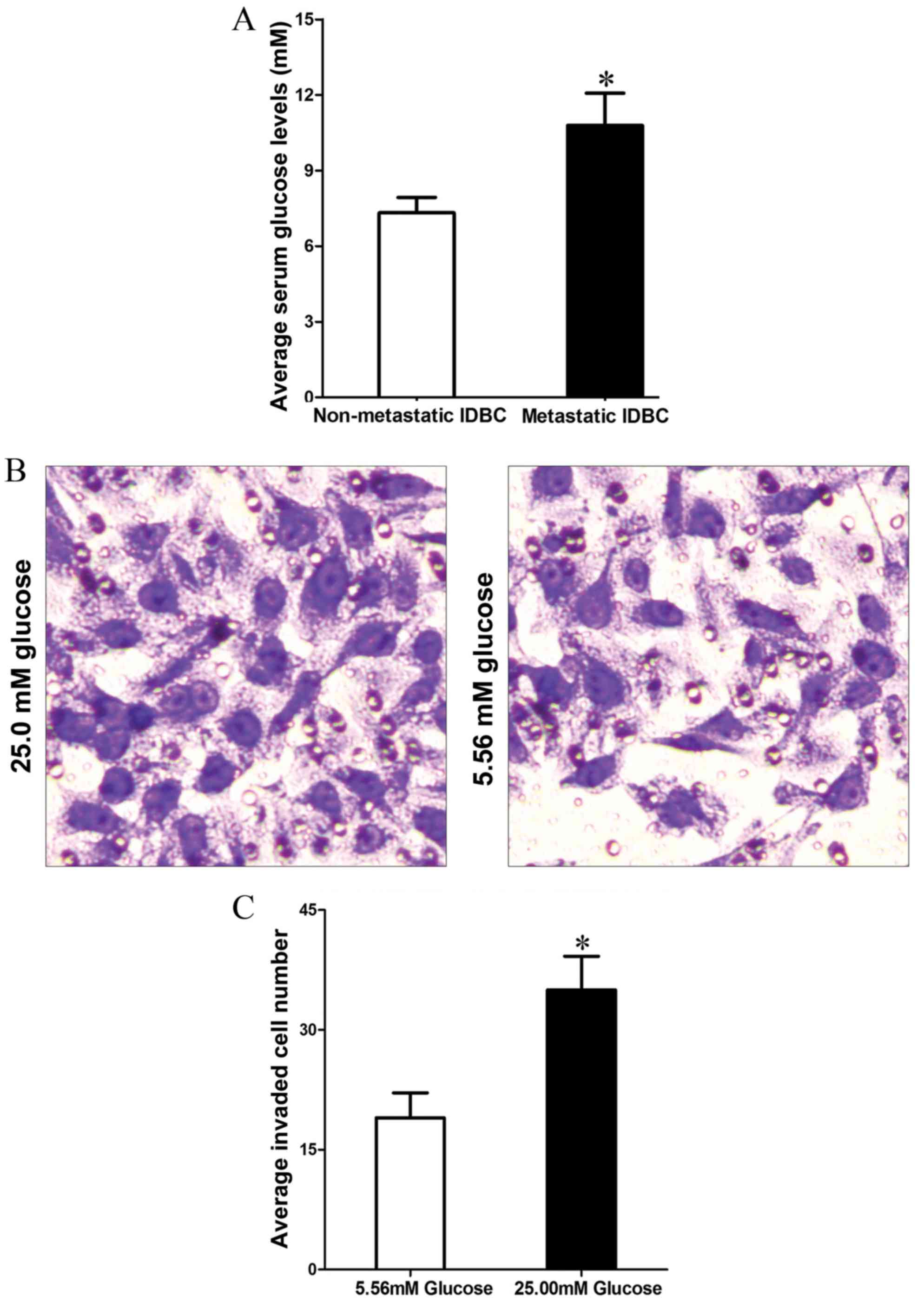
High glucose microenvironment enhances
MCF-7 cell invasion
Hyperglycemia is one of the most direct changes in
T2DM patients. In order to investigate the effect of hyperglycemia
in tumor metastasis, the present study first detected the fasting
blood glucose (FBG) level in IDBC patients with T2DM. Compared to
non-metastatic IDBC (nmIDBC) patients, the FBG level was increased
in metastatic IDBC (mIDBC) patients (7.32±0.62 vs. 10.79±1.28 mM;
P=0.0310; Fig. 1A). In addition,
Transwell assay demonstrated that MCF-7 cells cultured in high
glucose concentration (25 mM) medium acquired stronger invasion
ability compared with those cultured in low glucose concentration
(5.56 mM) medium (35.74±4.03 vs. 19.48±3.12; P=0.030; Fig. 1B and C).
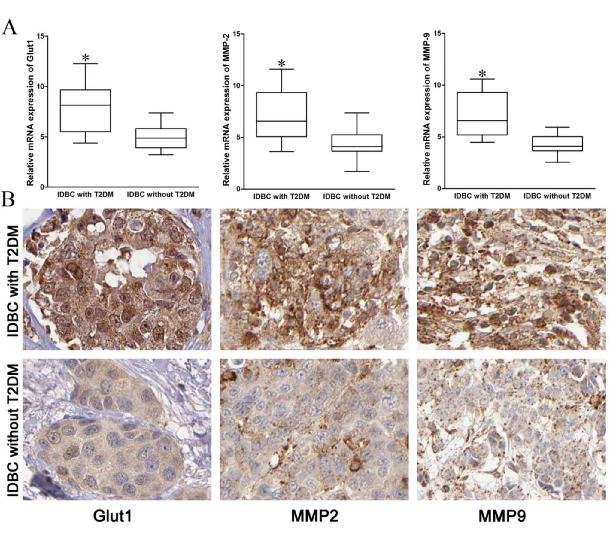
Expression of Glut1, MMP2 and MMP9 in
IDBC tissues
In order to investigate the mechanisms of IDBC
metastasis enhanced by T2DM, the present study detected the mRNA
and protein expressions of Glut1, MMP2 and MMP9 in IDBC tissues.
The mRNA and protein expression of Glut1, MMP2 and MMP9 in IDBC
with T2DM tissues were significantly higher compared with IDBC
without T2DM tissues (Fig. 2; P=0.02,
P=0.031, P=0.010, respectively).
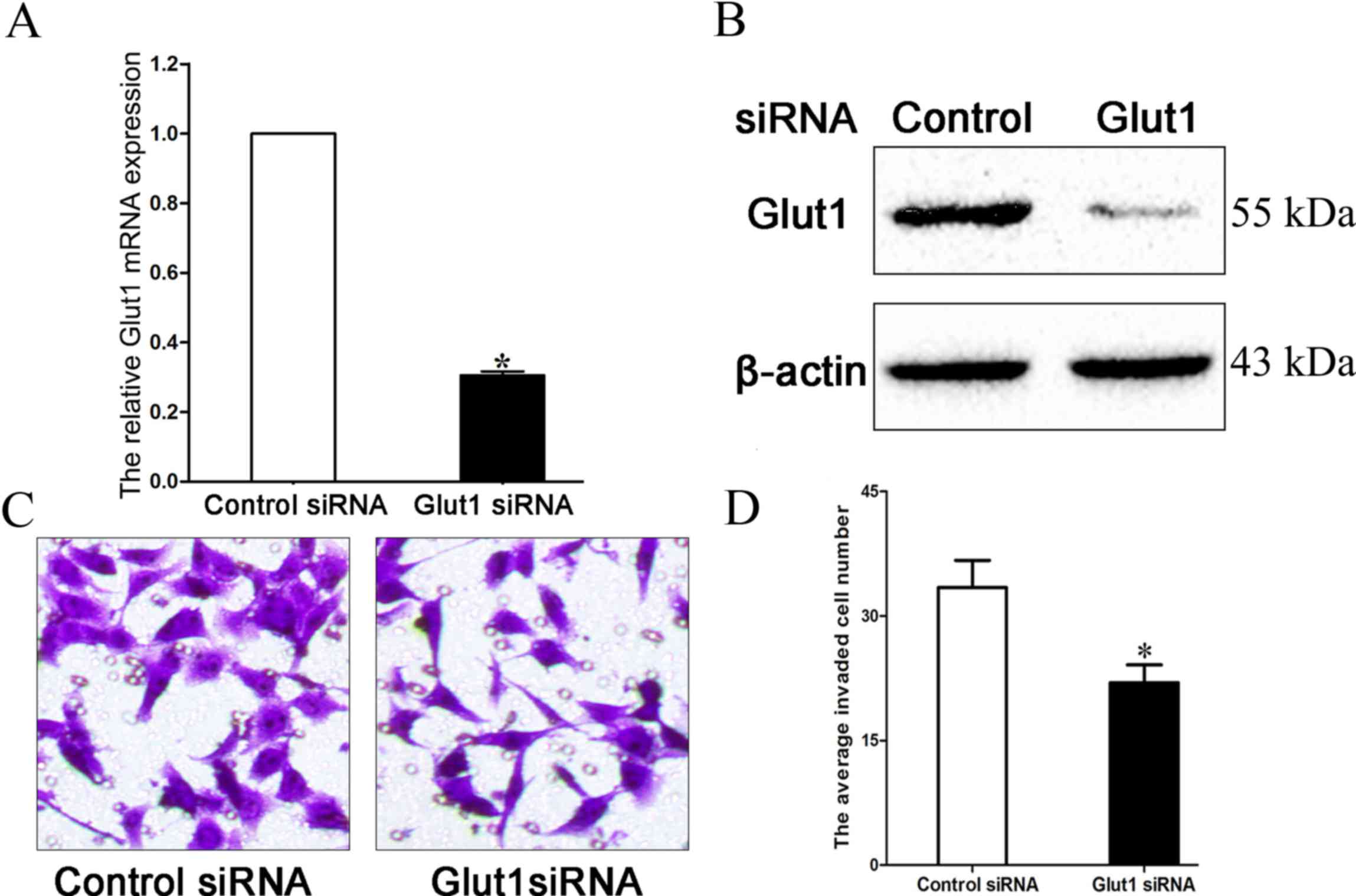
Downregulating Glut1 inhibits MCF-7
cell invasion in a high glucose microenvironment
The present study transfected Glut1-specific siRNA
into MCF-7 cells, which were cultured in high glucose (25 mM)
medium, and demonstrated that the transfection markedly inhibited
Glut1 expression (Fig. 3A and B;
P=0.033). Transwell assay additionally verified that downregulation
of Glut1 may suppress the invasion ability of MCF-7 cells cultured
in high glucose medium (33.43±3.28 vs. 21.96±2.15; Fig. 3C and D; P=0.027).
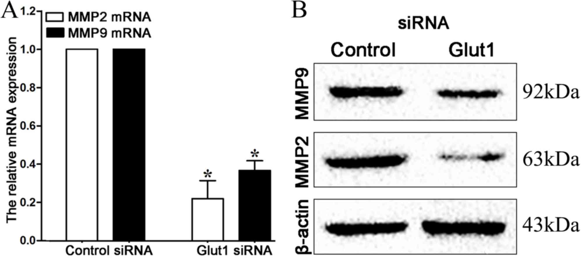
Downregulating Glut1 inhibits MMP2 and
MMP9 expression in high glucose microenvironment
MMPs, particularly MMP2 and MMP9, are crucial for
cancer cell invasion, and their expression may be regulated by
Glut1 (18). Therefore, the present
study detected the expression of MMP2 and MMP9 by RT-qPCR and
western blot analysis in MCF-7 cells subsequent to transfection. It
can be observed in Fig. 4 that mRNA
and protein expression of MMP2 and MMP9 are repressed subsequent to
the downregulation of Glut1 (P=0.021, P=0.034, respectively).
Discussion
The incidence of breast cancer accounts for 20% of
all female cancers (19,20), and numerous patients present with one
or more metabolic disease, such as T2DM. Studies have found that
T2DM is an important risk factor for gastric cancer, endometrial
cancer and numerous other cancer prognoses (21).
Recently, Liao et al (22) evaluated the blood glucose level of
2,048 patients with pancreatic cancer. A dose-response
meta-analysis demonstrated that every 0.56 mmol/l increase in FBG
was associated with a 14% increase in the rate of pancreatic
cancer. Hosokawa et al (23)
reviewed the prognosis of 344 patients with hepatocellular
carcinoma (HCC) subsequent to radiofrequency ablation (RFA), and
found the status of hyperglycemia is positively correlated with
cancer recurrence. The 3-year recurrent rate of HCC with T2DM
subsequent to RFA treatment was 93.8%, while only 73.0% for HCC
patients without T2DM. In the present study, it was similarly found
that IDBC patients with T2DM often had a larger tumor size, and
increased metastasis. Therefore, T2DM may promote tumor growth and
invasion in breast cancer. Additionally, it was found that,
compared with non-metastatic IDBC patients, the serum FBG level was
higher in metastatic IDBC patients. Additional in vitro
assays also confirmed that high glucose levels in the tumor
microenvironment may enhance MCF-7 cell invasion. Consequently, as
a systematic disease, T2DM can affect the glucose concentration of
the tumor microenvironment, which eventually changes cell
biological behaviors.
Overexpression of Glut1 has been confirmed in
numerous types of cancer (24);
however, the function of Glut1 in IDBC patients combined with T2DM
remains unknown. The extracellular matrix can be destroyed by
matrix metalloproteinases, which is the key process of cancer
metastasis (25). In pancreatic
cancer, Glut1 may promote the expression of MMP2 and enhance
MIAPaCa-2 and PANC-1 cell invasion (26). Xu et al (27) reported that the downregulation of
Glut1 by antisense oligodeoxynucleotide significantly inhibits MMP2
expression in laryngeal carcinoma Hep-2 cells. To investigate the
functions and mechanisms of Glut1 in IDBC, the present study first
tested the mRNA and protein levels of Glut1, MMP2 and MMP9 in IDBC
tissues, and demonstrated that the expression of these 3 factors is
higher in IDBC tissues with T2DM. Suppressing Glut1 expression in
MCF-7 cells cultured in high glucose medium markedly reduced the
number of invaded cells, this may be the result of lower expression
of MMP2 and MMP9, which were confirmed by RT-qPCR and western blot
analysis.
In conclusion, IDBC patients with T2DM experienced
an increased amount of malignant clinicopathological features in
comparison to patients with IDBC alone. IDBC cells cultured in a
high glucose microenvironment are more invasive, and this may be a
result of the excessive activation of the Glut1/MMP2/MMP9 axis.
Downregulation of Glut1 may suppress IDBC progression by impairing
cell invasion for T2DM patients.
Acknowledgements
The present study was supported by a grant from the
Key Science and Technology Project of Science and Technology
Department of Henan Province (grant no. 122102310535).
References
|
1
|
Ozyalvacli G, Yesil C, Kargi E, Kizildag
B, Kilitci A and Yilmaz F: Diagnostic and prognostic importance of
the neutrophil lymphocyte ratio in breast cancer. Asian Pac J
Cancer Prev. 15:10363–10366. 2014. View Article : Google Scholar : PubMed/NCBI
|
|
2
|
Joung KH, Jeong JW and Ku BJ: The
association between type 2 diabetes mellitus and women cancer: The
epidemiological evidences and putative mechanisms. Biomed Res Int.
2015:9206182015. View Article : Google Scholar : PubMed/NCBI
|
|
3
|
Nie SP, Chen H, Zhuang MQ and Lu M:
Anti-diabetic medications do not influence risk of lung cancer in
patients with diabetes mellitus: A systematic review and
meta-analysis. Asian Pac J Cancer Prev. 15:6863–6869. 2014.
View Article : Google Scholar : PubMed/NCBI
|
|
4
|
Wintrob ZA, Hammel JP, Khoury T, Nimako
GK, Fu HW, Fayazi ZS, Gaile DP, Forrest A and Ceacareanu AC:
Insulin use, adipokine profiles and breast cancer prognosis.
Cytokine. 89:45–61. 2017. View Article : Google Scholar : PubMed/NCBI
|
|
5
|
Ma FJ, Liu ZB, Qu L, Hao S, Liu GY, Wu J
and Shao ZM: Impact of type 2 diabetes mellitus on the prognosis of
early stage triple-negative breast cancer in People's Republic of
China. OncoTargets Ther. 7:2147–2154. 2014. View Article : Google Scholar
|
|
6
|
Chen Z, Meng Z, Jia L and Cui R: The tumor
microenvironment and cancer. Biomed Res Int. 2014:5739472014.
View Article : Google Scholar : PubMed/NCBI
|
|
7
|
Adham SA, Al Rawahi H, Habib S, Al
Moundhri MS, Viloria-Petit A and Coomber BL: Modeling of
hypo/hyperglycemia and their impact on breast cancer progression
related molecules. PLoS One. 9:e1131032014. View Article : Google Scholar : PubMed/NCBI
|
|
8
|
Yang W and Lu Z: Nuclear PKM2 regulates
the Warburg effect. Cell Cycle. 12:3154–3158. 2013. View Article : Google Scholar : PubMed/NCBI
|
|
9
|
Xu Q, Liu X, Zheng X, Yao Y and Liu Q:
PKM2 regulates Gli1 expression in hepatocellular carcinoma. Oncol
Lett. 8:1973–1979. 2014.PubMed/NCBI
|
|
10
|
Govers R: Cellular regulation of glucose
uptake by glucose transporter GLUT4. Adv Clin Chem. 66:173–240.
2014. View Article : Google Scholar : PubMed/NCBI
|
|
11
|
Tang Z, Wang J, Zhang H, Sun L, Tang F,
Deng Q and Yu J: Associations between diabetes and quality of life
among breast cancer survivors. PLoS One. 11:e01577912016.
View Article : Google Scholar : PubMed/NCBI
|
|
12
|
Heilig CW, Deb DK, Abdul A, Riaz H, James
LR, Salameh J and Nahman NS Jr: GLUT1 regulation of the
pro-sclerotic mediators of diabetic nephropathy. Am J Nephrol.
38:39–49. 2013. View Article : Google Scholar : PubMed/NCBI
|
|
13
|
McKinnon B, Bertschi D, Wotzkow C,
Bersinger NA, Evers J and Mueller MD: Glucose transporter
expression in eutopic endometrial tissue and ectopic endometriotic
lesions. J Mol Endocrinol. 52:169–179. 2014. View Article : Google Scholar : PubMed/NCBI
|
|
14
|
Osugi J, Yamaura T, Muto S, Okabe N,
Matsumura Y, Hoshino M, Higuchi M, Suzuki H and Gotoh M: Prognostic
impact of the combination of glucose transporter 1 and ATP citrate
lyase in node-negative patients with non-small lung cancer. Lung
cancer. 88:310–318. 2015. View Article : Google Scholar : PubMed/NCBI
|
|
15
|
Nam SO, Yotsumoto F, Miyata K, Fukagawa S,
Yamada H, Kuroki M and Miyamoto S: Warburg effect regulated by
amphiregulin in the development of colorectal cancer. Cancer Med.
4:575–587. 2015. View
Article : Google Scholar : PubMed/NCBI
|
|
16
|
Zhang J, Tu K, Yang W, Li C, Yao Y, Zheng
X and Liu Q: Evaluation of Jagged2 and Gli1 expression and their
correlation with prognosis in human hepatocellular carcinoma. Mol
Med Rep. 10:749–754. 2014.PubMed/NCBI
|
|
17
|
Livak KJ and Schmittgen TD: Analysis of
relative gene expression data using real-time quantitative PCR and
the 2(−Delta Delta C(T)) Method. Methods. 25:402–408. 2001.
View Article : Google Scholar : PubMed/NCBI
|
|
18
|
Liao H, Wang Z, Deng Z, Ren H and Li X:
Curcumin inhibits lung cancer invasion and metastasis by
attenuating GLUT1/MT1-MMP/MMP2 pathway. Int J Clin Exp Med.
8:8948–8957. 2015.PubMed/NCBI
|
|
19
|
Zeng H, Zheng R, Guo Y, Zhang S, Zou X,
Wang N, Zhang L, Tang J, Chen J, Wei K, et al: Cancer survival in
China, 2003–2005: A population-based study. Int J Cancer.
136:1921–1930. 2015. View Article : Google Scholar : PubMed/NCBI
|
|
20
|
Janssen S, Holz-Sapra E, Rades D, Moser A
and Studer G: Nipple-sparing mastectomy in breast cancer patients:
The role of adjuvant radiotherapy (Review). Oncol Lett.
9:2435–2441. 2015.PubMed/NCBI
|
|
21
|
Liu X, Hemminki K, Forsti A, Sundquist K,
Sundquist J and Ji J: Cancer risk in patients with type 2 diabetes
mellitus and their relatives. Int J Cancer. 137:903–910. 2015.
View Article : Google Scholar : PubMed/NCBI
|
|
22
|
Liao WC, Tu YK, Wu MS, Lin JT, Wang HP and
Chien KL: Blood glucose concentration and risk of pancreatic
cancer: Systematic review and dose-response meta-analysis. BMJ.
349:g73712015. View Article : Google Scholar : PubMed/NCBI
|
|
23
|
Hosokawa T, Kurosaki M, Tsuchiya K,
Matsuda S, Muraoka M, Suzuki Y, Tamaki N, Yasui Y, Nakata T,
Nishimura T, et al: Hyperglycemia is a significant prognostic
factor of hepatocellular carcinoma after curative therapy. World J
Gastroenterol. 19:249–257. 2013. View Article : Google Scholar : PubMed/NCBI
|
|
24
|
Labak CM, Wang PY, Arora R, Guda MR,
Asuthkar S, Tsung AJ and Velpula KK: Glucose transport: Meeting the
metabolic demands of cancer, and applications in glioblastoma
treatment. Am J Cancer Res. 6:1599–1608. 2016.PubMed/NCBI
|
|
25
|
Pal S, Moulik S, Dutta A and Chatterjee A:
Extracellular matrix protein laminin induces matrix
metalloproteinase-9 in human breast cancer cell line mcf-7. Cancer
Microenviron. 7:71–78. 2014. View Article : Google Scholar : PubMed/NCBI
|
|
26
|
Ito H, Duxbury M, Zinner MJ, Ashley SW and
Whang EE: Glucose transporter-1 gene expression is associated with
pancreatic cancer invasiveness and MMP-2 activity. Surgery.
136:548–556. 2004. View Article : Google Scholar : PubMed/NCBI
|
|
27
|
Xu YY, Bao YY, Zhou SH and Fan J: Effect
on the expression of MMP-2, MT-MMP in laryngeal carcinoma Hep-2
cell line by antisense glucose transporter-1. Arch Med Res.
43:395–401. 2012. View Article : Google Scholar : PubMed/NCBI
|