Introduction
Esophageal cancer occurs worldwide, and Eastern Asia
has one of the highest incidence rates (1). Esophageal squamous cell cancer (ESCC),
the predominant histological type, is the most common subtype of
esophageal cancer in Eastern Asia, including China (2). Owing to the aggressive invasion, early
metastasis, therapeutic resistance and other malignant biological
behaviors of ESCC, the prognosis is poor, despite numerous efforts
to develop effective therapeutic interventions, such as early
detection, advanced surgical techniques and chemoradiotherapy
(3). Therefore, it is important to
identify novel molecular biomarkers to allow for the identification
of high-risk patients that require prompt and tailored treatment
(4). However, efforts to identify
specific molecules associated with ESCC progression and prognosis
have thus far been unsuccessful (5),
to the best of our knowledge.
MicroRNAs (miRNAs/miRs) are small, endogenous
non-coding RNAs that are highly conserved across a variety of
eukaryotic species (6,7). Since the first report to highlight the
roles of miRNAs in chronic lymphocytic leukemia (8), several studies have demonstrated that
the deregulation of miRNA expression has a profound impact on human
biological and pathological processes through a number of molecular
mechanisms (9–11). Abnormal miR-1469 expression has been
reported in several human malignancies (12–15).
However, the role miR-1469 serves in ESCC has not yet been
reported, to the best of our knowledge. In the present study, the
expression of miR-1469 in ESCC tissues, and the value of miR-1469
in predicting disease progression, relapse and prognosis, was
investigated.
Materials and methods
Patients and follow-up
The present study recruited patients who underwent
the surgical resection of a primary ESCC between September 2009 and
June 2010 at Shandong Provincial Hospital Affiliated to Shandong
University and Jinan Central Hospital Affiliated to Shandong
University (Jinan, China). The 129 patients included in the present
study had not received preoperative chemotherapy and/or
radiotherapy, had undergone R0 resection and esophagogastrostomy
without perioperative death, and also had complete
clinicopathological and follow-up data. Clinicopathological data
for the recruited patients is illustrated in Table I.
 | Table I.Association between of miR-1469
expression and the clinicopathological characteristics of patients
with esophageal squamous cell cancer. |
Table I.
Association between of miR-1469
expression and the clinicopathological characteristics of patients
with esophageal squamous cell cancer.
|
|
| miR-1469 expression
(no. of patients) |
|
|---|
|
|
|
|
|
|---|
| Clinicopathological
characteristic | No. of patients | Low | High | P-valuea |
|---|
| Age (years) |
|
|
| 0.829 |
|
<60 | 53 | 22 | 31 |
|
| ≥60 | 76 | 33 | 43 |
|
| Gender |
|
|
| 0.124 |
| Male | 102 | 47 | 55 |
|
|
Female | 27 | 8 | 19 |
|
| Tumor length
(cm) |
|
|
| 0.224 |
|
<5 | 85 | 33 | 52 |
|
| ≥5 | 44 | 22 | 22 |
|
| Tumor
differentiation |
|
|
| 0.082 |
| High | 31 | 8 | 23 |
|
|
Moderate | 63 | 29 | 34 |
|
| Low | 35 | 18 | 17 |
|
| Tumor invasion
depth |
|
|
| 0.026 |
| T1 | 23 | 6 | 17 |
|
| T2 | 40 | 13 | 27 |
|
| T3 | 51 | 26 | 25 |
|
| T4 | 15 | 10 | 5 |
|
| Lymph node
metastasis stage |
|
|
| <0.001 |
| N0 | 65 | 13 | 52 |
|
| N1 | 29 | 19 | 10 |
|
| N2 | 23 | 14 | 9 |
|
| N3 | 12 | 9 | 3 |
|
| Pathological tumor
stage |
|
|
| <0.001 |
| I | 18 | 3 | 15 |
|
| II | 58 | 17 | 41 |
|
|
III | 53 | 35 | 18 |
|
Tumor samples were collected immediately following
surgical resection, snap-frozen in liquid nitrogen and then stored
at −80°C until required for the extraction of total RNA. The
histopathologic diagnosis of ESCC was verified based on sections
stained with hematoxylin and eosin, according to the classification
system of the World Health Organization (16). Tumor-node-metastasis (TNM) staging was
determined based on the 7th classification guidelines of the
American Joint Committee on Cancer (17). Written informed consent was obtained
from each patient, and the study design was approved by the
Institutional Review Boards of Shandong Provincial Hospital
Affiliated to Shandong University and Jinan Central Hospital
Affiliated to Shandong University, (Shandong, China).
All patients were followed up at 3–6 month
intervals, using a number of medical procedures, including physical
examination, upper gastrointestinal barium meal examination,
ultrasound examination, computed tomography and gastroscopy, until
patient mortality (all causes) or the end of the study period (June
2015). The median follow-up period was 35 months, with a range of
6–68 months.
Real-time quantitative reverse
transcription-polymerase chain reaction (RT-qPCR)
Total RNA was extracted from each frozen tissue
sample using TRIzol (Invitrogen; Thermo Fisher Scientific, Inc.,
Waltham, MA, USA) according to the manufacturer's instructions. The
purity and concentration of all RNA samples was measured using a
spectrophotometer (NanoDrop; Thermo Fisher Scientific, Wilmington,
DE, USA).
cDNA was reverse transcribed from 100 ng RNA using
SYBR® Premix Ex Taq™ (Takara Biotechnology
Co., Ltd., Dailan, China) in a final volume of 20 µl, according to
the manufacturer's instructions. The RT conditions used are as
follows: 37°C for 60 min, 85°C for 5 sec and 4°C for 60 min.
Subsequently, quantification of miR-1469 expression was performed
by RT-qPCR using SYBR® Premix Ex Taq™ II on a
MX3000P instrument (Agilent Technologies, Inc., Santa Clara, CA,
USA). The qPCR consisted of 2 µl (10 ng) cDNA, 12.5 µl SYBR Premix
Ex Taq II, 0.5 µl Dye II, 2 µl 5 µM forward primer, 1 µl 10 µM
Uni-miR RT-qPCR primer and 7 µl of ddH2O. The qPCR
conditions used were as follows: 95°C for 30 sec, 45 cycles of 95°C
for 5 sec and 57°C for 34 sec. The sequences of the primers are as
follows: miR-1469 forward, 5′-GGGCTGAGCTGAAGCAGTG-3′, and reverse,
5′-CAGTGCGTGTCGTGGAGT-3′, U6 forward,
5′-GCTTCGGCAGCACATATACTAAAAT-3′, and reverse primer,
5′-CGCTTCACGAATTTGCGTGTCAT-3′. The small nuclear RNA U6, which is
highly conserved and universally expressed, was used as the
endogenous reference. Relative changes of miR-1469 expression were
calculated using the comparative threshold cycle
(2−ΔΔCq) method (18) and
normalized to U6 expression. All RT-qPCR reactions were performed
in triplicate. High or low miR-1469 expression was defined based on
the median expression cut-off value, which was calculated
separately for each cohort (19).
Statistical analysis
All statistical analyses were performed using SPSS
software (version 18.0; SPSS Inc., Chicago, IL, USA). A
χ2 test was used to determine the association between
miR-1469 expression levels and clinicopathological characteristics.
The Kaplan-Meier method was used to calculate survival curves and
the log-rank test for the univariate analysis was performed to
determine the statistical significance of differences between the
survival curves of patient subgroups. Disease-free survival (DFS)
was calculated from the date of surgery to the date of tumor
recurrence or the last follow-up. Overall survival (OS) was
calculated from the date of surgery to the date of death or the
last follow-up. Multivariate analysis was performed using the Cox
proportional hazards regression model to identify independent
prognostic factors for predicting survival. P<0.05 was
considered to indicate a statistically significant difference.
Results
Association between miR-1469
expression and clinicopathological characteristics
The expression levels of miR-1469 in each sample
were determined by RT-qPCR, and compared with clinicopathological
characteristics using a χ2 test (Table I). Low miR-1469 expression was
significantly associated with tumor invasion depth (26.1% in T1,
32.5% in T2, 51.0% in T3 and 66.7% in T4 cases; P=0.026), lymph
node metastases (20.0% in N0, 65.5% in N1, 60.9% in N2, and 75.0%
in N3 cases; P<0.001) and pathological stage (16.7% in I, 29.3%
in II and 66.0% in III cases; P<0.001). However, no association
was identified between miR-1469 expression and patient age or
gender. Additionally, no association was identified between
miR-1469 expression and tumor length or differentiation.
Association between miR-1469
expression and tumor relapse
The location and time of tumor relapse was recorded
during the follow-up period. A total of 87 patients suffered tumor
relapse and the 5-year DFS rate was 32.6%. Of the 87 patients, 45
(51.7%) had low tumor miR-1469 expression and 42 cases (48.3%) had
high tumor miR-1469 expression (P=0.003; data not shown). A
univariate analysis demonstrated that the 5-year DFS was
significantly decreased for patients with low tumor miR-1469
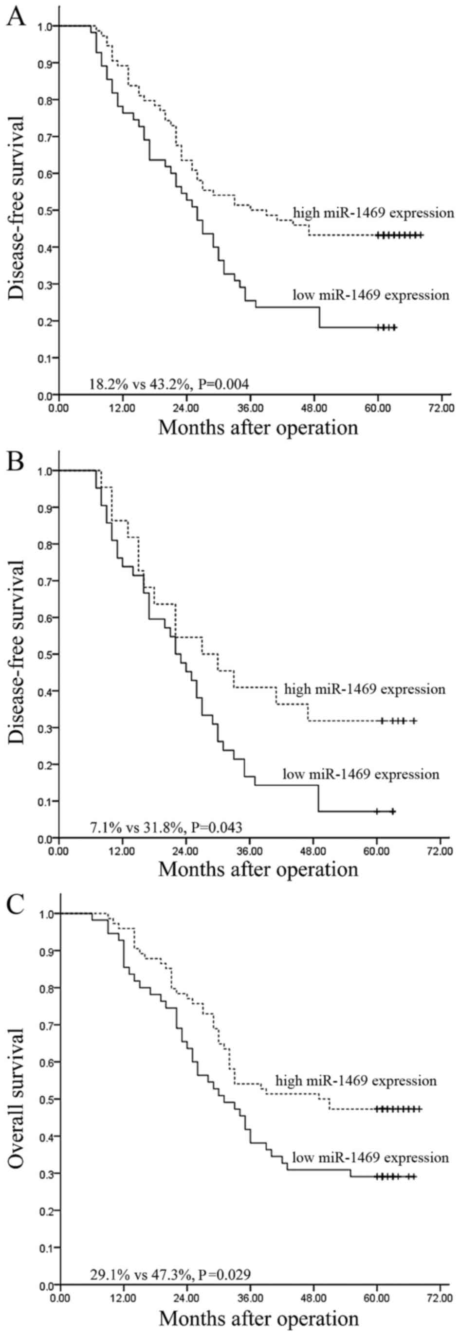
expression than for those with high tumor miR-1469 expression (18.2
vs. 43.2%; P=0.004; Fig. 1A). In
addition, multivariate analysis demonstrated that only lymph node
metastasis status retained its significance as an independent
predictor for unfavorable DFS [P=0.031; 95% confidence interval
(CI), 1.035–2.089; Table II].
| Table II.Results of univariate and
multivariate analyses for the association between
clinicopathological characteristics and survival in patients with
esophageal squamous cell cancer. |
Table II.
Results of univariate and
multivariate analyses for the association between
clinicopathological characteristics and survival in patients with
esophageal squamous cell cancer.
|
| Disease-free
survival | Overall
survival |
|---|
|
|
|
|
|---|
|
|
| Multivariate
analysis |
| Multivariate
analysis |
|---|
|
|
|
|
|
|
|---|
| Clinicopathological
characteristic | Univariate analysis
P-value | P-value | 95.0% CI | Univariate analysis
P-value | P-value | 95.0% CI |
|---|
| Age | 0.598 | 0.352 | 0.516–1.516 | 0.809 | 0.539 | 0.534–1.534 |
| Gender | 0.802 | 0.761 | 0.636–1.636 | 0.847 | 0.783 | 0.518–1.518 |
| Tumor length | 0.665 | 0.947 | 0.614–1.614 | 0.617 | 0.935 | 0.621–1.621 |
| Tumor
differentiation | 0.095 | 0.512 | 0.803–1.803 | 0.280 | 0.812 | 0.741–1.741 |
| Tumor invasion
depth | 0.033 | 0.512 | 0.785–1.785 | 0.009 | 0.361 | 0.815–1.815 |
| Lymph node
metastasis stage | <0.001 | 0.031 | 1.035–2.035 | <0.001 | 0.020 | 1.070–2.070 |
| Pathological tumor
stage | <0.001 | 0.742 | 0.552–2.552 | <0.001 | 0.582 | 0.582–2.582 |
| miR-1469
expression | 0.004 | 0.322 | 0.488–1.488 | 0.029 | 0.977 | 0.595–1.595 |
The predictive significance of miR-1469 expression
in selective patient subgroups was stratified according to lymph
node metastasis status and pathological stage, respectively.
Univariate analysis demonstrated that the 5-year DFS of patients
with low tumor miR-1469 expression was significantly lower compared
with that of the remaining cases with high miR-1469 expression
among N1-3 patients (7.1 vs. 31.8%; P=0.043; Fig. 1B). However, this was not the case for
patients with N0 (53.8 vs. 48.1%; P=0.751), pathological stage I–II
(40.0 vs. 48.2%; P=0.582) or pathological stage III (5.7 vs. 27.8%;
P=0.128) tumors with low miR-1469 tumor expression compared with
the corresponding groups with high miR-1469 tumor expression (data
not shown).
Association between miR-1469
expression and prognosis
Of the 129 patients, 78 succumbed to mortality
within the follow-up period and the 5-year OS rate was 39.5% (data
not shown). Univariate analysis revealed that low miR-1469
expression was associated with an unfavorable 5-year OS compared
with high miR-1469 expression (29.1 vs. 47.3%; P=0.029; Fig. 1C). Multivariate analysis revealed that
only lymph node metastasis retained its significance as an
independent predictor for unfavorable OS (P=0.020; 95% CI,
1.070–2.202; Table II).
A further stratified survival analysis demonstrated
that miR-1469 expression was not associated with an unfavorable
5-year OS for subgroups of patients grouped according to lymph node
metastasis status (N0, 69.2 vs. 51.9%; P=0.353; N1-3, 16.7 vs.
36.4%; P=0.145) and pathological stage (I–II, 65.0 vs. 53.6%,
P=0.566; III, 8.6 vs. 27.8%; P=0.223) (data not shown).
Discussion
Esophageal cancer is one of the most aggressive and
deadly malignancies of the digestive tract, with patients typically
presenting with bulky tumors and metastasis at the time of clinical
diagnosis. The prognosis of patients with esophageal cancer remains
poor and only a subset of patients respond well to treatment
(4,20). The current TNM classification cannot
accurately predict clinical outcome for patients with ESCC
(21), even for patients with
identical clinicopathological risk factors. Clearly distinguishing
between subgroups of patients who have a high or low risk of
relapse, treatment resistance and metastasis remains a challenge
(22), therefore there is an urgent
need to identify biomarkers that can be used to identify patients
that require more aggressive postoperative treatment.
miRNAs are deregulated in numerous human cancers
(23,24). Previous studies have demonstrated that
miRNAs are reliable and cost-effective biomarkers that can be
utilized in the diagnosis and assessment of the progression,
therapeutic response and prognosis of a wide spectrum of malignant
tumors, including esophageal cancer (7,25–27). A better understanding of the miRNAs
associated with particular cancers would provide valuable insights
for treatment. Previous studies have demonstrated that miR-1469 may
function as an oncogene (12,13) or a tumor suppressor gene (14,15) in
malignant tumors originating from different tissues. However, to
the best of our knowledge, the effects of miR-1469 expression on
the progression and clinical outcome of human ESCC has not yet been
reported.
In the present study, the expression of miR-1469 in
tumor samples from 129 ESCC patients was analyzed by RT-qPCR and
compared with clinicopathological characteristic and survival data.
Low miR-1469 expression was significantly associated with invasion
depth, lymph node metastasis status and pathological stage,
suggesting that miR-1469 acts as a tumor suppressor. Low expression
of miR-1469 was significantly associated with a poor prognosis; a
univariate analysis revealed a correlation between low miR-1469
expression and a poor 5-year DFS and OS. A multivariate analysis
demonstrated that only lymph node metastasis status could predict
tumor relapse and prognosis independently of other
clinicopathological parameters.
To explore whether miR-1469 expression levels could
be employed as a predictor to improve the current risk
stratification for ESCC based on the conventional staging system, a
further stratified survival analysis split by lymph node metastasis
status and pathological stage was performed. The results
demonstrated that low miR-1469 expression only significantly
predicted an unfavorable 5-year DFS among the subgroup of patients
with N1-3 disease. Although statistically significant differences
were not detected, low miR-1469 expression was associated with a
poorer DFS and OS in the subgroup of patients with pathologic stage
III disease. Therefore, the data from the present study indicate
that miR-1469 expression can predict the clinical outcome of ESCC,
particularly in patients with advanced disease. Measuring miR-1469
expression levels in patients with ESCC may allow clinicians to
more accurately identify high-risk patients who would benefit from
more aggressive therapeutic interventions.
Previous studies have identified an association
between dysregulated miRNA expression and cancer diagnosis
(28,29). In addition, it has been demonstrated
that miRNAs modulate numerous signaling pathways and regulate the
expression of thousands of genes (7,30). The
current gold standard of cancer diagnosis is a pathological
diagnosis, due to the heterogeneous nature of tumors. Future
studies will need to be performed to ascertain whether miR-1469 can
be reliably used as biomarker for the diagnosis of ESCC.
In conclusion, to the best of our knowledge, the
data in the present study provides the first evidence for the role
of miR-1469 in predicting disease progression and post-surgical
clinical outcomes in patients with ESCC. These data are consistent
with previous reports evaluating the tumor suppressor roles of
miR-1469 in human cancer (14,15).
However, multicenter and large-scale studies are required to verify
this data prior to the clinical application of miR-1469, in
addition to in vitro and in vivo functional studies
to identify the target genes and signaling pathways of miR-1469.
The results of these studies may allow for the identification and
selection of high-risk patients that would benefit from specific
treatments following surgery, therefore improving clinical
outcomes.
Glossary
Abbreviations
Abbreviations:
|
ESCC
|
esophageal squamous cell cancer
|
|
TNM
|
tumor-node-metastasis
|
|
DFS
|
disease-free survival
|
|
OS
|
overall survival
|
References
|
1
|
Torre LA, Bray F, Siegel RL, Ferlay J,
Lortet-Tieulent J and Jemal A: Global cancer statistics, 2012. CA
Cancer J Clin. 65:87–108. 2015. View Article : Google Scholar : PubMed/NCBI
|
|
2
|
Chen W, He Y, Zheng R, Zhang S, Zeng H,
Zou X and He J: Esophageal cancer incidence and mortality in China,
2009. J Thorac Dis. 5:19–26. 2013.PubMed/NCBI
|
|
3
|
Pennathur A, Gibson MK, Jobe BA and
Luketich JD: Oesophageal carcinoma. Lancet. 381:400–412. 2013.
View Article : Google Scholar : PubMed/NCBI
|
|
4
|
Kim T, Grobmyer SR, Smith R, Ben-David K,
Ang D, Vogel SB and Hochwald SN: Esophageal cancer-the five year
survivors. J Surg Oncol. 103:179–183. 2011. View Article : Google Scholar : PubMed/NCBI
|
|
5
|
Hong L, Han Y, Zhang H and Fan D:
Prognostic markers in esophageal cancer: From basic research to
clinical use. Expert Rev Gastroenterol Hepatol. 9:887–889. 2015.
View Article : Google Scholar : PubMed/NCBI
|
|
6
|
Lee RC, Feinbaum RL and Ambros V: The C.
elegans heterochronic gene lin-4 encodes small RNAs with antisense
complementarity to lin-14. Cell. 75:843–854. 1993. View Article : Google Scholar : PubMed/NCBI
|
|
7
|
Naidu S, Magee P and Garofalo M:
MiRNA-based therapeutic intervention of cancer. J Hematol Oncol.
8:682015. View Article : Google Scholar : PubMed/NCBI
|
|
8
|
Calin GA, Dumitru CD, Shimizu M, Bichi R,
Zupo S, Noch E, Aldler H, Rattan S, Keating M, Rai K, et al:
Frequent deletions and down-regulation of micro-RNA genes miR15 and
miR16 at 13q14 in chronic lymphocytic leukemia. Proc Natl Acad Sci
USA. 99:15524–15529. 2002. View Article : Google Scholar : PubMed/NCBI
|
|
9
|
Croce CM: Causes and consequences of
microRNA dysregulation in cancer. Nat Rev Genet. 10:704–714. 2009.
View Article : Google Scholar : PubMed/NCBI
|
|
10
|
Di Leva G, Garofalo M and Croce CM:
MicroRNAs in cancer. Annu Rev Pathol. 9:287–314. 2014. View Article : Google Scholar : PubMed/NCBI
|
|
11
|
Ohtsuka M, Ling H, Doki Y, Mori M and
Calin GA: MicroRNA processing and human cancer. J Clin Med.
4:1651–1667. 2015. View Article : Google Scholar : PubMed/NCBI
|
|
12
|
Fix LN, Shah M, Efferth T, Farwell MA and
Zhang B: MicroRNA expression profile of MCF-7 human breast cancer
cells and the effect of green tea polyphenon-60. Cancer Genomics
Proteomics. 7:261–277. 2010.PubMed/NCBI
|
|
13
|
White NM, Khella HW, Grigull J, Adzovic S,
Youssef YM, Honey RJ, Stewart R, Pace KT, Bjarnason GA, Jewett MA,
et al: miRNA profiling in metastatic renal cell carcinoma reveals a
tumour-suppressor effect for miR-215. Br J Cancer. 105:1741–1749.
2011. View Article : Google Scholar : PubMed/NCBI
|
|
14
|
Yang B, Jing C, Wang J, Guo X, Chen Y, Xu
R, Peng L, Liu J and Li L: Identification of microRNAs associated
with lymphangiogenesis in human gastric cancer. Clin Transl Oncol.
16:374–379. 2014. View Article : Google Scholar : PubMed/NCBI
|
|
15
|
Xu C, Zhang L, Li H, Liu Z, Duan L and Lu
C: MiRNA-1469 promotes lung cancer cells apoptosis through
targeting STAT5a. Am J Cancer Res. 5:1180–1189. 2015.PubMed/NCBI
|
|
16
|
Hamilton SR and Aaltonen LA: Pathology and
genetics of tumours of the digestive system, World Health
Organization classification of tumours. IARC Press; Lyon: pp.
10–25. 2000
|
|
17
|
Edge SF, Byrd DR, Compton CC, Fritz AG,
Greene FL and Trotti A: Esophagus and esophagogastric junctionAJCC
Cancer Staging Manual. 7th edition. Springer; New York, NY: pp.
103–111. 2010
|
|
18
|
Zhou H, Tang K, Xiao H, Zeng J, Guan W,
Guo X, Xu H and Ye Z: A panel of eight-miRNA signature as a
potential biomarker for predicting survival in bladder cancer. J
Exp Clin Cancer Res. 34:532015. View Article : Google Scholar : PubMed/NCBI
|
|
19
|
Mathé EA, Nguyen GH, Bowman ED, Zhao Y,
Budhu A, Schetter AJ, Braun R, Reimers M, Kumamoto K, Hughes D, et
al: MicroRNA expression in squamous cell carcinoma and
adenocarcinoma of the esophagus: Associations with survival. Clin
Cancer Res. 15:6192–6200. 2009. View Article : Google Scholar : PubMed/NCBI
|
|
20
|
Enzinger PC and Mayer RJ: Esophageal
Cancer. N Engl J Med. 349:2241–2252. 2003. View Article : Google Scholar : PubMed/NCBI
|
|
21
|
Li SH, Tian H, Yue WM, Li L, Gao C, Li WJ,
Hu WS and Hao B: Metastasis-associated protein 1 nuclear expression
is closely associated with tumor progression and angiogenesis in
patients with esophageal squamous cell cancer. World J Surg.
36:623–631. 2012. View Article : Google Scholar : PubMed/NCBI
|
|
22
|
Guo Y, Chen Z, Zhang L, Zhou F, Shi S,
Feng X, Li B, Meng X, Ma X, Luo M, et al: Distinctive microRNA
profiles relating to patient survival in esophageal squamous cell
carcinoma. Cancer Res. 68:26–33. 2008. View Article : Google Scholar : PubMed/NCBI
|
|
23
|
Pipan V, Zorc M and Kunej T: MicroRNA
polymorphisms in cancer: A literature analysis. Cancers (Basel).
7:1806–1814. 2015. View Article : Google Scholar : PubMed/NCBI
|
|
24
|
Lin S and Gregory RI: MicroRNA biogenesis
pathways in cancer. Nat Rev Cancer. 15:321–333. 2015. View Article : Google Scholar : PubMed/NCBI
|
|
25
|
Lu J, Getz G, Miska EA, Alvarez-Saavedra
E, Lamb J, Peck D, Sweet-Cordero A, Ebert BL, Mak RH, Ferrando AA,
et al: MicroRNA expression profiles classify human cancers. Nature.
435:834–838. 2005. View Article : Google Scholar : PubMed/NCBI
|
|
26
|
Garzon R, Calin GA and Croce CM: MicroRNAs
in Cancer. Annu Rev Med. 60:167–179. 2009. View Article : Google Scholar : PubMed/NCBI
|
|
27
|
Raza U, Zhang JD and Sahin O: MicroRNAs:
Master regulators of drug resistance, stemness, and metastasis. J
Mol Med (Berl). 92:321–336. 2014. View Article : Google Scholar : PubMed/NCBI
|
|
28
|
Tricoli JV and Jacobson JW: MicroRNA:
Potential for cancer detection, diagnosis, and prognosis. Cancer
Res. 67:4553–4555. 2007. View Article : Google Scholar : PubMed/NCBI
|
|
29
|
Kosaka N, Iguchi H and Ochiya T:
Circulating microRNA in body fluid: A new potential biomarker for
cancer diagnosis and prognosis. Cancer Sci. 101:2087–2092. 2010.
View Article : Google Scholar : PubMed/NCBI
|
|
30
|
Ye Y, Wang KK, Gu J, Yang H, Lin J, Ajani
JA and Wu X: Genetic variations in microRNA-related genes are novel
susceptibility loci for esophageal cancer risk. Cancer Prev Res
(Phila). 1:460–469. 2008. View Article : Google Scholar : PubMed/NCBI
|