Introduction
Unlike αβT cells, γδ T cells represent a minor
subset of T cells that does not require
self-majorhistocompatibility complex (MHC)-restricted priming
(1). γδ T cells are believed to serve
as a bridge, connecting the innate and adaptive immune responses
(1). Following infection by microbial
pathogens, γδ T cells appear to be the first T cells tomigrateto
the lung (2). Previous studies have
identified that γδ T cells can mediate antitumor activity (3).
In humans, γδ T cells are primarily identified by
the V(D)J recombination of the γ and δ chain genes, particularly
their Vδ chain usage (4). Vδ2 cells
(mainly Vγ9/Vδ2 cells) are the predominant γδ T-cell subset
(50–75%) in the peripheral blood (PB) of healthy individuals
(5). Uniquely, this subset of γδ T
cells responds to phosphoantigens via polymorphic γδ T-cell antigen
receptors (TCR) and undergoes rapid activation (6,7). Owing to
the relative abundance in the PB and ease of expansion, Vγ9/Vδ2
T-cell-based adoptive immunotherapy has been intensively
investigated as the treatment for a variety of malignancies
(8–10). Vδ1 T cells represent <30% of γδ T
cells in the PB, but have been identified as an enriched subset in
the thymus and in epithelial tissues, including the dermis, gut
epithelium and spleen (11). Unlike
Vδ2 cells, Vδ1 cells do not respond to phosphoantigens, but are
able to recognize the MHC class I chain-related molecules A and B
(12). A recent study suggested that
Vδ1+ T cells derived from the PB of normal donors are
able to undergo ex vivo expansion by administration of
phytohemagglutinin (PHA) and interleukin-7 (IL-7) (13); moreover, Vδ1 T cells exhibit more
favorable antitumor activity than Vδ2 T cells in colon cancer
(13). Ex vivo-expanded
circulating γδ T cells represent a promising prospect in antitumor
activities and are a potential candidate treatment for various
malignancies, including non-small cell lung cancer (NSCLC). A phase
I clinical study is currently being conducted using adoptive γδ T
cell therapy in patients with advanced or recurrent NSCLC (14). However, the characterization of γδ T
cells remains poorly understood in advanced NSCLC.
In the present study, the absolute count of
lymphocytes and monocytes, and the subtypes and characteristics of
γδ T cells in the PB and pleural effusion of advanced NSCLC
patients were analyzed in order to investigate which subsets (Vδ1
or Vδ2) should be expanded ex vivo and used as the source
from which these γδ T cells should be generated for adoptive γδ T
cell therapy.
Materials and methods
Subject recruitment and sample
preparation
The complete blood cell count of 102 patients with
stage I–IV NSCLC using seventh edition of the TNM classification
for lung cancer (15) (51 male, 51
female; 64.45±1.55 years of age; range, 48–86 years) who were
admitted to the Department of Oncology, The Second Affiliated
Hospital of Jiaxing College (Jiaxing, China) between January 2011
and December 2015, was retrospectively analyzed. Blood cell count
data was also obtained from 114 cases of aged-matched healthy
controls. Of the NSCLC patients and controls, 35 patients (stage
III and IV) and 25 age-matched healthy individuals underwent γδ
T-cell analysis in this study. Of these 35 patients, 10 were
diagnosed as having stage IV NSCLC with malignant pleural effusion.
Analysis of the characteristics of the γδ T cells in the pleural
effusion of these 10 patients, together with another 2 elderly
NSCLC patients (>75 years of age) was conducted. The studies
were approved by the Ethics Committee of The Second Affiliated
Hospital of Jiaxing College and written informed consent was
obtained from each individual that provided a specimen. Study
subjects did not have infectious diseases and had not undergone
chemotherapy or radiotherapy in the previous week; however, certain
patients and healthy donors did have chronic conditions, including
hypertension, high cholesterol and diabetes.
Isolation of mononuclear cells from
pleural effusion
Following collection of a 50-ml specimen of pleural
effusion from 12 patients, mononuclear cells were isolated by
centrifugation at 1,000 × g over a Ficoll-Paque (Beijing Solarbio
Science & Technology Co., Ltd., Beijing, China) density
gradient.
Blood cell count
A BC-5200 Hematology Analyzer (Beckman Coulter,
Inc., Brea, CA, USA) was used to examine the absolute number of
lymphocytes and monocytes in the present study.
Flow cytometry staining
To determine the identity of the biomarkers on the
surface of γδ T cells, multicolored immunofluorescence staining was
conducted using freshly collected blood samples and mononuclear
cells isolated from the pleural effusion of the subjects. The
antibodies were conjugated to fluorescent markers as follows:
CD3-PE-Cy5.5 (cat. no. 340949), TCR γδ-APC (cat. no. 555718),
TCRγδ-FITC (cat. no. 559878), Vδ2-PE (cat. no. 3345652), CD27-PE
(cat. no. 555441) and CD28-APC (cat. no. 559770). These antibodies,
as well as isotype-matched control antibodies, were purchased from
BD Pharmingen (dilution, ready to use; BD Biosciences, San Jose,
CA, USA). Vδ1-FITC antibodies (cat. no. TCR2730) were purchased
from Thermo Fisher Scientific, Inc., (Waltham, MA, USA). For
extracellular staining, 50 µl of each blood sample, and the
mononuclear cells isolated from the pleural effusion which were in
1X PBS with 1% bovine serum albumin, were incubated with different
combinations of fluorochrome-coupled antibodies (10 µl of each
antibody). After a 20-min incubation at room temperature, cells
were washed twice with 1X PBS and flow cytometry was performed
using a BD FACSCanto II flow cytometer (BD Biosciences). Data were
collected and analyzed with DIVA software (version 6.1.3; BD
Biosciences, San Jose, CA, USA).
Statistical analysis
Data are presented as the mean ± standard error of
the mean. Comparisons between groups were made using an unpaired
Student's t-test. P-values <0.05 were considered to indicate
statistical significance. GraphPad Prism version 5 (GraphPad
Software, Inc., La Jolla, CA, USA) was used for all statistical
calculation and figure generation.
Results
Absolute number of lymphocytes and
monocytes in the PB of NSCLC patients
The complete blood cell counts of 102 patients (51
male, 51 female; 64.45±1.55 years) with stage I–IV NSCLC were
retrospectively analyzed. The clinicopathological features of
patients are provided in Table I. The
blood cell count data were obtained from 114 cases of aged-matched
healthy controls (51 male, 63 female; mean age, 63.40±1.1 years).
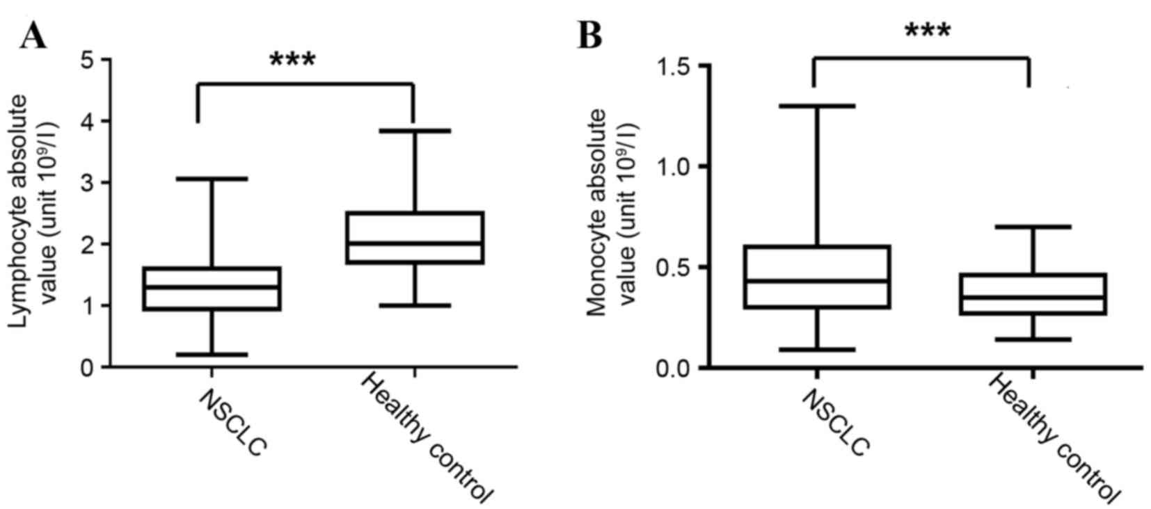
The absolute number of lymphocytes and monocytes was assessed using
an automatic hematology analyzer. The absolute value of lymphocytes
(normal range, 1.10–3.20×10⁹/l) in the PB was
1.285×10⁹±0.049×10⁹/l in the NSCLC group,
significantly lower than that of the healthy controls, where it was
2.065×10⁹±0.051×10⁹/l (P<0.001) (Fig. 1A); however, the absolute value of
monocytes (normal range is 0.10–0.60×10⁹/l) was
0.484×10⁹±0.022×10⁹/l in the NSCLC group,
significantly higher than that in the healthy controls, where it
was 0.363±0.011 (P<0.001) (Fig.
1B).
 | Table I.Clinicopathological features of lung
cancer patients in the present study (n=102). |
Table I.
Clinicopathological features of lung
cancer patients in the present study (n=102).
| Category | Value |
|---|
| Gender, n (%) |
|
|
Male | 51 (50.0) |
|
Female | 51 (50.0) |
| Mean age ± SEM,
years | 64.45±1.55 |
| Histology, n |
|
|
Adenocarcinoma | 69 |
|
Squamous cell carcinoma | 16 |
| NSCLC
(not specified) | 17 |
| TNM stage, n |
|
|
I–II | 9 |
|
III | 12 |
| IV | 81 |
Frequency of
CD3+γδ+T cells and Vδ1Vδ2 subtypes in
circulation of NSCLC patients
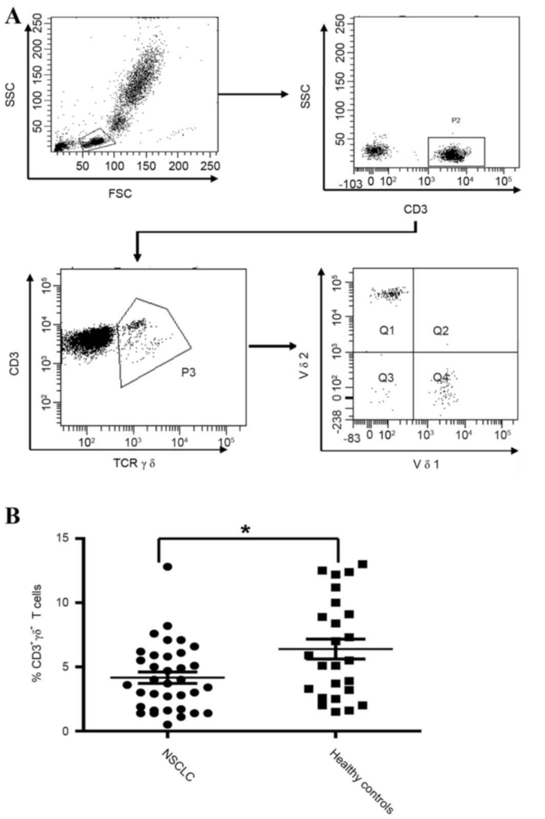
Flow cytometry was performed to determine the
proportion of CD3+γδ+T cells in the PB of
NSCLC patients. A total of 35 (stage III–IV) NSCLC patients (23
male, 12 female; mean age, 65.7±1.3 years) were examined in this
study (2 NSCLC patients with pleural effusion were excluded owing
to their old age, >75 years old). The clinicopathological
features of the patients are provided in Table II. In order to make a comparison, the
frequency of γδ T cells were examined in 25 cases of age-matched
healthy controls (17 male, 8 female; mean age, 63.6±4.2 years).
Among these subjects, 20 patients with NSCLC and 11 healthy donors
were further analyzed for the percentage of Vδ1 and Vδ2 T cells,
two major subsets of γδ T cells, and the expression of the
co-stimulatory markersCD27 and CD28. To assess the frequency of γδ
T cells present in the sample, lymphocytes were first gated on the
basis of a forward scatter/side scatter (FSC/SSC) profile, followed
by γδ T-cell analysis in a gated population with CD3+
staining (Fig. 2A). A pan-TCRγδ
antibody was used to identify γδ T cells, and Vδ1/Vδ2 antibodies
were further used to determine Vδ1 and Vδ2 subsets (Fig. 2A). Fig.
2B shows that in NSCLC patients, the mean frequency of
CD3+γδ+T cells was 4.16±0.44% (n=35), whereas
the value in healthy individuals was 6.40±0.77% (n=25); the two
groups have a statistically significant difference (P<0.05). In
parallel with a previous report (16), Vδ2 T cells represented a major subset,
and the Vδ2/Vδ1 ratio was >1 in the PB of all normal donors
(100.0%) (11/11); however, in NSCLC patients, the Vδ2+ T
cells only represented a major subtype in 35.0% (7/20) of the
population of γδ T cells; Vδ1+ cells were counted as the
priority γδ T cells in 35.0% (7/20) of patients, and in the rest of
patients, Vδ1−Vδ2− cells represented the main
subset (6/20).
| Table II.Clinicopathological features of stage
III and IV lung cancer patients. |
Table II.
Clinicopathological features of stage
III and IV lung cancer patients.
| Category | Value |
|---|
| Gender, n (%) |
|
|
Male | 23 (65.7) |
|
Female | 12 (34.3) |
| Mean age ± SEM,
years | 65.7±1.3 |
| Histology |
|
|
Adenocarcinoma | 19 |
|
Squamous cell carcinoma | 9 |
| NSCLC
(not specified) | 7 |
| TNM stage, n |
|
|
III | 9 |
| IV | 26 |
Frequency of
CD3+γδ+T cells and Vδ1Vδ2 subtypes in the PB
and in the pleural effusion of advanced NSCLC patients
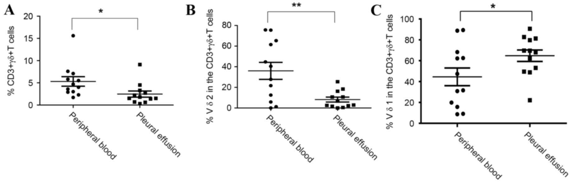
To compare the difference between γδ T cells and
Vδ1Vδ2 subtypes in the PB and pleural effusion in stage IV NSCLC
patients, 12 patients (6 male, 6 female; mean age, 70.2±9.2 years)
were recruited. The clinicopathological features of the patients
are provided in Table III. The
paired blood and pleural effusion samples were collected at the
same time. The mean frequency of CD3+γδ+T
cells was higher in the PB of the two groups (5.32±1.08%) than that
in the pleural effusion of the two groups (2.47±0.69%), with a
statistically significant difference (P<0.05) (Fig. 3A).
| Table III.Clinicopathological features of
advanced lung cancer patients with pleural effusion. |
Table III.
Clinicopathological features of
advanced lung cancer patients with pleural effusion.
| Category | Value |
|---|
| Gender, n (%) |
|
|
Male | 6 (50.0) |
|
Female | 6 (50.0) |
| Mean age ± SEM,
years | 70.2±9.2 |
| Histology, n |
|
|
Adenocarcinoma | 7 |
|
Squamous cell carcinoma | 2 |
| NSCLC
(not specified) | 3 |
In the 12 NSCLC patients recruited for the current
experiment, the Vδ2+γδ T cell was the predominant
subtype in the PB in 5 patients (41.7%), whereas Vδ1 or
Vδ1−Vδ2− was the major
CD3+γδ+T cell subtype in the remaining 7
patients (58.3%). In the pleural effusion, Vδ1 or
Vδ1−Vδ2− was the predominant subtype of
CD3+γδ T cells in all 12 patients (100.0%). In
comparison to the frequency of Vδ1+ and Vδ2+T
cells between the PB and the pleural effusion, data from the
present study showed that the percentage of Vδ2 T cells was
significant lower in the pleural effusion, with 36.01±8.25% of
cells found to be T cells in the PB vs. 8.16±2.38% in the pleural
effusion (P<0.01) (Fig. 3B).
However, the percentage of Vδ1 T cells in the pleural effusion was
significantly higher, with 44.54±8.49 of cells in the blood found
to be T cells vs. 64.78±5.50% in the pleural effusion (P<0.05)
(Fig. 3C).
Vδ1 and Vδ2 have different mean
fluorescence intensity (MFI) for CD3 and TCR γδ
Next, the expression of CD3 and TCRγδ on the surface
of the Vδ1 and Vδ2 T-cell population was analyzed. During the study
of circulating Vδ1 and Vδ2 cells in the 20 NSCLC patients and 11
healthy donors, further analysis was performed, investigating the
MFI of CD3 in Vδ2 and Vδ1 γδ T cells. This analysis found that the
MFI of CD3 was significant higher in Vδ2 cells than in Vδ1 cells in
NSCLC patients and healthy individuals; a CD3 MFI of 4,776±691.2 in
Vδ2+cells vs. 2,612±319.4 in Vδ1+cells (n=20;
P<0.01) was observed in NSCLC patients (Fig. 4A), and a CD3 MFI 9,689±1,270 in
Vδ2+ cells vs. 3,454±592.6 in Vδ1+ cells in
healthy controls (n=11; P<0.001) (Fig.
4B). The MFI of TCRγδ was significantly lower in
Vδ2+ cells than in Vδ1+ cells in NSCLC
(858±62.49 vs. 2,614±313.10, respectively; n=20; P<0.001)
(Fig. 4C), and in healthy controls
(1,351±182.8 vs. 3,724±725.20, respectively; n=11; P<0.01)
(Fig. 4D). Therefore, Vδ2 cells
should be considered to be a population of
CD3brightTCRγδdim T cells, and by contrast,
Vδ1 cells should be considered to be a population of
CD3dimTCRγδbright T cells.
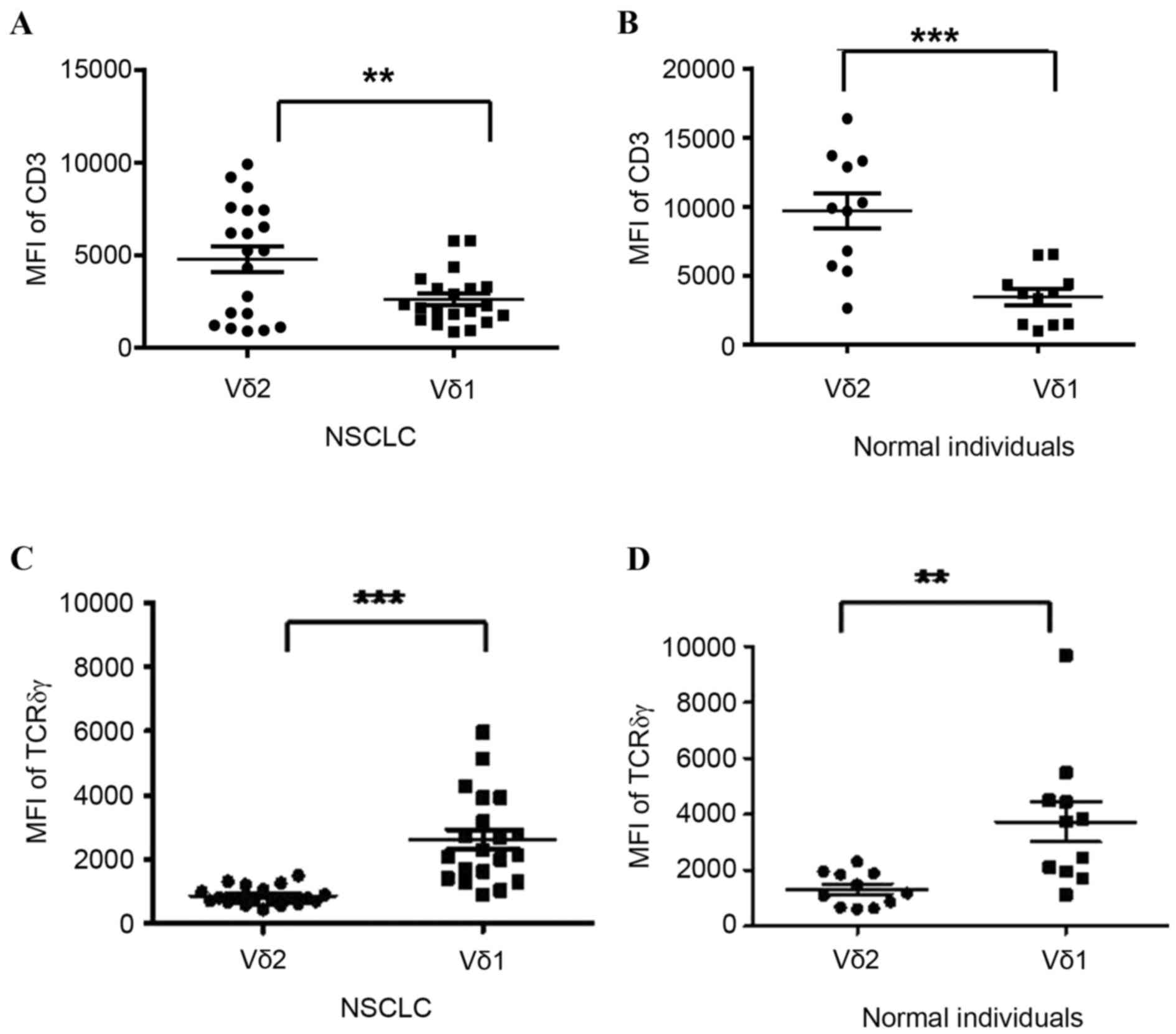 | Figure 4.Expression of CD3 and TCRγδ molecules
on the surface of Vδ1 and Vδ2 T cell subsets in the circulation of
NSCLC patients and healthy individuals. (A) The MFI of CD3 was
decreased in Vδ1 cells compared with that in Vδ2 cells in the group
of NSCLC patients (2,612±319.4 vs. 4,776±691.2 AU, respectively;
P<0.01). (B) The MFI of CD3 in Vδ1+ cells was
decreased compared with that in Vδ2 cells in the group of normal
individuals (3,454±592.6 vs. 9,689±1,270 AU, respectively;
P<0.001). (C) The MFI of TCRγδ in Vδ1 cells was increased
compared with Vδ2 in the group of NSCLC patients (2,614±313.10 vs.
858±62.49 AU, respectively; P<0.001). (D) The MFI of TCRγδ in
Vδ1 T cells was increased compared with that in Vδ2 cells in the
group of normal individuals (3,724±725.20 vs. 1,351±182.8,
respectively; P<0.01).**P<0.01. ***P<0.001. NSCLC,
non-small cell lung cancer; MFI, mean fluorescence intensity; TCR,
T-cell receptor; CD, cluster of differentiation. |
Expression of co-stimulatory
markersCD27 and CD28 is decreased on the surface of
CD3+γδ+T cells in patients with advanced
NSCLC
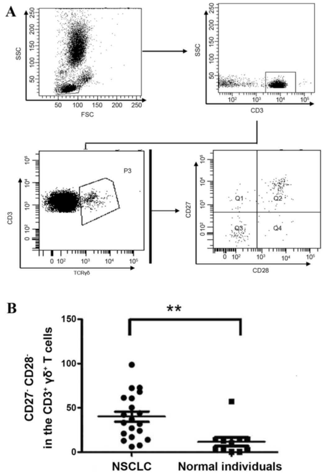
The TNF receptor family member CD27 is widely
expressed on natural killer cells, and on CD4+ and
CD8+ T lymphocytes, as well as on γδ T cells (17). CD27 and its ligand, CD70, are involved
in a signaling pathway that promotes the survival of primed T cells
(18). CD28 is a well-documented
co-receptor of αβT cells, and activation of the CD3/CD28 pathway
will enhance the proliferation of αβT cells (19). Therefore, the expression of CD27 and
CD28 on the surface of γδ T cells was investigated in the PB of the
same 20 NSCLC patients and 11 healthy donors. The gating strategy
is shown in Fig. 5A. In brief,
lymphocytes were first gated based on their FSC/SSC profile,
followed by analysis of CD27/CD28 expression in a gated population
of CD3+γδ+ cells (Fig. 5A). There was a significantly increased
frequency of
CD27−CD28−CD3+γδ+ T
cells in the PB of NSCLC patients (40.2±5.7%) compared with the PB
of the healthy control group (11.9±4.8%) (P<0.01) (Fig. 5B). An increased population of
CD27−CD28−CD3+γδ+T
cells was also observed in the pleural effusion of NSCLC patients
compared with that in the PB, but the difference was not
statistically significant (data not shown).
Discussion
In the present study, decreased numbers of
lymphocytes and increased numbers of monocytes were observed in the
PB of NSCLC patients. This observation supports previous reports
concerning the systemic immune defects that exist in malignancies,
which include low numbers of circulating T cells and low chemokine
levels, and enhance the number of anti-tumor T cells that are
likely to partially reverse immunosuppressive activities (20). A low lymphocyte-to-monocyte ratio
(LMR) has been shown to be an independent unfavorable prognostic
factor for predicting survival in SCLC patients (21). A lower LMR most likely have decreased
cytotoxic T lymphocytes and higher tumor-associated macrophages
(22). A previous study reported that
the monocyte count in the peripheral blood was higher in the
patients with cervical and endometrial cancer compared with a
control group of healthy blood donors (23). An elevated number of peripheral
monocytes has also been associated with a poor prognosis for
patients with lung adenocarcinoma (24).
The number of γδ T cells is lower in elderly
individuals than in the young population (25). The present study also observed a
decrease in the frequency of γδ T cells in the PB of NSCLC
patients. Circulating Vδ2 γδ T cells appeared to lose their
predominance in NSCLC patients compared with healthy individuals;
Vδ1, and to a lesser extent Vδ1−Vδ2−γδ, T
cells became a major subset and the ratio of Vδ2:Vδ1 T cells
inverted in one-third of all studied patients; a similar
observation has been reported in a study of γδ T cells in gastric
cancer (26). On the basis of
previous studies, Vδ1 T cells are less susceptible to
activation-induced cell death and exhaustion than Vδ2 T cells
(27,28). Vδ1 T cells are also preferentially
persistent in vivo; this could be the reason why Vδ1 T cells
are predominant in the PB of patients with lung cancer.
In the present study, γδ T cells were also detected
in the pleural effusion of patients with advanced NSCLC, in whom
the frequency of Vδ2+ T cells was even lower than in the
PB, and Vδ1 or Vδ1−Vδ2−γδ T cells acted as
the predominant subsets in all subjects studied. To the best of our
knowledge, no previous study has reported on the characteristics of
γδ T cells and their subsets in the pleural effusion of NSCLC.
Tumor-infiltrating leukocytes (TILs) represent a wide range of
variety of immune cells that have been found in invading solid
tumors, and the extent of infiltration of γδ T cells is reported to
enable a variety of prognostic predictions, for example; in the
earlier stages of melanoma there are higher percentages of
intra-tumoral γδ T cells, particularly V2γδ T cells, compared with
advanced stage melanoma, which serves as a biomarker of good
prognoses; in contrast, a higher level of intra-tumoral V1 γδ T
cells has been reported as a poor prognostic factor in breast
cancer (29,30). In a study of γδ T cells in the TILs of
melanoma, γδ T cells were detected in 76% of tumor specimens using
immunohistochemistry. Vδ1 was the major subset presenting in 52% of
samples (31,32). In a study of γδ T cells in human colon
cancer, it was found that the majority (80%) of IL-17+γδ
T cells expressed Vδ1 (γδ17), and those cells thatinfiltratedγδ17 T
cells in the tumor were associated with advanced tumor grades and
progression. As a result, these T cells may serve as prognostic
markers in human colon cancer (33).
However, Peng et al (34)
reported that the Vδ1 subtype was observed to be the dominant
subtype among tumor infiltrating lymphocytes in a group of breast
cancer patients, and that it appeared to exhibit a broad range of
immune suppression activities, via suppressing the production of
IL-2 by CD4+ and CD8+ T cells, and the
maturation of dendritic cells. The present study indicates that γδ
T cells derived from the pleural effusion could be a source of
adoptive γδ T cell immunotherapy in lung cancer. Unlike for Vδ2 T
cells, which respond to phosphoantigens, there is no protocol
concerning the expansion of Vδ1+ populations ex
vivo, although a recent study indicated that PHA and IL-7 may
be candidates to facilitate expansion of the Vδ1 subtype in
circulating γδ T cells (13).
However, the present authors have been unable to obtain a
satisfactory result on the ex vivo expansion of Vδ1 or Vδ2
cells, either using PHA and IL-7 stimulation or zoledronic acid
when using the γδ T cells isolated from pleural effusion in
advanced NSCLC (Bao et al, unpublished data). This is likely
to be due to the low frequency of γδ T cells in the pleural
effusion, and the fact that the majority γδ T cells are Vδ1 T
cells. Moreover, it remains unclear whether there are similar
antitumor effects of Vδ1 T cells derived from PB or from pleural
effusion in lung cancer patients.
In the present study, Vδ2 T cells appeared to be a
population of γδ T cells with a
CD3brightTCRγδdim phenotype; by contrast, Vδ1
T cells were characterized by a
CD3dimTCRγδbright phenotype. A previous study
suggested that TCRγδ-deficient mice display reduced tumor growth
compared with wild-type animals (35). The different levels of CD3 and TCRγδ
expression may indicate that in these two subsets, a variety of
co-stimulatory signaling pathways could be involved in the
regulation of activation and proliferation.
The present study found a decrease in the expression
of CD27 and CD28 molecules on the surface of circulating γδ T cells
in NSCLC patients. A previous study showed a majority of Vδ2 cells
in PB are CD27+γδ T cells (36). Another prior study reported evidence
supporting an absolute requirement of CD27 for the expansion of γδ
T cells by employing a CD27-deficient mice model. CD27 and its
ligand CD70 are involved in a signaling pathway that is required to
promote the survival of primed T cells (17,37). In
the thymus of mice, CD27 is likely to regulate γδ T cell
differentiation, and CD27+γδ T cells produce interferon
(IFN)-γ, whereas CD27−γδ T cells produce IL-17 (38). In αβ T cells, ‘signal 1’, which refers
to the mature T cells recognizing and binding to a major
histocompatibility complex (MHC) molecule carrying a peptide
antigen through their antigen-specific receptors (TCR), together
with a co-stimulatory pathway, CD28/B7, provides a ‘second signal’
that is required for the activation and proliferation of T cells
(39); this CD28/B7 pathway involves
the interaction of co-stimulatory molecule CD28 with its ligands
B7-1 (CD80) and B7-2 (CD86) on the antigen presenting cells, and is
critical for T-cell activation, proliferation, and survival
(40); however, the requirement of
the additional CD28/B7 signal for the activation of γδ T cells
remains controversial (41). In
studying lymphocytes isolated from the lymph node, it was apparent
that CD28 is expressed on the surface of γδ T cells and has the
capability to promote the activation of γδ T cells, as well as
their proliferation and survival, mainly by stimulating γδ T cells
to produce and secrete IL-2 (42,43). Data
obtained from a study of CD28-deficient mice suggested that the
population of IFN-γ+ and IL-17+γδ T cells
failed to expand during infection (43,44),
indicating that CD28 co-stimulation is also necessary for γδ T cell
expansion. Downregulation of CD27 and CD28 molecules in circulating
γδ T cells in NSCLC patients suggests an impaired capacity to
activate and proliferate γδ T cells in those patients, and
indicates that they have a different cytokine profile to healthy
individuals (42).
The present data suggested the presence of a
dysregulated repertoire of γδ T cells, including impaired
activation and a reformed cytokine releasing profile of γδ T cells
in NSCLC. Although the ex vivo expansion of γδ T cells may
be a prospective therapeutic strategy in NSCLC patients, it remains
necessary to clarify which subsets (Vδ1 or Vδ2) should be expanded
and the sources from which these γδ T cells should be
generated.
Acknowledgements
The authors thank Dr Bing Chen (University of
Liverpool, Liverpool, UK) for providing revisions to the
manuscript. This study was supported by grants from the Chinese
National Science Fund for Young Scholars (Beijing, China; no.
81101707), the Natural Science Foundation of Zhejiang Province
(Hangzhou, China; no. LY16H070007), the Health Bureau of Zhejiang
Province (Hangzhou, China; no. 2016KYB292) and the Zhejiang
Traditional Chinese Medicine Foundation Project (Hangzhou, China;
no. 2014ZB119).
References
|
1
|
Urban EM, Chapoval AI and Pauza CD:
Repertoire development and the control of cytotoxic/effector
function in human gammadelta T cells. Clin Dev Immunol.
2010:7328932010. View Article : Google Scholar : PubMed/NCBI
|
|
2
|
Dieli F, Ivanyi J, Marsh P, Williams A,
Naylor I, Sireci G, Caccamo N, Di Sano C and Salerno A:
Characterization of lung gamma delta T cells following intranasal
infection with Mycobacterium bovis bacillus Calmette-Guérin. J
Immunol. 170:463–469. 2003. View Article : Google Scholar : PubMed/NCBI
|
|
3
|
Paul S and Lal G: Regulatory and effector
functions of gamma-delta (γδ) T cells and their therapeutic
potential in adoptive cellular therapy for cancer. Int J Cancer.
139:976–985. 2016. View Article : Google Scholar : PubMed/NCBI
|
|
4
|
Legut M, Cole DK and Sewell AK: The
promise of γδ T cells and the γδ T cell receptor for cancer
immunotherapy. Cell Mol Immunol. 12:656–668. 2015. View Article : Google Scholar : PubMed/NCBI
|
|
5
|
Porcelli S, Brenner MB and Band H: Biology
of the human gamma delta T-cell receptor. Immunol Rev. 120:137–183.
1991. View Article : Google Scholar : PubMed/NCBI
|
|
6
|
Constant P, Davodeau F, Peyrat MA, Poquet
Y, Puzo G, Bonneville M and Fournié JJ: Stimulation of human gamma
delta T cells by nonpeptidic mycobacterial ligands. Science.
264:267–270. 1994. View Article : Google Scholar : PubMed/NCBI
|
|
7
|
Tanaka Y, Morita CT, Tanaka Y, Nieves E,
Brenner MB and Bloom BR: Natural and synthetic non-peptide antigens
recognized by human gamma delta T cells. Nature. 375:155–158. 1995.
View Article : Google Scholar : PubMed/NCBI
|
|
8
|
Meraviglia S, Eberl M, Vermijlen D, Todaro
M, Buccheri S, Cicero G, La Mendola C, Guggino G, D'Asaro M,
Orlando V, et al: In vivo manipulation of Vgamma9Vdelta2 T cells
with zoledronate and low-dose interleukin-2 for immunotherapy of
advanced breast cancer patients. Clin Exp Immunol. 161:290–297.
2010.PubMed/NCBI
|
|
9
|
Abe Y, Muto M, Nieda M, Nakagawa Y, Nicol
A, Kaneko T, Goto S, Yokokawa K and Suzuki K: Clinical and
immunological evaluation of zoledronate-activated Vgamma9gammadelta
T-cell-based immunotherapy for patients with multiple myeloma. Exp
Hematol. 37:956–968. 2009. View Article : Google Scholar : PubMed/NCBI
|
|
10
|
Kobayashi H, Tanaka Y, Yagi J, Minato N
and Tanabe K: Phase I/II study of adoptive transfer of γδ T cells
in combination with zoledronic acid and IL-2 to patients with
advanced renal cell carcinoma. Cancer Immunol Immunother.
60:1075–1084. 2011. View Article : Google Scholar : PubMed/NCBI
|
|
11
|
Bonneville M, O'Brien RL and Born WK:
Gammadelta T cell effector functions: A blend of innate programming
and acquired plasticity. Nat Rev Immunol. 10:467–478. 2010.
View Article : Google Scholar : PubMed/NCBI
|
|
12
|
Wu J, Groh V and Spies T: T cell antigen
receptor engagement and specificity in the recognition of
stress-inducible MHC class I-related chains by human epithelial
gamma delta T cells. J Immunol. 169:1236–1240. 2002. View Article : Google Scholar : PubMed/NCBI
|
|
13
|
Wu D, Wu P, Wu X, Ye J, Wang Z, Zhao S, Ni
C, Hu G, Xu J, Han Y, et al: Ex vivo expanded human circulating Vδ1
γδT cells exhibit favorable therapeutic potential for colon cancer.
Oncoimmunology. 4:e9927492015. View Article : Google Scholar : PubMed/NCBI
|
|
14
|
Kakimi K, Matsushita H, Murakawa T and
Nakajima J: γδ T cell therapy for the treatment of non-small cell
lung cancer. Transl Lung Cancer Res. 3:23–33. 2014.PubMed/NCBI
|
|
15
|
Rusch VW, Asamura H, Watanabe H, Giroux
DJ, Rami-Porta R and P; Members of IASLC Goldstraw: The IASLC lung
cancer staging project: A proposal for a new international lymph
node map in the forthcoming seventh edition of the TNM
classification for lung cancer. J Thorac Oncol. 4:568–577. 2009.
View Article : Google Scholar : PubMed/NCBI
|
|
16
|
Casorati G, De Libero G, Lanzavecchia A
and Migone N: Molecular analysis of human gamma/delta+ clones from
thymus and peripheral blood. J Exp Med. 170:1521–1535. 1989.
View Article : Google Scholar : PubMed/NCBI
|
|
17
|
Born WK and O'Brien RL: γδ T cells
develop, respond and survive-with a little help from CD27. Eur J
Immunol. 41:26–28. 2011. View Article : Google Scholar : PubMed/NCBI
|
|
18
|
Borst J, Hendriks J and Xiao Y: CD27 and
CD70 in T cell and B cell activation. Curr Opin Immunol.
17:275–281. 2005. View Article : Google Scholar : PubMed/NCBI
|
|
19
|
Salomon B and Bluestone JA: Complexities
of CD28/B7: CTLA-4 costimulatory pathways in autoimmunity and
transplantation. Annu Rev Immunol. 19:225–252. 2001. View Article : Google Scholar : PubMed/NCBI
|
|
20
|
Frey AB and Monu N: Signaling defects in
anti-tumor T cells. Immunol Rev. 222:192–205. 2008. View Article : Google Scholar : PubMed/NCBI
|
|
21
|
Go SI, Kim RB, Song HN, Kang MH, Lee US,
Choi HJ, Lee SJ, Cho YJ, Jeong YY, Kim HC, et al: Prognostic
significance of the lymphocyte-to-monocyte ratio in patients with
small cell lung cancer. Med Oncol. 31:3232014. View Article : Google Scholar : PubMed/NCBI
|
|
22
|
Yang J, Liao D, Chen C, Liu Y, Chuang TH,
Xiang R, Markowitz D, Reisfeld RA and Luo Y: Tumor-associated
macrophages regulate murine breast cancer stem cells through a
novel paracrine EGFR/Stat3/Sox-2 signaling pathway. Stem cells.
31:248–258. 2013. View Article : Google Scholar : PubMed/NCBI
|
|
23
|
Jóźwik M, Okungbowa OE, Lipska A and
Jóźwik M, Smoktunowicz M, Semczuk A and Jóźwik M: Surface antigen
expression on peripheral blood monocytes in women with gynecologic
malignancies. BMC Cancer. 15:1292015. View Article : Google Scholar : PubMed/NCBI
|
|
24
|
Kumagai S, Marumo S, Shoji T, Sakuramoto
M, Hirai T, Nishimura T, Arima N, Fukui M and Huang CL: Prognostic
impact of preoperative monocyte counts in patients with resected
lung adenocarcinoma. Lung Cancer. 85:457–464. 2014. View Article : Google Scholar : PubMed/NCBI
|
|
25
|
Vasudev A, Ying CT, Ayyadhury S, Puan KJ,
Andiappan AK, Nyunt MS, Shadan NB, Mustafa S, Low I, Rotzschke O,
et al: γ/δ T cell subsets in human aging using the classical αβ T
cell model. J Leukoc Biol. 96:647–655. 2014. View Article : Google Scholar : PubMed/NCBI
|
|
26
|
Kuroda H, Saito H and Ikeguchi M:
Decreased number and reduced NKG2D expression of Vδ1 γδ T cells are
involved in the impaired function of Vδ1 γδ T cells in the tissue
of gastric cancer. Gastric Cancer. 15:433–439. 2012. View Article : Google Scholar : PubMed/NCBI
|
|
27
|
Siegers GM and Lamb LS Jr: Cytotoxic and
regulatory properties of circulating Vδ1+ γδ T cells: A new player
on the cell therapy field? Mol Ther. 22:1416–1422. 2014. View Article : Google Scholar : PubMed/NCBI
|
|
28
|
Godder KT, Henslee-Downey PJ, Mehta J,
Park BS, Chiang KY, Abhyankar S and Lamb LS: Long term disease-free
survival in acute leukemia patients recovering with increased
gammadelta T cells after partially mismatched related donor bone
marrow transplantation. Bone Marrow Transplant. 39:751–757. 2007.
View Article : Google Scholar : PubMed/NCBI
|
|
29
|
Dieli F, Stassi G, Todaro M, Meraviglia S,
Caccamo N and Cordova A: Distribution, function and predictive
value of tumor-infiltrating γδ T lymphocytes. Oncoimmunology.
2:e234342013. View Article : Google Scholar : PubMed/NCBI
|
|
30
|
Ma C, Zhang Q, Ye J, Wang F, Zhang Y,
Wevers E, Schwartz T, Hunborg P, Varvares MA, Hoft DF, et al:
Tumor-infiltrating γδ T lymphocytes predict clinical outcome in
human breast cancer. J Immunol. 189:5029–5036. 2012. View Article : Google Scholar : PubMed/NCBI
|
|
31
|
Zocchi MR, Ferrarini M and Rugarli C:
Selective lysis of the autologous tumor by delta TCS1+ gamma/delta+
tumor-infiltrating lymphocytes from human lung carcinomas. Eur J
Immunol. 20:2685–2689. 1990. View Article : Google Scholar : PubMed/NCBI
|
|
32
|
Zocchi MR, Ferrarini M, Migone N and
Casorati G: T-cell receptor V delta gene usage by tumour reactive
gamma delta T lymphocytes infiltrating human lung cancer.
Immunology. 81:234–209. 1994.PubMed/NCBI
|
|
33
|
Wu P, Wu D, Ni C, Ye J, Chen W, Hu G, Wang
Z, Wang C, Zhang Z, Xia W, et al: γδT17 cells promote the
accumulation and expansion of myeloid-derived suppressor cells in
human colorectal cancer. Immunity. 40:785–800. 2014. View Article : Google Scholar : PubMed/NCBI
|
|
34
|
Peng G, Wang HY, Peng W, Kiniwa Y, Seo KH
and Wang RF: Tumor-infiltrating gammadelta T cells suppress T and
dendritic cell function via mechanisms controlled by a unique
toll-like receptor signaling pathway. Immunity. 27:334–348. 2007.
View Article : Google Scholar : PubMed/NCBI
|
|
35
|
Rei M, Gonçalves-Sousa N, Lanca T,
Thompson RG, Mensurado S, Balkwill FR, Kulbe H, Pennington DJ and
Silva-Santos B: Murine CD27(−) Vγ6(+) γδ T cells producing IL-17A
promote ovarian cancer growth via mobilization of protumor small
peritoneal macrophages. Proc Natl Acad Sci USA. 111:pp.
E3562–E3570. 2014; View Article : Google Scholar : PubMed/NCBI
|
|
36
|
Dieli F, Poccia F, Lipp M, Sireci G,
Caccamo N, Di Sano C and Salerno A: Differentiation of
effector/memory Vdelta2 T cells and migratory routes in lymph nodes
or inflammatory sites. J Exp Med. 198:391–397. 2003. View Article : Google Scholar : PubMed/NCBI
|
|
37
|
DeBarros A, Chaves-Ferreira M, d'Orey F,
Ribot JC and Silva-Santos B: CD70-CD27 interactions provide
survival and proliferative signals that regulate T cell
receptor-driven activation of human γδ peripheral blood
lymphocytes. Eur J Immunol. 41:195–201. 2011. View Article : Google Scholar : PubMed/NCBI
|
|
38
|
Ribot JC, deBarros A, Pang DJ, Neves JF,
Peperzak V, Roberts SJ, Girardi M, Borst J, Hayday AC, Pennington
DJ and Silva-Santos B: CD27 is a thymic determinant of the balance
between interferon-gamma- and interleukin 17-producing gammadelta T
cell subsets. Nat Immunol. 10:427–436. 2009. View Article : Google Scholar : PubMed/NCBI
|
|
39
|
Smith-Garvin JE, Koretzky GA and Jordan
MS: T cell activation. Annu Rev Immunol. 27:591–619. 2009.
View Article : Google Scholar : PubMed/NCBI
|
|
40
|
Lenschow DJ, Walunas TL and Bluestone JA:
CD28/B7 system of T cell costimulation. Annu Rev Immunol.
14:233–258. 1996. View Article : Google Scholar : PubMed/NCBI
|
|
41
|
Ribot JC, debarros A and Silva-Santos B:
Searching for ‘signal 2’: Costimulation requirements of γδ T cells.
Cell Mol Life Sci. 68:2345–2355. 2011. View Article : Google Scholar : PubMed/NCBI
|
|
42
|
Ribot JC and Silva-Santos B:
Differentiation and activation of γδ T Lymphocytes: Focus on CD27
and CD28 costimulatory receptors. Adv Exp Med Biol. 785:95–105.
2013. View Article : Google Scholar : PubMed/NCBI
|
|
43
|
Ribot JC, Debarros A, Mancio-Silva L,
Pamplona A and Silva-Santos B: B7-CD28 costimulatory signals
control the survival and proliferation of murine and human γδ T
cells via IL-2 production. J Immunol. 189:1202–1208. 2012.
View Article : Google Scholar : PubMed/NCBI
|
|
44
|
Ribot JC, Chaves-Ferreira M, d'Orey F,
Wencker M, Goncalves-Sousa N, Gonçalves-Sousa N, Decalf J, Simas
JP, Hayday AC and Silva-Santos B: Cutting edge: Adaptive versus
innate receptor signals selectively control the pool sizes of
murine IFN-γ- or IL-17-producing γδ T cells upon infection. J
Immunol. 185:6421–6425. 2010. View Article : Google Scholar : PubMed/NCBI
|