Introduction
Angiomyolipoma (AML) is the most common type of
kidney mesenchymal tumor and accounts for between 1 and 3% of
kidney tumors (1,2). There are two clinical subtypes of AML:
Sporadic AML and tuberous sclerosis complex (TSC)-associated AML
(in patients with TSC) (3). The
allelic loss of the TSC2 gene is a common genetic event in AML
(4,5).
There are three histological subtypes of AML: Triphasic, monophasic
and epithelioid (3). Triphasic AML,
the most common subtype of AML, is a benign tumor containing
various proportions of mature fat, thick-walled and poorly
organized blood vessels and smooth muscle (3,5).
Monophasic and epithelioid AML (EAML) are classified as atypical
AML (3). EAML is a rare subtype of
AML which was first described by Mai et al (6) and is characterized by the proliferation
of atypical epithelioid cells, with abundant eosinophilic and
granular cytoplasm (5). Triphasic and
EAML co-express melanocytic markers [human melanoma black-45
(HMB45), melanoma antigen recognized by T cells 1 (Mart1/Melan A),
etc.] with variable intensity in smooth muscle markers, including
smooth muscle actin (SMA), and progesterone receptor (PR) (5). In contrast with triphasic AML, ~33% of
EAML cases exhibit malignant potential with metastasis to the lymph
nodes, liver, lungs or spine (5).
However, there are no definite histopathological features to
determine the prognosis of EAML. A number of genitourinary
pathologists previously agreed that EAMLs should be divided into
low, intermediate and high risk of malignant behavior (7–9).
Currently, the majority of studies on malignant EAML have focused
on the p53 gene and the protein Ki-67 (10–15).
One of the characteristic differences between EAML
and triphasic AML is in the cellular morphology. The former is
characterized by the appearance of epithelioid cells, which may
exhibit the potency of metastasis, whereas the latter presents with
spindle cells, which do not get the potency of metastasis (5). This difference may be similar to the
process of epithelial-to-mesenchymal transition (EMT) and
mesenchymal-to-epithelial transition (MET) to a certain extent
(16,17). Epithelial (E-)cadherin was reported as
one of the key factors in EMT (16–18). A
previous study conducted on renal epithelial tumor cell lines
demonstrated that loss-of-function mutations of the TSC2 gene
resulted in cytoplasmic localization of E-cadherin, which led to a
decrease in cell-cell adhesion and the development of EMT (19). This may indicate that E-cadherin is an
important factor in the progression of kidney tumors. In addition,
the expression of E-cadherin is reported as a common event in AML,
and, generally, the membrane immunoreactivity is more marked in
epithelioid tumor cells compared with that in spindle tumor cells
(20,21). These results raise the question of
whether EAML expresses E-cadherin with distinct subcellular
localization, compared with triphasic AML.
In the present study, a preliminary case-control
study was conducted to detect the expression of E-cadherin in EAML
compared with triphasic AML (the most common AML subtype), as a
control. The results of the present study demonstrated that the
subcellular localization of E-cadherin in EAML was distinct from
that in triphasic AML. It was additionally revealed that E-cadherin
and melanocytic markers may be co-expressed in distinct subtypes of
AML.
Materials and methods
Patients and tissues
The present study was part of a series of
retrospective studies which was approved by the National Cancer
Center Ethics Committee/Institutional Review Board (IRB; Chinese
Academy of Medical Sciences and Peking Union Medical College,
Beijing, China). All identifying information of patients was
anonymized, and there were no risks anticipated to the
participants. It was difficult to acquire patient consent from each
participant in a retrospective study and, with the permission of
the National Cancer Center Ethics Committee/IRB, patient consent
was waived in this series of studies. There were 9 males and 13
females recruited into the present study. The median age was 42.8,
and ranged from 23–58 years old.
In the present study, 11 cases of EAML from the
National Cancer Center/Cancer Hospital, Chinese Academy of Medical
Sciences and Peking Union Medical College, between January 2005 and
December 2011. Another 11 cases of triphasic AML, during the same
period, were selected as the control. The World Health Organization
classification of renal tumors (2004) criteria were applied to all
EAML and classic AML cases examined in the present study (5). EAML followed these diagnostic criteria,
as follows: i) Polygonal larger cells with abundant granular
cytoplasm; ii) enlarged vesicular nuclei, prominent nucleoli,
multinucleated or enlarged ganglion-like cells; iii) nuclear
atypia; iv) expression of one or both melanocytic markers (HMB-45,
Mart1/Melan A); v) positive or negative for SMA or PR; and vi)
negative immunoreaction for the epithelial markers pan-cytokeratin
(CK), CK8, CK18 and epithelial membrane antigen (EMA). The cases of
triphasic AML followed these diagnostic criteria, as follows: i) A
variable mixture of fat, blood vessels and smooth muscle; ii) the
smooth cells emanated from blood vessel walls in a radical fashion
with or without atypia; iii) mature adipose tissue and thick-walled
poorly organized blood vessels were observed in the tumor; and iv)
immunoprofile was similar to that of EAML (as aforementioned).
In the EAML group, the epithelioid component was
required to be ≥5% in the tumors, which used the same criteria as a
previous study (7). The nuclear
atypia was also required to be at least moderate in EAML. The
nuclear atypia criteria followed the criteria of Brimo et al
(7). The nuclear atypia was defined
as vesicular nuclei, prominent nucleoli and nuclear size, which was
at least twice the size of adjacent nuclei. Moderate atypia
described epithelioid cells that were intermediate in size and
exhibited enlarged nuclei with moderate pleomorphism and prominent
nucleoli. Identifying carcinoma-like growth patterns was conducted
following the definition of Nese et al (8): Tumor cells arranged as cohesive nests,
broad alveoli and compartmentalized sheets. All EAMLs were divided
into three groups of malignant behaviors (low, intermediate and
high risk) on the basis of five adverse prognostic parameters:
Associated TSC of concurrent AML, necrosis, tumor size >7 cm,
extrarenal extension and/or renal vein involvement and a
carcinoma-like growth pattern (8,9). Tumors
with <2 adverse prognostic parameters were classified as low
risk, tumors with between 2 and 3 adverse prognostic parameters
were classified as intermediate risk and tumors with ≥4 adverse
prognostic parameters were classified as high risk of exhibiting
malignant behavior.
Three pathologists (Y.Z., H.T.Z. and S.Z.) from the
Department of Pathology, National Cancer Center/Cancer Hospital,
Chinese Academy of Medical Sciences and Peking Union Medical
College (Beijing, China) reviewed all the hematoxylin and eosin
slices of every case to evaluate the proportion of epithelioid
cells (ECs), nuclear atypia and growth pattern. The demographic and
clinical information including age, sex, history of TSC, smoking
and alcohol consumption was obtained from medical records. Tumor
size was obtained according to the gross description of the
specimen following formalin fixation.
Immunohistochemistry (IHC)
IHC staining was performed in all 22 cases using 9
antibodies (Table I) according to the
manufacturer's protocol for each antibody. Tissue blocks with tumor
and normal tissue were selected for the IHC staining. Prior to IHC,
sections of 4 µm from each block were deparaffinized by xylene,
rehydrated by gradient alcohol and then steamed in 10 mM sodium
citrate buffer, pH 6.0 (except SMA), for 2 min in a pressure
cooker. SMA did not require any antigen retrieval according to the
manufacturer's protocol. Endogenous peroxidase was blocked by
incubating the sections at room temperature with ultraView
Universal diaminobenzidine (DAB) inhibitor (3%
H2O2; Ventana Medical Systems, Inc., Tucson,
AZ, USA) for 5 min. Sections were subsequently incubated with
primary antibodies for between 20 and 30 min according to the
antibody (Table I). Following this,
sections were incubated in the ultraView Universal horseradish
peroxidase Multimer for 15 min and then visualized using ultraView
Universal DAB Chromogen (0.2% DAB), ultraView Universal DAB
H2O2 and ultraView Universal DAB Copper (all
Ventana Medical Systems, Inc.).
 | Table I.Antibody information. |
Table I.
Antibody information.
| Antibody | Clone | Catalogue
numbers | Dilution | Incubation time,
min | Source |
|---|
| CK8 | TS1 | Kit-0034 | Working
solution | 30 | Fuzhou Maixin
Biotech Co., Ltd., Fuzhou, China |
| CK18 | DC10 | MAB-0182 | Working
solution | 20 | Fuzhou Maixin
Biotech Co., Ltd. |
| E-cadherin | 4A2C7 | MAB-0589 | Working
solution | 30 | Fuzhou Maixin
Biotech Co., Ltd. |
| EMA | E29 | IR62961 | Working
solution | 30 | Dako; Agilent
Technologies, Inc., Santa Clara, CA, USA |
| Pan-keratin | AE1/AE3 | Z2061 | 1:120 | 30 | Zeta Corporation,
Arcadia, CA, USA |
| Mart1/Melan A | A103 | IR63361 | Working
solution | 20 | Dako; Agilent
Technologies, Inc. |
| Melanoma | HMB45 | MAB-0098 | Working
solution | 30 | Fuzhou Maixin
Biotech Co., Ltd. |
| PR | 1E2 | 790–4296 | Working
solution | 30 | Ventana Medical
Systems, Inc., Tucson, AZ, USA |
| SMA | 1A4 | 202M-97 | Working
solution | 30 | Cell Marque,
Austin, TX, USA |
The results of staining were initially assigned an
immunoreactive score (IRS) (22,23).
Values for the proportion of positive cells were determined as
follows: Negative, 0; 1–10%, 1; 11–50%, 2; 51–80%, 3; and 81–100%,
4. Staining intensity was qualitatively evaluated as follows:
Negative, 0; weak, 1; moderate, 2; and strong, 3. The final IRS was
calculated by multiplying the value for the proportion of positive
cells by the value for staining intensity. The total range of
values was 0–12. Subsequently, the expression of each biomarker was
divided into four categories based on the IRS: IRS 0, negative (−);
IRS 1–4, weak staining (+); IRS 5–8, intermediate staining (++);
IRS 9–12, strong staining (+++). The slices were evaluated
independently by three pathologists (X.L.W., Q.W. and S.Z.). In
ambiguous cases, these three pathologists evaluated together using
a Leica Multiviews system (Leica Microsystems, Inc., Buffalo Grove,
IL, USA) to obtain the final result.
Statistical analysis
The mean and standard deviation (SD) were calculated
for continuous data and a Student's t-test was carried out.
Frequency and proportion values were reported for the categorical
data, including sex, surgical type, history of TSC, smoking and
alcohol history. Fisher's exact test was conducted. Spearman's rank
correlation was used to determine the association between
E-cadherin and other markers of interest, including HMB45,
Mart1/Melan A, SMA and PR. Odds ratios (ORs) and 95% confidence
intervals for risk were calculated. To estimate the adjusted OR,
the logistic regression models including age, sex and tumor size as
factors were used. All the statistical tests carried out were
two-tailed and P<0.05 was considered to indicate a statistically
significant difference. No adjustment for multiplicity was applied
since all analyses were to be exploratory. In EAML, only the
staining result of epithelial components was compared with that in
the control (triphasic AML) components. The data were analyzed
using SAS (version 9.2; SAS Institute Inc., Cary, NC, USA).
Results
Baseline characteristics between EAML
and control group of triphasic AML
There were a total of 22 cases in the EAML group and
the control (triphasic AML) group, some of which were reported
previously (24) (Table II). Only 1 case with EAML exhibited
metastases (24). In total, there
were 13 females and 9 males with a mean ± SD age of 42.8±9.8 years.
Tumor size ranged between 2.0 and 17.5 cm and the majority of cases
underwent radical nephrectomy. In the EAML group, 9 cases exhibited
≥90% ECs in tumors; the other two cases exhibited 10% ECs in
tumors. None of the patients had a history of TSC; however, 15 of
the patients had a history of smoking and/or alcohol consumption.
The important baseline characteristics between EAML and control
groups were comparable (P>0.05; Table III). In the EAML group, 9 cases were
classified as possessing a low risk of malignant behavior, 1 case
was classified as possessing an intermediate risk of malignant
behavior and 1 case was classified as possessing a high risk of
malignant behavior (Table IV).
| Table II.Detailed baseline characteristics of
EAML and triphasic AML. |
Table II.
Detailed baseline characteristics of
EAML and triphasic AML.
| Case no. | Pathological
diagnosis | Age/sex | Tumor size, cm | ECs, % | Carcinoma-like
growth pattern | Necrosis | Extrarenal
extension and/or renal vein involvement | Surgical type | TSC | Smoking
history | Alcohol
consumption |
|---|
| 1 | EAML | 40/F | 5.5 | 100 | − | − | − | Partial
nephrectomy | − | Never | Never |
| 2 | EAML | 41/F | 6.0 | 90 | − | + | − | Radical
nephrectomy | − | Never | Never |
| 3 | EAML | 49/M | 4.0 | 10 | − | + | − | Radical
nephrectomy | − | Current | Current |
| 4 | EAML | 23/M | 3.5 | 100 | − | − | + | Radical
nephrectomy | − | Current | Never |
| 5 | EAML | 41/M | 2.0 | 10 | − | − | − | Partial
nephrectomy | − | Current | Never |
| 6 | EAML | 46/M | 8.5 | 100 | − | − | − | Radical
nephrectomy | − | Current | Former |
| 7 | EAML | 25/F | 12.5 | 100 | + | + | + | Radical
nephrectomy | − | Never | Never |
| 8 | EAML | 51/F | 4.5 | 100 | − | + | − | Radical
nephrectomy | − | Former | Former |
| 9 | EAML | 56/F | 6.0 | 100 | + | − | − | Partial
nephrectomy | − | Never | Never |
| 10 | EAML | 58/F | 17.5 | 95 | + | − | − | Radical
nephrectomy | − | Never | Never |
| 11 | EAML | 47/M | 2.0 | 100 | + | − | − | Radical
nephrectomy | − | Never | Never |
| 12 | Triphasic AML | 57/M | 5.0 | − | − | − | − | Partial
nephrectomy | − | Current | Former |
| 13 | Triphasic AML | 47/F | 2.5 | − | − | − | − | Partial
nephrectomy | − | Never | Never |
| 14 | Triphasic AML | 33/F | 13.0 | − | − | − | − | Radical
nephrectomy | − | Never | Never |
| 15 | Triphasic AML | 55/M | 5.5 | − | − | − | − | Partial
nephrectomy | − | Current | Current |
| 16 | Triphasic AML | 39/F | 15.0 | − | − | − | − | Radical
nephrectomy | − | Never | Never |
| 17 | Triphasic AML | 31/F | 4.0 | − | − | − | − | Partial
nephrectomy | − | Never | Never |
| 18 | Triphasic AML | 35/F | 15.0 | − | − | + | − | Radical
nephrectomy | − | Never | Never |
| 19 | Triphasic AML | 41/F | 2.0 | − | − | − | − | Partial
nephrectomy | − | Never | Never |
| 20 | Triphasic AML | 49/M | 3.0 | − | − | − | − | Radical
nephrectomy | − | Current | Former |
| 21 | Triphasic AML | 38/M | 6.5 | − | − | + | − | Radical
nephrectomy | − | Current | Never |
| 22 | Triphasic AML | 40/F | 13.0 | − | − | − | − | Radical
nephrectomy | − | Never | Never |
| Table III.Comparison of baseline
characteristics between EAML and triphasic AML. |
Table III.
Comparison of baseline
characteristics between EAML and triphasic AML.
| Characteristic | EAML | Triphasic AML | Total |
|---|
| Age, years |
|
|
|
| n | 11 | 11 | 22 |
| Mean
(SD) | 43.4 (11.2) | 42.3 (8.7) | 42.8 (9.8) |
|
Range | 23–58 | 31–57 | 23–58 |
| Sex |
|
|
|
| Female,
n (%) | 6 (54.5) | 7 (63.6) | 13 (59.1) |
| Male, n
(%) | 5 (45.5) | 4 (36.4) | 9
(40.9) |
| Tumor size, cm |
|
|
|
| Mean
(SD) | 6.5 (4.7) | 7.7 (5.2) | 7.0 (4.9) |
|
Range | 2.0–17.5 | 2.0–15.0 | 2.0–17.5 |
| Surgical type |
|
|
|
| Radical
nephrectomy, n (%) | 8 (72.7) | 6 (54.5) | 14
(63.6) |
| Partial
nephrectomy, n (%) | 3 (27.3) | 5 (45.5) | 8
(36.4) |
| History of TSC |
|
|
|
| Yes, n
(%) | 0 (0) | 0 (0) | 0 (0) |
| No, n
(%) | 11 (100) | 11 (100) | 22 (100) |
| Smoking
history |
|
|
|
| Never,
n (%) | 6 (54.5) | 7 (63.6) | 13 (59.1) |
| Former
or current, n (%) | 5 (45.5) | 4
(36.4) | 9
(40.9) |
| Alcohol
consumption |
|
|
|
| Never,
n (%) | 8 (72.7) | 8 (72.7) | 16 (72.7) |
| Former
or current, n (%) | 3 (27.3) | 3 (27.3) | 6
(27.3) |
| Table IV.Pathological characters and
expression of biomarkers in EAML and triphasic AML. |
Table IV.
Pathological characters and
expression of biomarkers in EAML and triphasic AML.
|
|
|
| E-cadherin |
|
|
|
|
|
|
|
|
|---|
|
|
|
|
|
|
|
|
|
|
|
|
|
|---|
| Case no. | Pathological
diagnosis | Risk of malignant
behavior | Expression
category | Subcellular
localization | HMB45 | Mart1/Melan A | SMA | PR |
Pan-cytokeratin | CK18 | CK8 | EMA |
|---|
| 1 | EAML | Low | ++ | M+P | + | + | ++ | − | − | − | − | − |
| 2 | EAML | Low | +++ | M+P | + | + | ++ | + | − | − | − | − |
| 3 | EAML | Low | ++ | M+P | + | + | ++ | + | − | − | − | − |
| 4 | EAML | Low | ++ | M+P | + | + | + | + | − | − | − | − |
| 5 | EAML | Low | +++ | M+P | + | + | ++ | ++ | − | − | − | − |
| 6 | EAML | Low | ++ | M+P | + | + | − | + | − | − | − | − |
| 7 | EAML | High | ++ | M+P | + | − | + | +++ | − | − | − | − |
| 8 | EAML | Low | + | P | + | + | +++ | ++ | − | − | − | − |
| 9 | EAML | Low | ++ | M+P | + | + | − | + | − | − | − | − |
| 10 | EAML | Intermediate | − | − | − | + | − | + | − | − | − | − |
| 11 | EAML | Low | + | P | + | + | + | + | − | − | − | − |
| 12 | Triphasic AML | − | + | P | + | + | +++ | ++ | − | − | − | − |
| 13 | Triphasic AML | − | + | M+P | + | + | ++ | ++ | − | − | − | − |
| 14 | Triphasic AML | − | ++ | P | + | + | − | + | − | − | − | − |
| 15 | Triphasic AML | − | + | P | + | + | ++ | + | − | − | − | − |
| 16 | Triphasic AML | − | + | P | + | − | ++ | + | − | − | − | − |
| 17 | Triphasic AML | − | + | P | + | + | + | ++ | − | − | − | − |
| 18 | Triphasic AML | − | ++ | P | + | + | − | + | − | − | − | − |
| 19 | Triphasic AML | − | ++ | M | + | + | + | + | − | − | − | − |
| 20 | Triphasic AML | − | + | P | + | + | +++ | − | − | − | − | − |
| 21 | Triphasic AML | − | + | P | + | + | + | − | − | − | − | − |
| 22 | Triphasic AML | − | + | P | + | − | +++ | + | − | − | − | − |
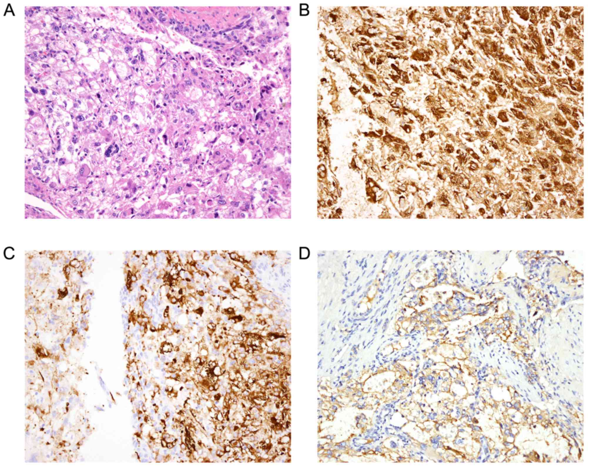
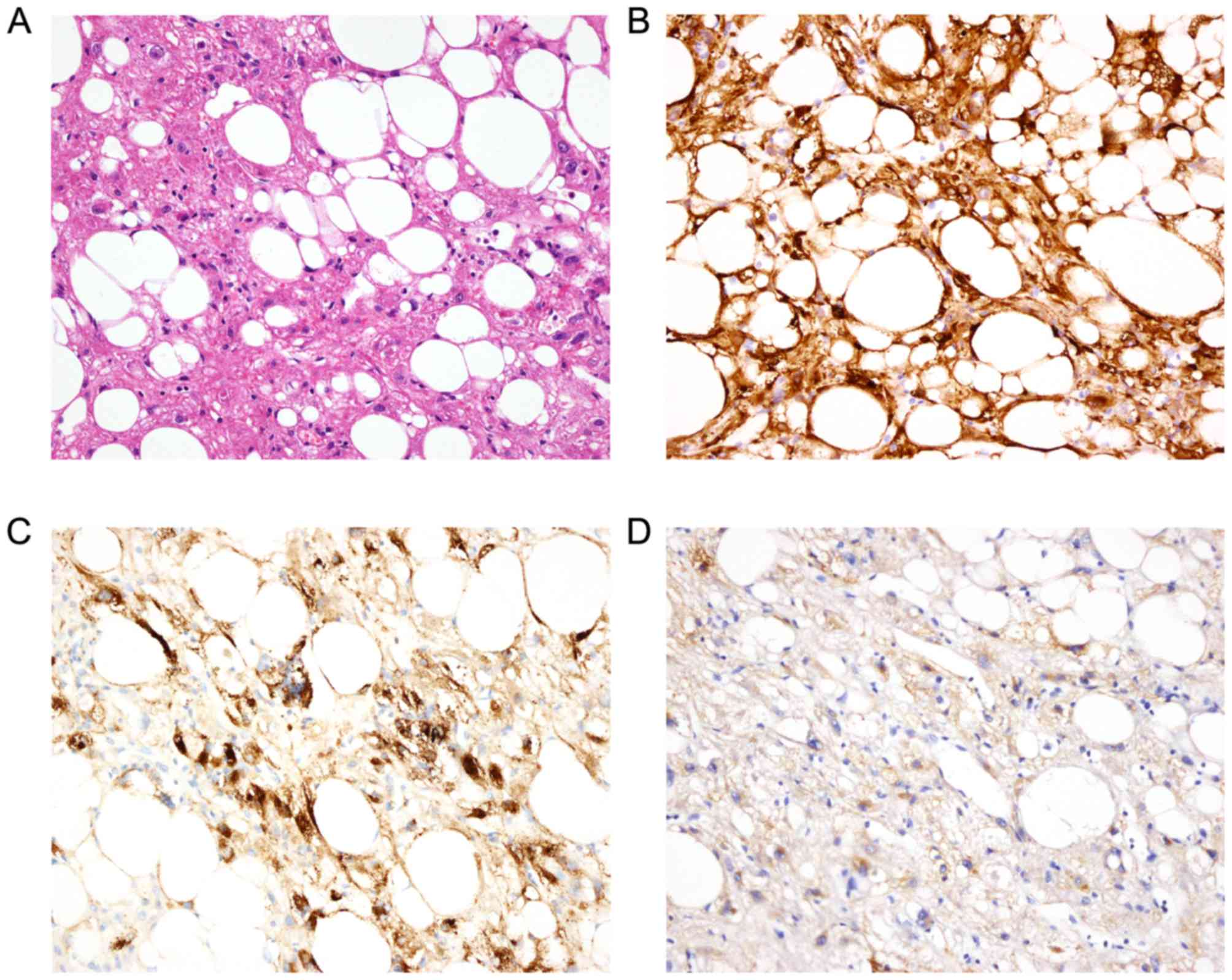
Expression of E-cadherin and other
biomarkers in all AML
First, the expression of 9 biomarkers in the two AML
groups were identified (Figs. 1 and
2). In all 22 cases, the proportion
of cases expressing E-cadherin, HMB45, Mart1/Melan A, SMA and PR
was 95.5 (21/22), 95.5 (21/22), 86.4 (19/22), 77.3 (17/22) and
86.4% (19/22), respectively. All biomarkers, with the exception of
PR, exhibited positive subcellular localization in the cell
membrane and/or plasma. The latter exhibited nuclear staining. The
majority of EAML cases (8/11, 72.7%) demonstrated intermediate
E-cadherin expression, whereas that of the control cases (8/11,
72.7%) demonstrated weak E-cadherin expression. In all cases,
results demonstrated that E-cadherin and HMB45 were expressed;
however, E-cadherin and Mart1/Melan A were expressed in 86.4%
(19/22) of cases (Table IV).
AE1/AE3, CK18, CK8 and EMA were not expressed in any of the cases
(Table IV).
The association between E-cadherin and other
biomarkers was subsequently identified. The results from the
present study revealed that the expression of E-cadherin
demonstrated a positive association with the expression of HMB45
(P=0.0044; Table V) and Mart1/Melan A
(P=0.0049; Table V). Additionally,
the results of the present study revealed negative associations
between E-cadherin and SMA or PR. However, none of these results
were considered to indicate a statistical significance.
| Table V.Spearman's rank correlation between
E-cadherin and other biomarkers. |
Table V.
Spearman's rank correlation between
E-cadherin and other biomarkers.
| Variable | Biomarker | n | Sample
correlation | P-value |
|---|
| E-cadherin | HMB45 | 21 | 0.5956 | 0.0044 |
|
| Mart1/Melan A | 22 | 0.5777 | 0.0049 |
|
| SMA | 22 | −0.0904 | 0.6893 |
|
| PR | 22 | −0.1879 | 0.4023 |
Distinct subcellular localization of
E-cadherin in EAML and control group of triphasic AML
The subcellular localization and staining categories
of E-cadherin were investigated between EAML and the control group
of triphasic AML to observe any differences. The majority of cases
in the EAML group demonstrated cell membrane and cell plasma
staining of E-cadherin (8/11), whereas the majority of cases in the
triphasic AML group demonstrated cell plasma staining only (9/11).
The subcellular localization of E-cadherin was identified to
demonstrate differences between the EAML group and the control
group (P=0.0089). The OR was calculated to be 20.0, indicating that
cases with cell membrane E-cadherin staining (with or without cell
plasma staining) had a 20-fold increased likelihood of exhibiting
EAML rather than triphasic AML (Table
VI). However, no statistically significant difference in
staining categories of E-cadherin between these two groups was
identified (P=0.0950) following the adjustment for age, sex and
tumor size (data not shown).
| Table VI.Fisher's exact test, OR for
E-cadherin score and localization between EAML and triphasic
AML. |
Table VI.
Fisher's exact test, OR for
E-cadherin score and localization between EAML and triphasic
AML.
| E-cadherin
expression | EAML, n (%) | Triphasic AML, n
(%) | P-value (Fisher's
exact test) | OR (95% CI),
P-value |
|---|
| E-cadherin
localization |
|
|
|
|
|
Membrane staining with or
without plasma | 8 (80.0) | 2 (18.2) | 0.0089 | 20.0 (2.0, 159.1),
0.0093 |
|
Cytoplasm only | 2 (20.0) | 9 (81.8) |
|
|
| E-cadherin
expression category |
|
|
|
|
|
++/+++ | 8 (72.7) | 4 (36.4) | 0.1984 | 4.7 (0.77, 28.5),
0.0950 |
|
−/+ | 3 (27.3) | 7 (63.6) |
|
|
The majority of cases classified as possessing a low
risk of malignant behavior exhibited at least intermediate
E-cadherin staining (7/9, 77.8%), whereas the case classified as
possessing an intermediate risk of exhibiting malignant behavior
exhibited negative E-cadherin staining. The case classified as
exhibiting a high risk of malignant behavior group also exhibited
intermediate E-cadherin staining; however, due to the limited
number of cases, the significance of this remains unclear.
Discussion
E-cadherin belongs to the classical cadherin
superfamily; it has five repetitive extracellular cadherin domains
and a conserved cytoplasmic domain (25,26).
E-cadherin was first reported as a cell adhesion protein in chicken
liver cells and mice (27,28), and Takeichi and colleagues named this
Ca2+-dependent cell-cell adhesion molecule E-cadherin in
the early 1980s (29,30). Since then, the role E-cadherin serves
in cell architecture and tissue formation of normal epithelium has
been extensively investigated (25,26,31,32).
It has been identified previously that E-cadherin is a key factor
in EMT and MET (33). EAML exhibits
epithelioid cells with metastatic potential, while triphasic AML
presents with spindle cells without metastatic potential (5). These differences between EAML and
triphasic AML were similar to the EMT process to a certain extent
(16,17). Hence, the factors associated with the
EMT process, including E-cadherin (33), may be a reason for these differences.
Previous studies have revealed E-cadherin to be expressed in tumors
of the urinary system (34,35), soft tissue sarcoma (36,37) and
AML (20,21). E-cadherin may be associated with the
differences in cellular morphology between EAML and triphasic AML.
The present preliminary case-control study was conducted to detect
the expression pattern of E-cadherin in EAML compared with the
common subtype of AML, triphasic AML.
The results of the present study indicated that
E-cadherin was expressed commonly in EAML and triphasic AML. All
but one case expressed E-cadherin and half of the cases
demonstrated at least intermediately stained E-cadherin. The
proportion of cases demonstrating E-cadherin expression in EAML was
slightly decreased compared with that in triphasic AML. The results
of the present study were similar to those from a previous study by
Wang et al (20) which
revealed a slight increase in the proportion of cases demonstrating
E-cadherin expression (98%), and the majority of cases (71%)
demonstrated moderate or strong E-cadherin staining. The slight
difference between the two studies may be due to the differences in
case selection. In the present study, renal EAML and control
(triphasic AML) cases were selected at the constituent ratio 1:1.
Conversely, the study by Wang et al (20) collected a variety of AML types
(including renal, hepatic and retroperitoneal), with only 2 cases
of EAML (5%). The differences in the proportion of epithelioid cell
and antibody clone of E-cadherin may be another reason for the
differences between the results of the two studies. Additionally,
the present study investigated the association between E-cadherin
and other biomarkers which have been identified in AML (5,38). The
results of the present study demonstrated that the expression of
E-cadherin may be positively associated with the expression of
HMB45 and Mart1/Melan A which are common biomarkers of melanomas
(P<0.05). Tumors which expressed E-cadherin exhibited a
decreased level of SMA and PR; however, no statistically
significant difference was identified (P>0.05). In a previous
study, Barnes et al (19)
revealed that loss of E-cadherin expression may upregulate the
expression of SMA in TSC2 gene (−/-) cell lines and is the only
study, to the best of our knowledge, which has focused on the
association between E-cadherin and other biomarkers. These cell
lines were confirmed to exhibit a loss of the wild-type allele for
the Tsc2 locus. In the present study, the association between
E-cadherin and other biomarkers was observed at the tissue level;
however, the underlying molecular mechanism and significance
remains unknown. To elucidate this, a mechanistic study based on
cell lines is required.
Secondly, the subcellular localization and staining
category of E-cadherin in EAML and its control (triphasic AML) were
investigated in the present study. The subcellular localization of
E-cadherin was demonstrated to be significantly different between
EAML and triphasic AML. Staining of the former demonstrated
membranous (with or without cytoplasmic) E-cadherin, whereas
triphasic AML revealed cytoplasmic E-cadherin staining only. In
previous studies, E-cadherin exhibited a different staining
intensity, a distinct localization between spindle and epithelioid
cells in AML and staining identified cell membrane E-cadherin
(20,21), which is consistent with the results of
the present study. In addition, the results of the study by Barnes
et al (19) were consistent
with those of the present study at the cell line level. The study
by Barnes et al (19)
demonstrated that wild-type rat renal epithelial cells exhibited
plasma membrane staining of E-cadherin which aided cells to
maintain the epithelioid characteristics. In contrast, the TSC2
(−/−) renal epithelial tumor-derived cells demonstrated a paucity
of membrane E-cadherin and cells lost epithelioid morphology. In a
previous study, Yang et al (39) hypothesized that increased expression
of E-cadherin may be a critical step in MET. The results of the
present study indicated that the various subcellular localizations
of E-cadherin observed between EAML and triphasic AML may be one
reason for the differences in cellular morphology between these two
types of tumor.
Finally, the results of the present study identified
differences between EAML cases possessing a low risk of exhibiting
malignant behavior and EAML cases possessing intermediate and high
risk of exhibiting malignant behavior. The cases categorized as low
risk exhibited at least intermediate staining of E-cadherin,
whereas the cases categorized as intermediate and high risk
exhibited the opposite. Despite this, due to the limited number of
cases, the significance of the association between expression of
E-cadherin and malignant behavior remains unclear. Further studies
are required to investigate this potential association.
The present case-control study was conducted to
obtain more EAML cases retrospectively in a relatively short
period. On the basis of current knowledge, it is understood that
there are a number of risk factors for EAML; however, the only
accepted risk factor was TSC (4,5). All the
EAML cases had no history of TSC so only cases of triphasic AML
without TSC were selected as a control. Additionally, the present
study selected cases where patients also possessed common risk
factors for cancer including a history of smoking and alcohol
consumption. Although the cases with a strict condition were
matched to guarantee the comparability between EAML and triphasic
AML, there may be some confounding factors not identified in the
present study which cannot be resolved using a case-control study.
The small sample size and the heterogeneity in the proportion of
ECs in EAML group were other limitations of the present study.
However, EAML was a rare variant of AML and the present study
established relative strict criteria of EAML and triphasic AML, on
the basis of World Health Organization classification and the
literature. The present study aimed for more suitable cases of EAML
and triphasic AML to be recruited which resulted in all but 2 cases
of EAML demonstrating ≥90% of ECs, which led to minimal
heterogeneity. A study with a larger sample size from multiple
centers and a relative homogeneity of the proportion of epithelioid
cells in EAML, is required to increase the reliability of the
results.
E-cadherin may an important biomarker in AML. The
mechanisms of E-cadherin may be distinct between EAML and triphasic
AML, since the subcellular localization of E-cadherin may be
distinct between EAML and triphasic AML. A study with a large
sample size is required to validate the results of the present
study, followed by a mechanistic study based on cell lines to
determine any significance.
Acknowledgements
The authors thank Professor Xiaohong R. Yang from
the Genetic Epidemiology Branch, Division of Cancer Epidemiology
and Genetics, National Cancer Institute, National Institutes of
Health (Bethesda, MD, USA) for advice; Dr Zheng Yuan (Z.Y.) from
the Department of Pathology, National Cancer Center/Cancer
Hospital, Chinese Academy of Medical Sciences and Peking Union
Medical College for assistance with reviewing the hematoxylin and
eosin slides of the 22 cases; Dr Wei-Hua Li from the Department of
Pathology, National Cancer Center/Cancer Hospital, Chinese Academy
of Medical Sciences and Peking Union Medical College for assistance
with preparing figures; and Mr Jiang-Nan Shao from the Fourth
Educational Department of Beijing Li Li Changping Education
Technology Co., Ltd. (Beijing, China) for assistance in native
language revision. The present study was supported by the Beijing
Hope Run Special Fund (grant no. LC2011B35) and the Basic
Scientific Research Fund of China Central Public Welfare (grant no.
JK 2014B06).
Glossary
Abbreviations
Abbreviations:
|
AML
|
angiomyolipoma
|
|
CK
|
cytokeratin
|
|
EAML
|
epithelioid angiomyolipoma
|
|
EMA
|
epithelial membrane antigen
|
|
EMT
|
epithelial-to-mesenchymal
transition
|
|
ECs
|
epithelioid cells
|
|
IHC
|
immunohistochemistry
|
|
IRS
|
immunoreactive score
|
|
MET
|
mesenchymal-to-epithelial
transition
|
|
OR
|
odds ratio
|
|
PR
|
progesterone receptor
|
|
SD
|
standard deviation
|
|
SMA
|
smooth muscle actin
|
|
TSC
|
tuberous sclerosis complex
|
References
|
1
|
Hassan M, El-Hefnawy AS, Elshal AM, Mosbah
A, El-Baz M and Shaaban A: Renal epithelioid angiomyolipoma: A rare
variant with unusual behavior. Int Urol Nephrol. 46:317–322. 2014.
View Article : Google Scholar : PubMed/NCBI
|
|
2
|
Koo KC, Kim WT, Ham WS, Lee JS, Ju HJ and
Choi YD: Trends of presentation and clinical outcome of treated
renal angiomyolipoma. Yonsei Med J. 51:728–734. 2010. View Article : Google Scholar : PubMed/NCBI
|
|
3
|
Lane BR, Aydin H, Danforth TL, Zhou M,
Remer EM, Novick AC and Campbell SC: Clinical correlates of renal
angiomyolipoma subtypes in 209 patients: Classic, fat poor,
tuberous sclerosis associated and epithelioid. J Urol. 180:836–843.
2008. View Article : Google Scholar : PubMed/NCBI
|
|
4
|
Rakowski SK, Winterkorn EB, Paul E, Steele
DJ, Halpern EF and Thiele EA: Renal manifestations of tuberous
sclerosis complex: Incidence, prognosis, and predictive factors.
Kidney Int. 70:1777–1782. 2006. View Article : Google Scholar : PubMed/NCBI
|
|
5
|
Eble JN, Sauter G, Epstein J and
Sesterhenn IA: World Health Organization Classification of
TumoursPathology and Genetics of Tumours of the Urinary System and
Male Genital Organs. IARC Press; Lyon: pp. 65–69. 2004
|
|
6
|
Mai KT, Perkins DG and Collins JP:
Epithelioid cell variant of renal angiomyolipoma. Histopathology.
28:277–280. 1996. View Article : Google Scholar : PubMed/NCBI
|
|
7
|
Brimo F, Robinson B, Guo C, Zhou M, Latour
M and Epstein JI: Renal epithelioid angiomyolipoma with atypia: A
series of 40 cases with emphasis on clinicopathologic prognostic
indicators of malignancy. Am J Surg Pathol. 34:715–722.
2010.PubMed/NCBI
|
|
8
|
Nese N, Martignoni G, Fletcher CD, Gupta
R, Pan CC, Kim H, Ro JY, Hwang IS, Sato K, Bonetti F, et al: Pure
epithelioid PEComas (so-called epithelioid angiomyolipoma) of the
kidney: A clinicopathologic study of 41 cases: Detailed assessment
of morphology and risk stratification. Am J Surg Pathol.
35:161–176. 2011. View Article : Google Scholar : PubMed/NCBI
|
|
9
|
Srigley JR, Delahunt B, Eble JN, Egevad L,
Epstein JI, Grignon D, Hes O, Moch H, Montironi R, Tickoo SK, et
al: The international society of urological pathology (ISUP)
vancouver classification of renal neoplasia. Am J Surg Pathol.
37:1469–1489. 2013. View Article : Google Scholar : PubMed/NCBI
|
|
10
|
Martignoni G, Pea M, Rigaud G, Manfrin E,
Colato C, Zamboni G, Scarpa A, Tardanico R, Roncalli M and Bonetti
F: Renal angiomyolipoma with epithelioid sarcomatous transformation
and metastases: Demonstration of the same genetic defects in the
primary and metastatic lesions. Am J Surg Pathol. 24:889–894. 2000.
View Article : Google Scholar : PubMed/NCBI
|
|
11
|
Sato K, Ueda Y, Tachibana H, Miyazawa K,
Chikazawa I, Kaji S, Nojima T and Katsuda S: Malignant epithelioid
angiomyolipoma of the kidney in a patient with tuberous sclerosis:
An autopsy case report with p53 gene mutation analysis. Pathol Res
Pract. 204:771–777. 2008. View Article : Google Scholar : PubMed/NCBI
|
|
12
|
Bing Z, Yao Y, Pasha T, Tomaszewski JE and
Zhang PJ: p53 in pure epithelioid PEComa: An immunohistochemistry
study and gene mutation analysis. Int J Surg Pathol. 20:115–122.
2012. View Article : Google Scholar : PubMed/NCBI
|
|
13
|
Li J, Zhu M and Wang YL: Malignant
epithelioid angiomyoliopoma of the kidney with pulmonary metastases
and p53 gene mutation. World J Surg Oncol. 10:2132012. View Article : Google Scholar : PubMed/NCBI
|
|
14
|
Li W, Guo L, Bi X, Ma J and Zheng S:
Immunohistochemistry of p53 and Ki-67 and p53 mutation analysis in
renal epithelioid angiomyolipoma. Int J Clin Exp Pathol.
8:9446–9451. 2015.PubMed/NCBI
|
|
15
|
Ooi SM, Vivian JB and Cohen RJ: The use of
the Ki-67 marker in the pathological diagnosis of the epithelioid
variant of renal angiomyolipoma. Int Urol Nephrol. 41:559–565.
2009. View Article : Google Scholar : PubMed/NCBI
|
|
16
|
Thiery JP: Epithelial-mesenchymal
transitions in development and pathologies. Curr Opin Cell Biol.
15:740–746. 2003. View Article : Google Scholar : PubMed/NCBI
|
|
17
|
Yang J and Weinberg RA:
Epithelial-mesenchymal transition: At the crossroads of development
and tumor metastasis. Dev Cell. 14:818–829. 2008. View Article : Google Scholar : PubMed/NCBI
|
|
18
|
Thiery JP, Acloque H, Huang RY and Nieto
MA: Epithelial-mesenchymal transitions in development and disease.
Cell. 139:871–890. 2009. View Article : Google Scholar : PubMed/NCBI
|
|
19
|
Barnes EA, Kenerson HL, Jiang X and Yeung
RS: Tuberin regulates E-cadherin localization: Implications in
epithelial-mesenchymal transition. Am J Pathol. 177:1765–1778.
2010. View Article : Google Scholar : PubMed/NCBI
|
|
20
|
Wang Z, Gong Q and Fan Q: Expression of
E-cadherin in angiomyolipoma. Hum Pathol. 43:2348–2353. 2012.
View Article : Google Scholar : PubMed/NCBI
|
|
21
|
Konosu-Fukaya S, Nakamura Y, Fujishima F,
Kasajima A, McNamara KM, Takahashi Y, Joh K, Saito H, Ioritani N,
Ikeda Y, et al: Renal epithelioid angiomyolipoma with malignant
features: Histological evaluation and novel immunohistochemical
findings. Pathol Int. 64:133–141. 2014. View Article : Google Scholar : PubMed/NCBI
|
|
22
|
Alì G, Boldrini L, Capodanno A,
Pelliccioni S, Servadio A, Crisman G, Picchi A, Davini F, Mussi A
and Fontanini G: Expression of p-AKT and p-mTOR in a large series
of bronchopulmonary neuroendocrine tumors. Exp Ther Med. 2:787–792.
2011.PubMed/NCBI
|
|
23
|
Li N, Zhong M and Song M: Expression of
phosphorylated mTOR and its regulatory protein is related to
biological behaviors of ameloblastoma. Int J Clin Exp Pathol.
5:660–667. 2012.PubMed/NCBI
|
|
24
|
Zheng S, Bi XG, Song QK, Yuan Z, Guo L,
Zhang H and Ma JH: A suggestion for pathological grossing and
reporting based on prognostic indicators of malignancies from a
pooled analysis of renal epithelioid angiomyolipoma. Int Urol
Nephrol. 47:1643–1651. 2015. View Article : Google Scholar : PubMed/NCBI
|
|
25
|
Lecuit T and Yap AS: E-cadherin junctions
as active mechanical integrators in tissue dynamics. Nat Cell Biol.
17:533–539. 2015. View
Article : Google Scholar : PubMed/NCBI
|
|
26
|
Takeichi M: Dynamic contacts: Rearranging
adherens junctions to drive epithelial remodelling. Nat Rev Mol
Cell Biol. 15:397–410. 2014. View
Article : Google Scholar : PubMed/NCBI
|
|
27
|
Gallin WJ, Edelman GM and Cunningham BA:
Characterization of L-CAM, a major cell adhesion molecule from
embryonic liver cells. Proc Natl Acad Sci USA. 80:pp. 1038–1042.
1983; View Article : Google Scholar : PubMed/NCBI
|
|
28
|
Schuh R, Vestweber D, Riede I, Ringwald M,
Rosenberg UB, Jäckle H and Kemler R: Molecular cloning of the mouse
cell adhesion molecule uvomorulin: cDNA contains a B1-related
sequence. Proc Natl Acad Sci USA. 83:pp. 1364–1368. 1986;
View Article : Google Scholar : PubMed/NCBI
|
|
29
|
Ogou SI, Yoshida-Noro C and Takeichi M:
Calcium-dependent cell-cell adhesion molecules common to
hepatocytes and teratocarcinoma stem cells. J Cell Biol.
97:944–948. 1983. View Article : Google Scholar : PubMed/NCBI
|
|
30
|
Yoshida-Noro C, Suzuki N and Takeichi M:
Molecular nature of the calcium-dependent cell-cell adhesion system
in mouse teratocarcinoma and embryonic cells studied with a
monoclonal antibody. Dev Biol. 101:19–27. 1984. View Article : Google Scholar : PubMed/NCBI
|
|
31
|
Mertz AF, Che Y, Banerjee S, Goldstein JM,
Rosowski KA, Revilla SF, Niessen CM, Marchetti MC, Dufresne ER and
Horsley V: Cadherin-based intercellular adhesions organize
epithelial cell-matrix traction forces. Proc Natl Acad Sci USA.
110:pp. 842–847. 2013; View Article : Google Scholar : PubMed/NCBI
|
|
32
|
Ghahhari NM and Babashah S: Interplay
between microRNAs and WNT/β-catenin signalling pathway regulates
epithelial-mesenchymal transition in cancer. Eur J Cancer.
51:1638–1649. 2015. View Article : Google Scholar : PubMed/NCBI
|
|
33
|
Bhatt T, Rizvi A, Batta SP, Kataria S and
Jamora C: Signaling and mechanical roles of E-cadherin. Cell Commun
Adhes. 20:189–199. 2013. View Article : Google Scholar : PubMed/NCBI
|
|
34
|
Kobayashi N, Matsuzaki O, Shirai S, Aoki
I, Yao M and Nagashima Y: Collecting duct carcinoma of the kidney:
An immunohistochemical evaluation of the use of antibodies for
differential diagnosis. Hum Pathol. 39:1350–1359. 2008. View Article : Google Scholar : PubMed/NCBI
|
|
35
|
Muramaki M, Miyake H, Terakawa T, Kusuda Y
and Fujisawa M: Expression profile of E-cadherin and N-cadherin in
urothelial carcinoma of the upper urinary tract is associated with
disease recurrence in patients undergoing nephroureterectomy.
Urology. 78:1443.e7–e12. 2011. View Article : Google Scholar
|
|
36
|
Wang N, He YL, Pang LJ, Zou H, Liu CX,
Zhao J, Hu JM, Zhang WJ, Qi Y and Li F: Down-regulated E-cadherin
expression is associated with poor five-year overall survival in
bone and soft tissue sarcoma: Results of a meta-analysis. PLoS One.
10:e01214482015. View Article : Google Scholar : PubMed/NCBI
|
|
37
|
Yoo J, Park S, Kang CS, Kang SJ and Kim
BK: Expression of E-cadherin and p53 proteins in human soft tissue
sarcomas. Arch Pathol Lab Med. 126:33–38. 2002.PubMed/NCBI
|
|
38
|
Martignoni G, Pea M, Reghellin D, Zamboni
G and Bonetti F: PEComas: The past, the present and the future.
Virchows Arch. 452:119–132. 2008. View Article : Google Scholar : PubMed/NCBI
|
|
39
|
Yang J, Du X, Wang G, Sun Y, Chen K, Zhu
X, Lazar AJ, Hunt KK, Pollock RE and Zhang W: Mesenchymal to
epithelial transition in sarcomas. Eur J Cancer. 50:593–601. 2014.
View Article : Google Scholar : PubMed/NCBI
|