Introduction
Prostate cancer is the second leading cause of
cancer-associated mortality in men worldwide, and the incidence of
the disease is increasing year by year (1). Currently, the level of prostate-specific
antigen, the clinical stage and the grade of the tumor (Gleason
score) are the predominant methods for predicting the prognosis of
prostate cancer (2). However, the
epigenetic silencing of key tumor suppressors genes during prostate
cancer tumorigenesis and progression may be translated into a
promising clinical diagnostic marker (2). CpG islands at transcription initiation
sites are generally poorly methylated. However, in certain cancer
tissues, hypermethylation of CpG may silence gene transcription
(3). In prostate cancer tissues, a
number of large-scale genome-wide analyses have suggested that
>30 key genes may be regulated through promoter hypermethylation
(3). At present, there are still
relatively few biomarkers for the prediction of prostate cancer
(2).
Protocadherin 8 (PCDH8) is a member of the
cadherin family and has been identified to be a novel tumor
suppressor gene that is silenced in a number of cancer types,
including gastric cancer, breast cancer, bladder cancer and renal
cell carcinoma (4–7). Transcriptional silencing may occur via
genetic mutation (5,6) or epigenetic promoter hypermethylation
(4–7).
The present study analyzed the methylation status of
the promoter of the PCDH8 gene in prostate cancer tissues,
with the aim of identifying a potential association between the
methylation status and the diagnosis and prognosis of prostate
cancer.
Materials and methods
Patients and specimens
According to the prostate cancer diagnostic criteria
of the World Health Organization and National Cancer Institute for
prostate cancer (8), 82 patients with
prostate cancer and 30 hospitalized patients with benign prostatic
hyperplasia at Yantai Yuhuangding Hospital (Yantai, China) were
enrolled between May 2010 and May 2012. The inclusion criteria were
as follows: i) no radiation therapy, chemotherapy or hormone
therapy administered prior to surgery; ii) general information,
including clinical stage and histological grade, is available; iii)
diagnosis of primary prostate cancer; iv) confirmed prostate cancer
by pathological report. The exclusion criteria were as follows: i)
history of other malignant tumors; ii) incomplete clinical data;
iii) presence of remote metastatic or recurrent prostate cancer.
The prostate cancer patients were aged between 37 and 77 years,
with a mean age of 58.1±3.9 years. There were 34 cases with a
single tumor, and 48 cases with multiple tumors. With regard to
tumor size, 48 cases had a tumor diameter of ≤3 cm, and 34 cases
had a diameter of >3 cm. A total of 57 cases were papillary and
25 cases were non-papillary. There were 55 cases of grade (G)1-G2,
and 27 cases of G3. Regarding tumor stage, a total of 49 cases were
Ta-T1 and 33 cases were T2-T4 (9,10).
DNA extraction
All surgical specimens were placed immediately in
liquid nitrogen following the surgery. The specimens (~25 mg) were
ground in liquid nitrogen and DNA was extracted using QIAamp DNA
Mini Kit (Qiagen GmbH, Hilden, Germany; lot 130217) according to
the manufacturer's instructions. The purity and concentration of
DNA was measured using a DU800 ultraviolet (UV) spectrophotometer
(Beckman-Coulter, Inc., Brea, CA, USA).
Methylation-specific polymerase chain
reaction (MSP)
The methylation status of PCDH8 was
determined by MSP (11). A total of 1
µg genomic DNA was modified by methylation using an EZ DNA
Methylation-Gold Kit (UZymo Research Corporation, Irvine, CA, USA;
lot 130522) according to the manufacturer's instructions. In this
method, following the genomic DNA methylation reaction,
unmethylated cytosine is converted to uracil, whereas methylated
cytosine remains unchanged. Therefore, specific primers may be used
to differentiate the methylation status. The primers were designed
as previously described (12). All
primers were synthesized by BayGene Biotech Company Limited
(Beijing, China). The primers for the methylated reaction were as
follows: Sense, 5′-CGGTTATTGGTTATTCGGTTCC-3′; and antisense,
5′-ACGAACTCTAAAAACGCGCG-3′; product size, 94 bp. The primers for
the unmethylated reaction were as follows: Sense,
5′-GGTGGTTATTGGTTATTTGGTTT-3′; antisense,
5′-CCAACAAACTCTAAAAACACACA-3′; product size, 97 bp. Water was used
as a blank control in each assay. PCR products (10 µl) were
separated on a 2% agarose gel, stained with ethidium bromide and
visualized under UV illumination. Specimens were scored as
methylation-positive when a specific band was amplified by
methylated reaction-specific primers, and negative when amplified
only by unmethylated reaction-specific primers. The thermocycling
conditions were as follows for a total of 40 cycles: Denaturing at
95°C for 30 sec, annealing at 60°C for 5 sec and extension at 72°C
for 45 sec. The 2−ΔΔCq method (13) was used for the quantification of the
PCR results.
Western blotting
Western blotting was performed to study the protein
expression levels of PCDH8 in the clinical samples. A total of 1 mg
tissue was solubilized in 100 µl lysis buffer (30 mM Tris, 2 M
Thiourea, 4% CHAPS and 7 M urea; pH 8.5) on ice for 30 min. The
lysis buffer was subsequently centrifuged at 1,000 × g for
15 min and the supernatant was collected for further testing.
Primary antibodies were used for incubation at 4°C overnight and
secondary antibodies at room temperature for 1 h. Primary
antibodies targeting PCDH8 (1:1,000 dilution; catalog no. ab85561;
Abcam, Cambridge, UK) and actin (1:1,000; rabbit; polyclonal; cat.
no. ab8227; Abcam, Cambridge, UK) were used. The following
secondary antibodies were used: Anti-rabbit IgG-biotin (cat. no.
BA1020, Boster Biological Technology, Wuhan, China). Bands were
detected using the DAB Chromogenic Reagent kit (cat. no. AR1021;
Boster, Biological Technology).
Postoperative follow-up
Following surgery, patients were followed up for
6–24 months for the analysis of recurrence and survival.
Statistical analysis
All data were statistically analyzed using SPSS 18.0
(SPSS, Inc., Chicago, IL, USA). The different mRNA and protein
expression between cancer tissues and controls were analyzed with
the Student's t-test. For survival data, differences between two
groups were assessed with the log-rank test. Kaplan-Meier curves of
ovarall survival were constructed. The differences between
clinicopathological features and the status of methylation were
analyzed using the χ2 test. The data were presented as
the mean ± standard deviation. P<0.05 was considered to indicate
a statistically significant difference.
Results
Expression of PCDH8 is reduced in
prostate cancer tissues
As a tumor suppressor, the expression of
PCDH8 is reduced in various cancer types (4–7).
Therefore, the present study investigated the expression levels of
PCDH8 in prostate cancer tissues by quantitative polymerase
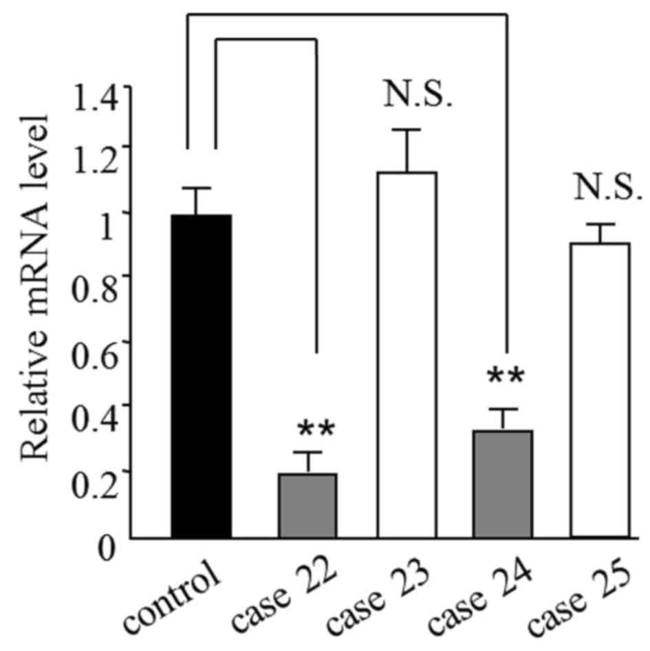
chain reaction (qPCR). The results revealed that the mRNA level of
PCDH8 was reduced to 0.30±0.10 in 70.7% (58/82) of prostate
cancer tissues (Table I). Cases 22–25
are shown as representative samples for the mRNA results in
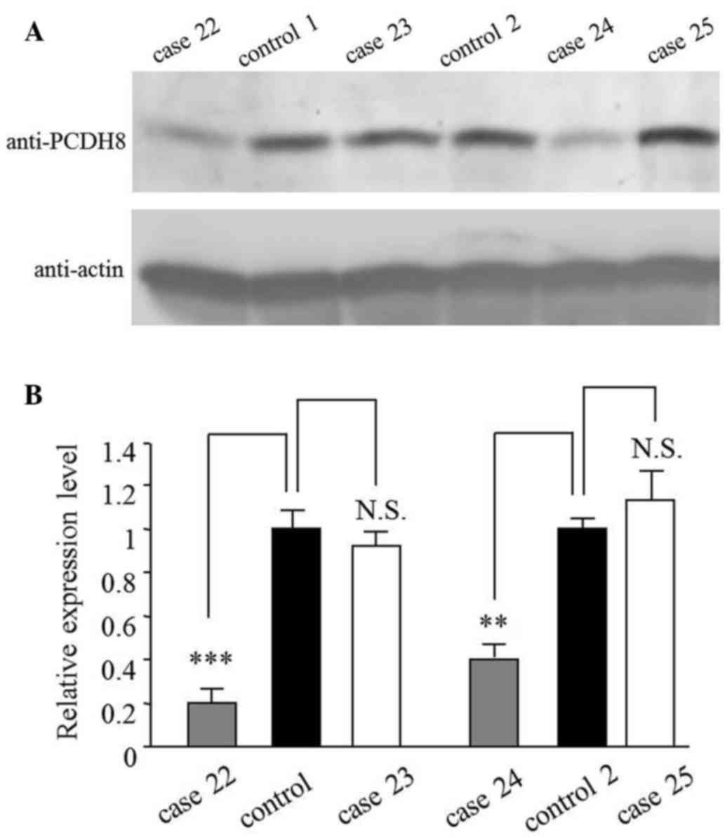
Fig. 1. This result was also verified
at the protein level by western blot analysis (Fig. 2). The mRNA and protein levels in cases
22 and 24, but not in cases 23 and 25, were reduced compared with
the control (Table I; Figs. 1 and 2).
 | Table I.Relative PCDH8 expression and
methylation summary for patients with prostate cancer (n=82) or
benign prostatic hyperplasia. |
Table I.
Relative PCDH8 expression and
methylation summary for patients with prostate cancer (n=82) or
benign prostatic hyperplasia.
|
|
| Relative mRNA
expression |
|
|
|---|
|
|
|
|
|
|
|---|
| Group | Effect | Mean | SEM | P-value | MSP-positive |
|---|
| Control (C1-C3) | – | 1.00 | 0.11 | – | – |
| Cases with no
PCDH8 reduction (n=24/82; 29.3%) |
|
|
|
|
|
| P3 | N | 1.03 | 0.21 | N.S. | n |
| P4 | N | 1.21 | 0.14 | N.S. | n |
|
P10 | N | 1.17 | 0.13 | N.S. | n |
|
P14 | N | 1.08 | 0.22 | N.S. | n |
|
P15 | N | 1.22 | 0.32 | N.S. | n |
|
P23 | N | 1.12 | 0.21 | N.S. | n |
|
P25 | N | 0.91 | 0.09 | N.S. | n |
|
P31 | N | 1.08 | 0.28 | N.S. | n |
|
P38 | N | 0.86 | 0.14 | N.S. | n |
|
P39 | N | 0.85 | 0.23 | N.S. | n |
|
P40 | N | 1.08 | 0.27 | N.S. | n |
|
P45 | N | 0.88 | 0.14 | N.S. | n |
|
P46 | N | 0.93 | 0.13 | N.S. | n |
|
P47 | N | 1.21 | 0.37 | N.S. | n |
|
P50 | N | 0.79 | 0.32 | N.S. | n |
|
P55 | N | 0.86 | 0.09 | N.S. | n |
|
P58 | N | 0.81 | 0.16 | N.S. | n |
|
P59 | N | 1.08 | 0.11 | N.S. | n |
|
P64 | N | 1.32 | 0.44 | N.S. | n |
|
P73 | N | 1.21 | 0.13 | N.S. | n |
|
P76 | N | 1.24 | 0.21 | N.S. | n |
|
P78 | N | 0.86 | 0.16 | N.S. | n |
|
P80 | N | 1.21 | 0.28 | N.S. | n |
|
P81 | N | 0.94 | 0.08 | N.S. | n |
| Cases with
PCDH8 reduction (n=58/82; 70.7%) |
|
|
|
|
|
| P1 | R | 0.27 | 0.03 | <0.005 | y |
| P2 | R | 0.33 | 0.18 | <0.050 | y |
| P5 | R | 0.41 | 0.13 | <0.050 | n |
| P6 | R | 0.39 | 0.18 | <0.050 | y |
| P7 | R | 0.35 | 0.11 | <0.005 | y |
| P8 | R | 0.31 | 0.13 | <0.005 | y |
| P9 | R | 0.25 | 0.18 | <0.050 | n |
|
P11 | R | 0.32 | 0.09 | <0.010 | y |
|
P12 | R | 0.28 | 0.07 | <0.050 | y |
|
P13 | R | 0.39 | 0.13 | <0.050 | n |
|
P16 | R | 0.33 | 0.13 | <0.050 | y |
|
P17 | R | 0.37 | 0.18 | <0.005 | y |
|
P18 | R | 0.17 | 0.04 | <0.010 | y |
|
P19 | R | 0.42 | 0.03 | <0.050 | n |
|
P20 | R | 0.40 | 0.18 | <0.050 | y |
|
P21 | R | 0.38 | 0.11 | <0.005 | y |
|
P22 | R | 0.18 | 0.08 | <0.010 | y |
|
P24 | R | 0.31 | 0.07 | <0.010 | y |
|
P26 | R | 0.24 | 0.18 | <0.005 | y |
|
P27 | R | 0.22 | 0.07 | <0.010 | n |
|
P28 | R | 0.39 | 0.07 | <0.050 | y |
| Cases with
PCDH8 reduction (n=58/82; 70.7%) |
|
|
|
|
|
|
P29 | R | 0.27 | 0.13 | <0.005 | y |
|
P30 | R | 0.40 | 0.02 | <0.010 | n |
|
P32 | R | 0.33 | 0.13 | <0.005 | y |
|
P33 | R | 0.30 | 0.05 | <0.005 | y |
|
P34 | R | 0.32 | 0.07 | <0.010 | y |
|
P35 | R | 0.39 | 0.08 | <0.050 | y |
|
P36 | R | 0.30 | 0.13 | <0.005 | y |
|
P37 | R | 0.17 | 0.04 | <0.005 | n |
|
P41 | R | 0.30 | 0.13 | <0.010 | y |
|
P42 | R | 0.21 | 0.13 | <0.010 | y |
|
P43 | R | 0.19 | 0.13 | <0.010 | y |
|
P44 | R | 0.24 | 0.02 | <0.005 | y |
|
P48 | R | 0.27 | 0.09 | <0.005 | y |
|
P49 | R | 0.36 | 0.18 | <0.010 | n |
|
P51 | R | 0.27 | 0.09 | <0.010 | y |
|
P52 | R | 0.39 | 0.04 | <0.050 | y |
|
P53 | R | 0.30 | 0.05 | <0.005 | y |
|
P54 | R | 0.33 | 0.11 | <0.010 | y |
|
P56 | R | 0.34 | 0.08 | <0.050 | y |
|
P57 | R | 0.24 | 0.04 | <0.010 | y |
|
P60 | R | 0.24 | 0.13 | <0.010 | n |
|
P61 | R | 0.40 | 0.05 | <0.005 | y |
|
P62 | R | 0.37 | 0.08 | <0.010 | y |
|
P63 | R | 0.22 | 0.09 | <0.050 | y |
|
P65 | R | 0.15 | 0.03 | <0.010 | y |
|
P66 | R | 0.34 | 0.14 | <0.050 | y |
|
P67 | R | 0.31 | 0.05 | <0.050 | y |
|
P68 | R | 0.21 | 0.05 | <0.010 | n |
|
P69 | R | 0.33 | 0.08 | <0.010 | y |
|
P70 | R | 0.17 | 0.07 | <0.005 | y |
|
P71 | R | 0.32 | 0.08 | <0.005 | y |
|
P72 | R | 0.24 | 0.18 | <0.010 | y |
|
P74 | R | 0.27 | 0.04 | <0.010 | n |
|
P75 | R | 0.15 | 0.03 | <0.050 | y |
|
P77 | R | 0.32 | 0.09 | <0.050 | y |
|
P79 | R | 0.24 | 0.13 | <0.005 | y |
|
P82 | R | 0.33 | 0.08 | <0.010 | y |
| Mean | – | 0.30 | 0.10 | – | – |
Methylation status of PCDH8 gene
promoter
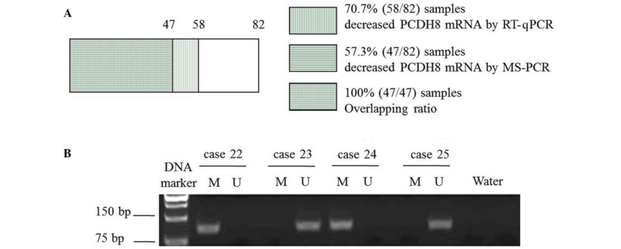
To identify the possible reason for the mRNA
downregulation, the methylation status of the PCDH8 promoter
was examined. The results revealed that the benign prostatic
hyperplasia and prostate cancer samples that did not exhibit
PCDH8 reduction were not methylated (Tables I and II). The 47 cases in which methylation was
detected were all prostate cancer samples that exhibited
PCDH8 reduction (Table I;
Fig. 3A). Representative results from
cases 22–25 are shown in Fig. 3B.
| Table II.Relative protocadherin 8 expression
and methylation summary for controls with benign prostatic
hyperplasia (n=30). |
Table II.
Relative protocadherin 8 expression
and methylation summary for controls with benign prostatic
hyperplasia (n=30).
|
|
| Relative mRNA
expression |
|
|
|---|
|
|
|
|
|
|
|---|
| Case no. | Effect | Mean | SEM | P-value | MSP-positive |
|---|
|
Controla | – | 1.00 | 0.11 | – | – |
| C1 | N | 1.07 | 0.13 | N.S. | n |
| C2 | N | 0.96 | 0.08 | N.S. | n |
| C3 | N | 1.17 | 0.12 | N.S. | n |
| C4 | N | 1.24 | 0.08 | N.S. | n |
| C5 | N | 1.22 | 0.22 | N.S. | n |
| C6 | N | 1.05 | 0.13 | N.S. | n |
| C7 | N | 1.21 | 0.21 | N.S. | n |
| C8 | N | 1.20 | 0.09 | N.S. | n |
| C9 | N | 1.06 | 0.16 | N.S. | n |
| C10 | N | 1.08 | 0.14 | N.S. | n |
| C11 | N | 1.17 | 0.14 | N.S. | n |
| C12 | N | 0.84 | 0.22 | N.S. | n |
| C13 | N | 1.04 | 0.28 | N.S. | n |
| C14 | N | 1.21 | 0.08 | N.S. | n |
| C15 | N | 1.24 | 0.21 | N.S. | n |
| C16 | N | 0.86 | 0.23 | N.S. | n |
| C17 | N | 1.21 | 0.19 | N.S. | n |
| C18 | N | 0.94 | 0.16 | N.S. | n |
| C19 | N | 1.10 | 0.11 | N.S. | n |
| C20 | N | 0.91 | 0.08 | N.S. | n |
| C21 | N | 1.23 | 0.20 | N.S. | n |
| C22 | N | 1.11 | 0.07 | N.S. | n |
| C23 | N | 0.89 | 0.19 | N.S. | n |
| C24 | N | 1.24 | 0.28 | N.S. | n |
| C25 | N | 1.20 | 0.10 | N.S. | n |
| C26 | N | 1.02 | 0.19 | N.S. | n |
| C27 | N | 0.98 | 0.05 | N.S. | n |
| C28 | N | 1.26 | 0.23 | N.S. | n |
| C29 | N | 1.14 | 0.16 | N.S. | n |
| C30 | N | 1.24 | 0.19 | N.S. | n |
Analysis of the association between
methylation status of PCDH8 and various clinicopathological
features
The detailed clinicopathological features of
patients with prostate cancer are shown in Table III. The results demonstrated that
PCDH8 methylation was significantly associated with a larger
tumor diameter (>3 vs. ≤3 cm; P=0.016), non-papillary shape
(P=0.023), advanced tumor stage (T2-T4 vs. Ta-T1;
P=0.016) and advanced pathological grade (G3 vs. G1-2;
P=0.009). By contrast, no significant associations were
identified between PCDH8 methylation and age (P=0.842) or
number of tumors (P=0.500).
| Table III.Association between protocadherin 8
methylation and pathological features of patients with prostate
cancer. |
Table III.
Association between protocadherin 8
methylation and pathological features of patients with prostate
cancer.
| Feature | Total, n | Unmethylated, n
(%) | Methylated, n
(%) | χ2
value | P-value |
|---|
| Age, years |
|
|
| 0.040 | 0.842 |
|
≤65 | 50 | 27 (54.00) | 23 (46.00) |
|
|
|
>65 | 32 | 18 (56.25) | 14 (43.75) |
|
|
| Tumor number |
|
|
| 0.455 | 0.500 |
|
Single | 34 | 16 (47.06) | 18 (52.94) |
|
|
|
Multiple | 48 | 19 (39.58) | 29 (60.42) |
|
|
| Tumor diameter |
|
|
| 5.828 | 0.016 |
| ≤3
cm | 46 | 25 (54.35) | 21 (45.65) |
|
|
| >3
cm | 36 | 10 (27.78) | 26 (72.22) |
|
|
| Tumor shape |
|
|
| 5.131 | 0.023 |
|
Papillary | 57 | 29 (50.88) | 28 (49.12) |
|
|
|
Non-papillary | 25 | 6
(24.00) | 19 (76.00) |
|
|
| Tumor stage |
|
|
| 5.803 | 0.016 |
|
Ta-T1 | 51 | 27 (52.94) | 24 (47.06) |
|
|
|
T2-T4 | 31 | 8
(25.81) | 23 (74.19) |
|
|
| Pathological
grade |
|
|
| 6.888 | 0.009 |
|
G1-G2 | 55 | 29 (52.73) | 26 (47.27) |
|
|
| G3 | 27 | 6
(22.22) | 21 (77.78) |
|
|
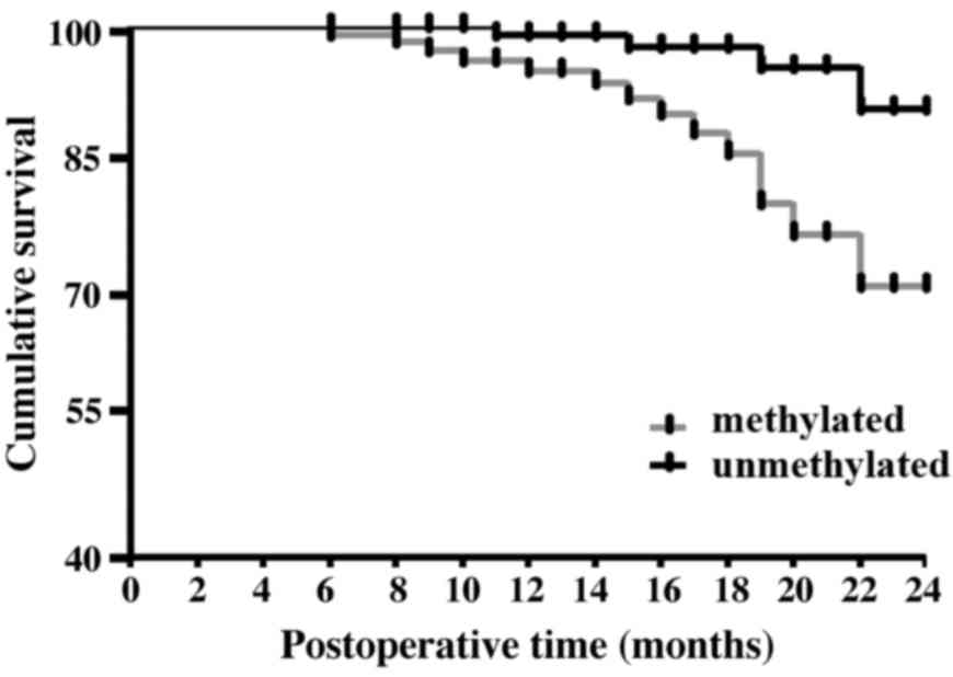
Postoperative follow-up to assess the
association between PCDH8 methylation and prognosis
Survival data were collected for 6–24 months after
surgery. The data revealed that the methylation status of
PCDH8 is associated with the overall survival time of
patients with prostate cancer; those with unmethylated PCDH8
have a better prognosis (recurrence rate, 14.29; and mortality
rate, 8.57%) compared with those with methylated PCDH8
(recurrence rate, 36.17%; mortality rate, 29.79%). These
differences were statistically significant (recurrence rate,
P=0.027; mortality rate, P=0.019) (Table
IV; Fig. 4).
| Table IV.Postoperative follow-up analysis
(n=82). |
Table IV.
Postoperative follow-up analysis
(n=82).
| Status | Total, n | Recurrence rate, n
(%) | Mortality rate, n
(%) |
|---|
| Methylated | 47 | 17 (36.17) | 14 (29.79) |
| Unmethylated | 35 | 5
(14.29) | 3
(8.57) |
| χ2
value |
| 4.894 | 5.495 |
| P-value |
| 0.027 | 0.019 |
Discussion
In recent years, the incidence of prostate cancer
has been increasing. At present, a comprehensive treatment program
consisting of radioactive seed implantation combined with radical
prostatectomy has achieved remarkable results (14), particularly in those patients
diagnosed early. However, a clinical survey found that distant
metastasis occurs in 17–51% of prostate cancer cases, and local
recurrence in 6–21% (15).
Furthermore, the treatment of patients with metastasis and
recurrence is often challenging, and the 5-year survival rate is
<77% for such patients (16).
Clinical studies have demonstrated that molecular markers,
including growth hormone, vascular endothelial growth factor and
transforming growth factor β1 may provide information relevant for
prostate cancer diagnosis and treatment (10,15). For
instance, studies have shown that patients with lymph node
metastases had significantly higher expression of VEGF-C than
patients without lymph node metastases (17). DNA methylation is a common form of
epigenetic modification and changes in its status have been
identified in multiple types of cancer. Hypermethylation of a gene
inactivates its expression, which is important in the process of
tumorigenesis (18). Therefore, the
study of DNA methylation is significant for the diagnosis and
prognosis of cancer.
PCDH8 is a recently identified member of the
cadherin family (19). The protein is
composed of an intracellular domain, a transmembrane domain and six
repeating extracellular regions. Previous studies have revealed
that PCDH8 is important in cytoskeleton formation,
intercellular signaling, cell growth and differentiation (20). Létoquart et al found that the
methylation of PCDH8 was higher in patients with breast
cancer than in those with breast hyperplasia, and that the
methylation status of PCDH8 is associated with the stage and
metastasis of breast cancer (21).
Heichman and Warren found that PCDH8 methylation occurs in
pancreatic cancer, whereas there is no occurrence in normal human
pancreatic tissues (22). These
findings indicate that PCDH8 gene methylation may be closely
associated with the occurrence and development of cancer (23). In the present study, the methylation
status of the PCDH8 gene promoter was examined and the
correlation between methylation status and prostate cancer
progression was analyzed. The results revealed that in 82 cases of
prostate cancer, PCDH8 gene promoter methylation was present
in 57.32%, while no such methylation was present in benign
prostatic hyperplasia samples; this is consistent with the results
of clinical studies (24). These
results demonstrate that the methylation of PCDH8 may be
associated with the occurrence of prostate cancer and could
potentially be used as a molecular marker for the early diagnosis
of prostate cancer.
PCDH8 gene is a tumor suppressor gene that is
downregulated in a variety of malignant tumors. To date, studies
have found that the methylation of the PCDH8 promoter is an
important factor for its downregulation (25,26).
Inactivation of PCDH8 weakens its ability to suppress
tumorigenesis. In the present study, in cases in which PCDH8
promoter hypermethylation was identified, a reduced mRNA level of
these gene was also found. However, in certain cases that exhibited
a reduced mRNA level, the promoter of PCDH8 was not
hypermethylated, indicating that other mechanisms may regulate the
expression of PCDH8.
PCDH8 was found to be methylated in prostate
cancer patients with a larger tumor diameter (>3 cm) and
advanced tumor stage (T2-T4) and pathological grade (G3) (27), indicating that PCDH8
methylation is significantly associated with the degree of
differentiation and depth of invasion of the cancer. Furthermore,
the data obtained in the present study revealed that the
methylation of PCDH8 is highly associated with tumor shape,
with the non-papillary form more common than the papillary form in
cases showing promoter methylation. This difference may be related
to the geographical location, test methods, sample size, individual
differences and other factors (28).
In the present study, postoperative follow-up of the
82 patients revealed that patients with methylated PCDH8 had
a poorer outcome compared with those with unmethylated PCDH8,
indicating that methylation status may be associated with the
prognosis of patients with prostate cancer. Considering the limited
sample size of this study, a large multi-center study is necessary
to confirm the findings.
In summary, the methylation of the PCDH8 gene
promoter in prostate cancer is associated with the development and
prognosis of prostate cancer and may be used as a molecular marker
to determine the early diagnosis and prognosis of prostate
cancer.
References
|
1
|
Javanmard B, Haddad A Hassanzadeh,
Yaghoobi M and Lotfi B: Diode laser ablation of prostate and
channel transurethral resection of prostate in patients with
prostate cancer and bladder outlet obstruction symptoms. Urol J.
11:1788–1792. 2014.PubMed/NCBI
|
|
2
|
Park JY: Promoter hypermethylation in
prostate cancer. Cancer Control. 17:245–255. 2010.PubMed/NCBI
|
|
3
|
Deaton AM and Bird A: CpG islands and the
regulation of transcription. Genes Dev. 25:1010–1022. 2011.
View Article : Google Scholar : PubMed/NCBI
|
|
4
|
Morris MR, Ricketts CJ, Gentle D, McRonald
F, Carli N, Khalili H, Brown M, Kishida T, Yao M, Banks RE, et al:
Genome-wide methylation analysis identifies epigenetically
inactivated candidate tumour suppressor genes in renal cell
carcinoma. Oncogene. 30:1390–1401. 2011. View Article : Google Scholar : PubMed/NCBI
|
|
5
|
Yu JS, Koujak S, Nagase S, Li CM, Su T,
Wang X, Keniry M, Memeo L, Rojtman A, Mansukhani M, et al: PCDH8,
the human homolog of PAPC, is a candidate tumor suppressor of
breast cancer. Oncogene. 27:4657–4665. 2008. View Article : Google Scholar : PubMed/NCBI
|
|
6
|
Zhang D, Zhao W, Liao X, Bi T, Li H and
Che X: !, !Frequent silencing of protocadherin 8 by promoter
methylation, a candidate tumor suppressor for human gastric cancer.
Oncol Rep. 28:1785–1791. 2012.PubMed/NCBI
|
|
7
|
Lin YL, Ma JH, Luo XL, Guan TY and Li ZG:
Clinical significance of protocadherin-8 (PCDH8) promoter
methylation in bladder cancer. J Int Med Res. 41:48–54. 2013.
View Article : Google Scholar : PubMed/NCBI
|
|
8
|
Welch HG and Albertsen PC: Prostate cancer
diagnosis and treatment after the introduction of prostate-specific
antigen screening: 1986–2005. J Ntl Cancer Inst. 101:1325–1329.
2009. View Article : Google Scholar
|
|
9
|
Partin AW, Mangold LA, Lamm DM, Walsh PC,
Epstein JI and Pearson JD: Contemporary update of prostate cancer
staging nomograms (Partin Tables) for the new millennium. Urology.
58:843–848. 2001. View Article : Google Scholar : PubMed/NCBI
|
|
10
|
Ugolkov AV, Eisengart LJ, Luan C and Yang
XJ: Expression analysis of putative stem cell markers in human
benign and malignant prostate. Prostate. 71:18–25. 2011. View Article : Google Scholar : PubMed/NCBI
|
|
11
|
Barfeld SJ, East P, Zuber V and Mills IG:
Meta-analysis of prostate cancer gene expression data identifies a
novel discriminatory signature enriched for glycosylating enzymes.
BMC Med Genomics. 7:5132014. View Article : Google Scholar : PubMed/NCBI
|
|
12
|
Tong SJ, Liu J, Wang X and Qu LX:
microRNA-181 promotes prostate cancer cell proliferation by
regulating DAX-1 expression. Exp Ther Med. 8:1296–1300.
2014.PubMed/NCBI
|
|
13
|
Livak KJ and Schmittgen TD: Analysis of
relative gene expression data using real-time quantitative PCR and
the 2−ΔΔCT method. Methods. 25:402–408. 2001. View Article : Google Scholar : PubMed/NCBI
|
|
14
|
Frank SJ, Levy LB, van Vulpen M, Crook J,
Sylvester J, Grimm P, Pugh TJ and Swanson DA: Outcomes after
prostate brachytherapy are even better than predicted. Cancer.
118:839–847. 2012. View Article : Google Scholar : PubMed/NCBI
|
|
15
|
Monma F, Hozumi Y, Ikematsu S, Kawaguchi
M, Kadomatsu K and Suzuki T: Expression of midkine in normal human
skin, dermatitis and neoplasms: Association with differentiation of
keratinocytes. J Dermatol. 40:980–986. 2013. View Article : Google Scholar : PubMed/NCBI
|
|
16
|
Tangen CM, Faulkner JR, Crawford ED,
Thompson IM, Hirano D, Eisenberger M and Hussain M: Ten-year
survival in patients with metastatic prostate cancer. Clin Prostate
Cancer. 2:41–45. 2003. View Article : Google Scholar : PubMed/NCBI
|
|
17
|
Jennbacken K, Vallbo C, Wang W and Damber
JE: Expression of vascular endothelial growth factor C (VEGF-C) and
VEGF receptor-3 in human prostate cancer is associated with
regional lymph node metastasis. Prostate. 65:110–116. 2005.
View Article : Google Scholar : PubMed/NCBI
|
|
18
|
Sheyhidin I, Hasim A, Zheng F and Ma H:
Epigenetic changes within the promoter regions of antigen
processing machinery family genes in Kazakh primary esophageal
squamous cell carcinoma. Asian Pac J Cancer Prev. 15:10299–10306.
2014. View Article : Google Scholar : PubMed/NCBI
|
|
19
|
Yu JS, Koujak S, Nagase S, Li CM, Su T,
Wang X, Keniry M, Memeo L, Rojtman A, Mansukhani M, et al: PCDH8,
the human homolog of PAPC, is a candidate tumor suppressor of
breast cancer. Oncogene. 27:4657–4665. 2008. View Article : Google Scholar : PubMed/NCBI
|
|
20
|
Bray NJ, Kirov G, Owen RJ, Jacobsen NJ,
Georgieva L, Williams HJ, Norton N, Spurlock G, Jones S, Zammit S,
et al: Screening the human protocadherin 8 (PCDH8) gene in
schizophrenia. Genes Brain Behav. 1:187–191. 2002. View Article : Google Scholar : PubMed/NCBI
|
|
21
|
Létoquart J, Huvelle E, Wacheul L,
Bourgeois G, Zorbas C, Graille M, Heurgué-Hamard V and Lafontaine
DL: Structural and functional studies of Bud23-Trm112 reveal 18S
rRNA N7-G1575 methylation occurs on late 40S precursor ribosomes.
Proc Natl Acad Sci USA. 111:pp. E5518–E5526. 2014; View Article : Google Scholar : PubMed/NCBI
|
|
22
|
Heichman KA and Warren JD: DNA methylation
biomarkers and their utility for solid cancer diagnostics. Clin
Chem Lab Med. 50:1707–1721. 2012. View Article : Google Scholar : PubMed/NCBI
|
|
23
|
Redshaw N, Huggett JF, Taylor MS, Foy CA
and Devonshire AS: Quantification of epigenetic biomarkers: An
evaluation of established and emerging methods for DNA methylation
analysis. BMC Genomics. 15:11742014. View Article : Google Scholar : PubMed/NCBI
|
|
24
|
LePage DP, Jernigan KK and Bordenstein SR:
The relative importance of DNA methylation and Dnmt2-mediated
epigenetic regulation on Wolbachia densities and cytoplasmic
incompatibility. PeerJ. 2:e6782014. View Article : Google Scholar : PubMed/NCBI
|
|
25
|
Peng L, Wei H and Liren L: Promoter
methylation assay of SASH1 gene in hepatocellular carcinoma. J
BUON. 19:1041–1047. 2014.PubMed/NCBI
|
|
26
|
Jeffries MA and Sawalha AH: Autoimmune
disease in the epigenetic era: How has epigenetics changed our
understanding of disease and how can we expect the field to evolve?
Expert Rev Clin Immunol. 11:45–58. 2015. View Article : Google Scholar : PubMed/NCBI
|
|
27
|
Sircar K, Huang H, Hu L, Cogdell D,
Dhillon J, Tzelepi V, Efstathiou E, Koumakpayi IH, Saad F, Luo D,
et al: Integrative molecular profiling reveals asparagine
synthetase is a target in castration-resistant prostate cancer. Am
J Pathol. 180:895–903. 2012. View Article : Google Scholar : PubMed/NCBI
|
|
28
|
Yi JM, Dhir M, Guzzetta AA,
Iacobuzio-Donahue CA, Heo K, Yang KM, Suzuki H, Toyota M, Kim HM
and Ahuja N: DNA methylation biomarker candidates for early
detection of colon cancer. Tumour Biol. 33:363–372. 2012.
View Article : Google Scholar : PubMed/NCBI
|