|
1
|
Pantanowitz L, Valenstein PN, Evans AJ,
Kaplan KJ, Pfeifer JD, Wilbur DC, Collins LC and Colgan TJ: Review
of the current state of whole slide imaging in pathology. J Pathol
Inform. 2:362011. View Article : Google Scholar : PubMed/NCBI
|
|
2
|
Goacher E, Randell R, Williams B and
Treanor D: The diagnostic concordance of whole slide imaging and
light microscopy: A systematic review. Arch Pathol Lab Med.
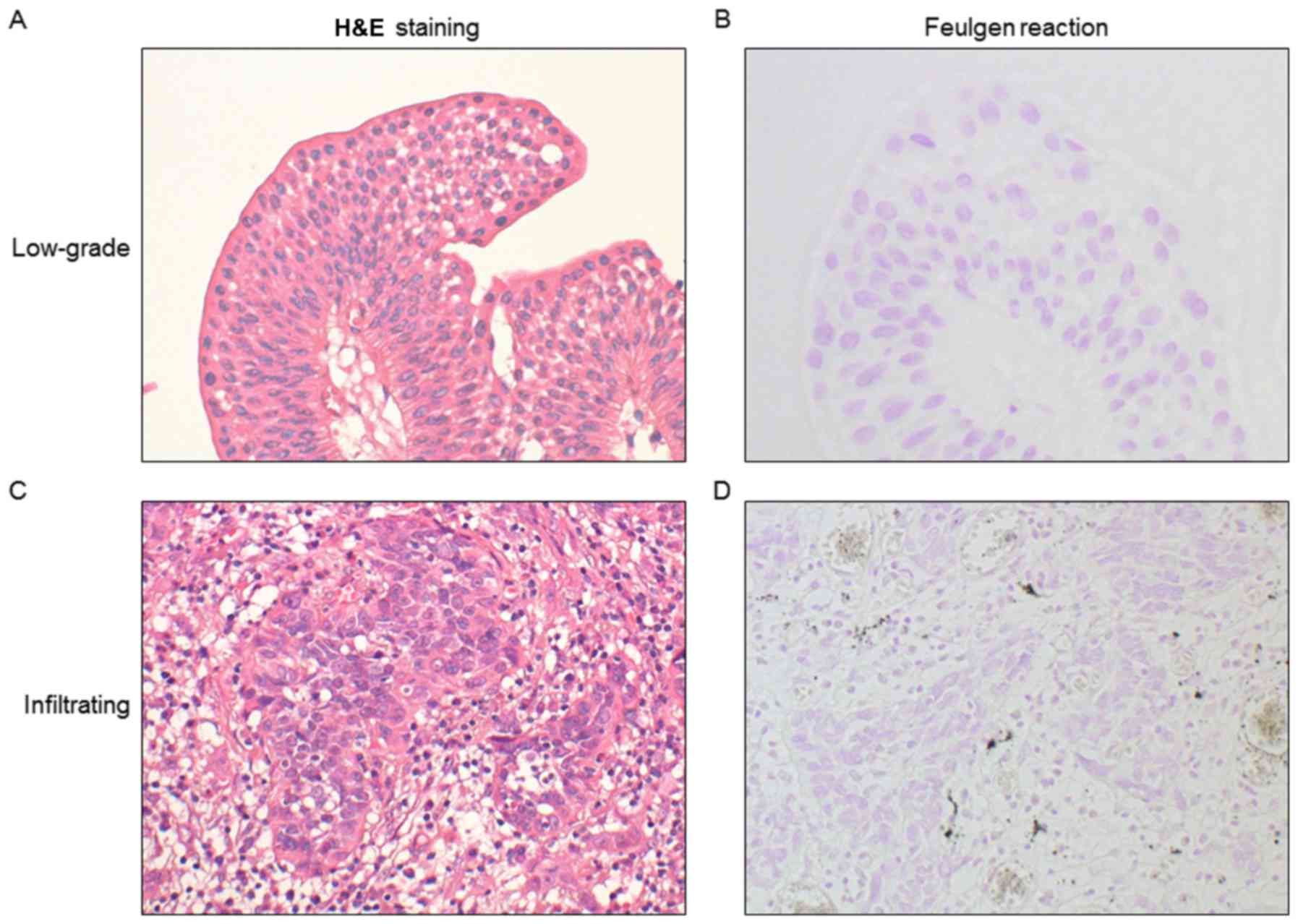
141:151–161. 2017. View Article : Google Scholar : PubMed/NCBI
|
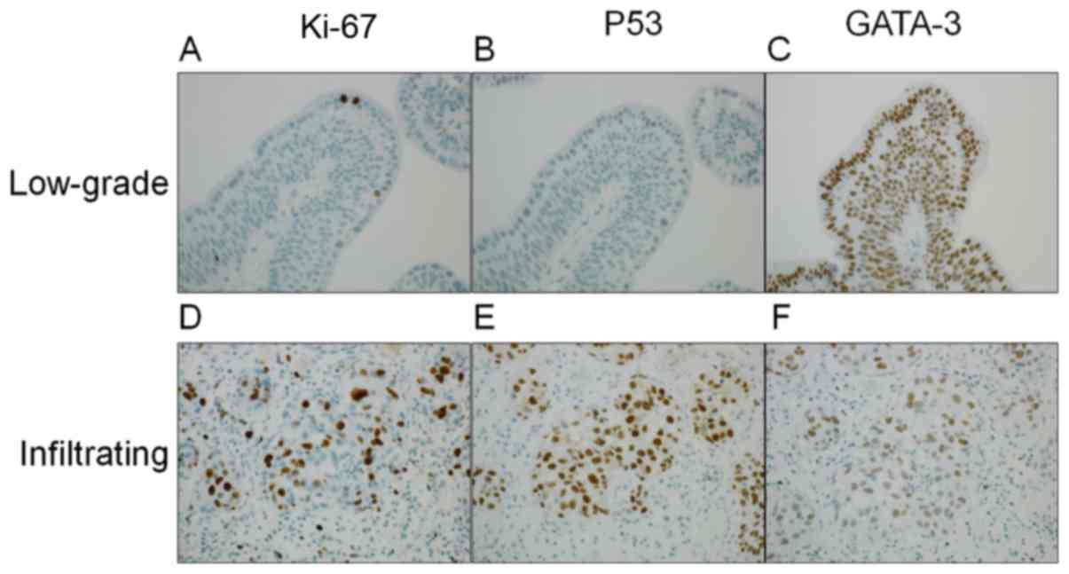
|
3
|
Calvaruso V, Burroughs AK, Standish R,
Manousou P, Grillo F, Leandro G, Maimone S, Pleguezuelo M,
Xirouchakis I, Guerrini GP, et al: Computer-assisted image analysis
of liver collagen: Relationship to Ishak scoring and hepatic venous
pressure gradient. Hepatology. 49:1236–1244. 2009. View Article : Google Scholar : PubMed/NCBI
|
|
4
|
Yamada M, Saito A, Yamamoto Y, Cosatto E,
Kurata A, Nagao T, Tateishi A and Kuroda M: Quantitative nucleic
features are effective for discrimination of intraductal
proliferative lesions of the breast. J Pathol Inform. 7:12016.
View Article : Google Scholar : PubMed/NCBI
|
|
5
|
Hipp J, Smith SC, Cheng J, Tomlins SA,
Monaco J, Madabhushi A, Kunju LP and Balis UJ: Optimization of
complex cancer morphology detection using the SIVQ pattern
recognition algorithm. Anal Cell Pathol (Amst). 35:41–50. 2012.
View Article : Google Scholar : PubMed/NCBI
|
|
6
|
Grignon DJ, Al-Ahmadie H, Algaba F, et al:
Infiltrating urothelial carcinomaWHO Classification of Tumors of
the Urinary Systems and Male Genital Organs. Moch H, Humphrey PA,
Ulbright TM and Reuter VE: IARC; Lyon: pp. 81–98. 2016
|
|
7
|
Reuter VE, Algaba F, Amin MB, et al:
Non-invasive urothelial lesionsWHO Classification of Tumors of the
Urinary Systems and Male Genital Organs. Moch H, Humphrey PA,
Ulbright TM and Reuter VE: IARC; Lyon: pp. 99–107. 2016
|
|
8
|
Loghavi S, Al-Ibraheemi A, Zuo Z,
Garcia-Manero G, Yabe M, Wang SA, Kantarjian HM, Yin CC, Miranda
RN, Luthra R, et al: TP53 overexpression is an independent adverse
prognostic factor in de novo myelodysplastic syndromes with
fibrosis. Br J Haematol. 171:91–99. 2015. View Article : Google Scholar : PubMed/NCBI
|
|
9
|
Fang JC, Xia ZX, Wang CN and Li Z:
Clinicopathologic and immunophenotypic features of primary
intestinal extranodal NK/T-Cell Lymphoma, Nasal Type. Int J Surg
Pathol. 23:609–616. 2015. View Article : Google Scholar : PubMed/NCBI
|
|
10
|
Watanabe G, Ishida T, Furuta A, Takahashi
S, Watanabe M, Nakata H, Kato S, Ishioka C and Ohuchi N: Combined
immunohistochemistry of PLK1, p21, and p53 for predicting TP53
status: An independent prognostic factor of breast cancer. Am J
Surg Pathol. 39:1026–1034. 2015. View Article : Google Scholar : PubMed/NCBI
|
|
11
|
Sobin LH, Gospodarowicz MK and Wittekind
C: TNM Classification of Malignant Tumours. 7th. Wiley-Blackwell;
Hoboken, NJ: 2009
|
|
12
|
Akobeng AK: Understanding diagnostic tests
3: Receiver operating characteristic curves. Acta Paediatr.
96:644–647. 2007. View Article : Google Scholar : PubMed/NCBI
|
|
13
|
Perkins NJ and Schisterman EF: The
inconsistency of ‘optimal’ cutpoints obtained using two criteria
based on the receiver operating characteristic curve. Am J
Epidemiol. 163:670–675. 2006. View Article : Google Scholar : PubMed/NCBI
|
|
14
|
Biesterfeld S, Beckers S, Del Carmen Villa
Cadenas M and Schramm M: Feulgen staining remains the gold standard
for precise DNA image cytometry. Anticancer Res. 31:53–58.
2011.PubMed/NCBI
|
|
15
|
Knowles MA and Hurst CD: Molecular biology
of bladder cancer: New insights into pathogenesis and clinical
diversity. Nat Rev Cancer. 15:25–41. 2015. View Article : Google Scholar : PubMed/NCBI
|
|
16
|
Enache M, Simionescu C and Lascu LC: Ki67
and Bcl-2 immunoexpression in primitive urothelial bladder
carcinoma. Rom J Morphol Embryol. 53:521–525. 2012.PubMed/NCBI
|
|
17
|
Miyamoto H, Izumi K, Yao JL, Li Y, Yang Q,
McMahon LA, Gonzalez-Roibon N, Hicks DG, Tacha D and Netto GJ: GATA
binding protein 3 is down-regulated in bladder cancer yet strong
expression is an independent predictor of poor prognosis in
invasive tumor. Hum Pathol. 43:2033–2040. 2012. View Article : Google Scholar : PubMed/NCBI
|
|
18
|
Rosenthal DL, McLatchie C, Stern E, White
BS and Castleman KR: Endocervical columnar cell atypia coincident
with cervical neoplasia characterized by digital image analysis.
Acta Cytol. 26:115–120. 1982.PubMed/NCBI
|
|
19
|
Stern E, Rosenthal DL, McLatchie C, White
BS and Castleman KR: An expanded cervical cell classification
system validated by automated measurements. Anal Quant Cytol.
4:110–114. 1982.PubMed/NCBI
|
|
20
|
Kriete A, Romen W, Schäffer R, Harms H,
Haucke M, Gerlach B, Aus HM and ter Meulen V: Computer analysis of
chromatin arrangement and nuclear texture in follicular thyroid
tumours. Histochemistry. 78:227–230. 1983. View Article : Google Scholar : PubMed/NCBI
|
|
21
|
Komitowski D and Zinser G: Quantitative
description of chromatin structure during neoplasia by the method
of image processing. Anal Quant Cytol Histol. 7:178–182.
1985.PubMed/NCBI
|
|
22
|
Saito A, Numata Y, Hamada T, Horisawa T,
Cosatto E, Graf HP, Kuroda M and Yamamoto Y: A novel method for
morphological pleomorphism and heterogeneity quantitative
measurement: Named cell feature level co-occurrence matrix. J
Pathol Inform. 7:362016. View Article : Google Scholar : PubMed/NCBI
|
|
23
|
Caie PD, Zhou Y, Turnbull AK, Oniscu A and
Harrison DJ: Novel histopathologic feature identified through image
analysis augments stage II colorectal cancer clinical reporting.
Oncotarget. 7:44381–44394. 2016. View Article : Google Scholar : PubMed/NCBI
|
|
24
|
Ali HR, Dariush A, Provenzano E, Bardwell
H, Abraham JE, Iddawela M, Vallier AL, Hiller L, Dunn JA, Bowden
SJ, et al: Computational pathology of pre-treatment biopsies
identifies lymphocyte density as a predictor of response to
neoadjuvant chemotherapy in breast cancer. Breast Cancer Res.
18:212016. View Article : Google Scholar : PubMed/NCBI
|
|
25
|
Keshtkar A, Keshtkar A and Lawford P:
Cellular morphological parameters of the human urinary bladder
(malignant and normal). Int J Exp Pathol. 88:185–190. 2007.
View Article : Google Scholar : PubMed/NCBI
|
|
26
|
Gschwendtner A and Mairinger T:
Quantitative assessment of bladder carcinoma by acid labile DNA
assay. Cancer. 86:105–113. 1999. View Article : Google Scholar : PubMed/NCBI
|
|
27
|
Krabbe LM, Bagrodia A, Lotan Y, Gayed BA,
Darwish OM, Youssef RF, John G, Harrow B, Jacobs C, Gaitonde M, et
al: Prospective analysis of Ki-67 as an independent predictor of
oncologic outcomes in patients with high grade upper tract
urothelial carcinoma. J Urol. 191:28–34. 2014. View Article : Google Scholar : PubMed/NCBI
|
|
28
|
Shariat SF, Karakiewicz PI, Ashfaq R,
Lerner SP, Palapattu GS, Cote RJ, Sagalowsky AI and Lotan Y:
Multiple biomarkers improve prediction of bladder cancer recurrence
and mortality in patients undergoing cystectomy. Cancer.
112:315–325. 2008. View Article : Google Scholar : PubMed/NCBI
|
|
29
|
Shariat SF, Chade DC, Karakiewicz PI,
Ashfaq R, Isbarn H, Fradet Y, Bastian PJ, Nielsen ME, Capitanio U,
Jeldres C, et al: Combination of multiple molecular markers can
improve prognostication in patients with locally advanced and lymph
node positive bladder cancer. J Urol. 183:68–75. 2010. View Article : Google Scholar : PubMed/NCBI
|
|
30
|
Shariat SF, Ashfaq R, Sagalowsky AI and
Lotan Y: Predictive value of cell cycle biomarkers in nonmuscle
invasive bladder transitional cell carcinoma. J Urol. 177:481–487.
2007. View Article : Google Scholar : PubMed/NCBI
|
|
31
|
Kalantari MR and Ahmadnia H: P53
overexpression in bladder urothelial neoplasms: New aspect of World
Health Organization/International Society of Urological Pathology
classification. Urol J. 4:230–233. 2007.PubMed/NCBI
|
|
32
|
Gao J, Chen YH and Peterson LC: GATA
family transcriptional factors: Emerging suspects in hematologic
disorders. Exp Hematol Oncol. 4:282015. View Article : Google Scholar : PubMed/NCBI
|
|
33
|
Zheng R and Blobel GA: GATA transcription
factors and cancer. Genes Cancer. 1:1178–1188. 2010. View Article : Google Scholar : PubMed/NCBI
|
|
34
|
Asselin-Labat ML, Sutherland KD, Barker H,
Thomas R, Shackleton M, Forrest NC, Hartley L, Robb L, Grosveld FG,
van der Wees J, et al: Gata-3 is an essential regulator of
mammary-gland morphogenesis and luminal-cell differentiation. Nat
Cell Biol. 9:201–209. 2007. View
Article : Google Scholar : PubMed/NCBI
|
|
35
|
Liang Y, Heitzman J, Kamat AM, Dinney CP,
Czerniak B and Guo CC: Differential expression of GATA-3 in
urothelial carcinoma variants. Hum Pathol. 45:1466–1472. 2014.
View Article : Google Scholar : PubMed/NCBI
|
|
36
|
Papathomas TG, Pucci E, Giordano TJ, Lu H,
Duregon E, Volante M, Papotti M, Lloyd RV, Tischler AS, van
Nederveen FH, et al: An international Ki67 reproducibility study in
adrenal cortical carcinoma. Am J Surg Pathol. 40:569–576. 2016.
View Article : Google Scholar : PubMed/NCBI
|