Introduction
Glioma is one of the most common types of primary
brain tumor in adults and represents >50% of all brain tumor
cases (1). Low-grade gliomas (LGGs)
generally referred to the three most common histological subtypes
of World Health Organization (WHO) 2007 grade II gliomas that occur
in the cerebral cortex, diffuse astrocytoma, oligoastrocytoma and
oligodendroglioma, and are not benign tumors (1). LGGs represent a heterogeneous group with
a 2016 WHO classification that includes astrocytomas and
oligodendrogliomas and specific molecular markers (1p/19q
codeletion, IDH1/2 mutation, and histone H3-K27 M mutation)
(2). LGGs are infiltrative and
slow-growing. Following the surgical resection of an LGG, residual
tumor cells at the periphery of the excised tumor core can give
rise to a recurrent tumor, and their natural history often
terminates with transformation into a high-grade glioma (1).
The treatment options for patients with infiltrating
LGGs remain controversial and the prognosis of patients with LGGs
vary. Patient prognosis and the optimal management of patients with
LGGs depend on numerous factors, including the following: Karnofsky
Performance Scale score; presence of preoperative neurological
deficits; presence of seizures; tumor diameter; velocity of tumor
diameter expansion; crossing of the midline of the brain; tumor
contrast enhancement; histological type; and the expression of
molecular markers (3–11). The therapeutic goals are aimed at
improving overall survival (OS) and progression-free survival
(PFS), in addition to minimizing morbidity and maximizing the
quality of life of patients with LGGs. Evidence over the last 2
decades, from the European and American literature, has noted that
the extent of resection in patients with LGGs is an important
factor affecting OS (12–15). However, prospective controlled studies
evaluating the role of surgery are lacking (14–16).
Observation appears to be appropriate for selected patients, and
the wait-and-see approach remains widely practiced and continues to
be a controversial approach (16).
Guidelines exist on the management of patients with LGGs, as
outlined by the European Federation of Neurological Society and the
European Association of Neuro-Oncology task force (17), however, the treatment of patients with
newly diagnosed or suspected LGGs is a controversial area in
neuro-oncology. Since numerous areas of controversy involve the
management at recurrence or evolution (18), the present retrospective study
analyzed the multidisciplinary management of adult patients with
supratentorial recurrent LGGs.
Patients and methods
Patients
Retrospective analyses were performed on the
follow-up (FU) care of 35 adult patients with histologically
confirmed supratentorial LGGs (7 with astrocytoma, 22 with
oligodendroglioma and 6 with oligoastrocytoma; WHO 2007
classification). Patients received their first surgery between
August 2004 and November 2010, and were regularly followed up by a
multidisciplinary neuro-oncological group at the Hôpital Erasme
(Brussels, Belgium). Tumor recurrence/progression was defined
through clinical, radiological and/or metabolic evolution.
Metabolic assessments with positron emission tomography (PET) have
previously been performed for the diagnosis and FU of patients with
LGGs (19).
The present study was approved by the research
ethics board of the Hôpital Erasme (ref Erasme P2016/231;
Université Libre de Bruxelles, Brussels, Belgium). The clinical
data collected included the following: Age, gender, clinical
presentation, contrast enhancement at magnetic resonance imaging
(MRI), MR perfusion, metabolic assessment using PET with C-11
methionine (Met), adjuvant therapies [external beam radiotherapy
(EBRT) and chemotherapy] received, PFS time and OS time.
Statistical analysis
Continuous data are presented as the median and
range. Categorical data are presented as numbers and percentages.
The OS and PFS were calculated using non-parametric Kaplan-Meier
estimates. All statistical analyses were performed using Statistix©
software (version 9.0; Analytical Software, Tallahassee, FL, USA).
P<0.05 was considered to indicate a statistically significant
difference.
Results
In the present study, clinical data from 35 patients
with histologically confirmed supratentorial LGGs (7 astrocytomas,
22 oligodendrogliomas and 6 oligoastrocytomas), who had surgery
between August 2004 and November 2010, were included. Demographic
data are shown in Table I.
 | Table I.Clinicopathological characteristics of
patients with supratentorial recurrent low-grade glioma. |
Table I.
Clinicopathological characteristics of
patients with supratentorial recurrent low-grade glioma.
| Clinicopathological
characteristic | At diagnosis | At recurrence |
|---|
| Number of
patients | 35 | 32 |
| Gender, n |
|
|
| Male | 20 | 18 |
|
Female | 15 | 14 |
| Age, years |
|
|
|
Median | 37 | 39 |
|
Range | 18–78 | 18–78 |
| Primary clinical
presentation, n |
|
|
|
Epilepsy | 30 | 8 |
|
Headache | 3 | 3 |
|
Aphasia | 1 | 2 |
|
Hazard/asymptomatic | 1 | 16 |
|
Neurocognitive impairment | 0 | 2 |
|
Hemiparesis | 0 | 1 |
| Contrast
enhancement at MRI, n |
|
|
|
Yes | 12 | 16 |
| No | 21 | 16 |
|
Unknown | 2 | 0 |
| Perfusion with MRI,
n |
|
|
|
Increased | 4 | 4 |
|
Decreased | 8 | 10 |
|
Unknown | 23 | 18 |
| PET-Met metabolism,
n |
|
|
|
Hyper | 28 | 30 |
|
Hypo | 1 | 2 |
|
Unknown | 6 | 0 |
| Tumor size,
cm3 | |
|
|
Median | 53 | – |
|
Range | 24–86 | – |
| Surgery received,
n |
|
|
|
Yes | 35 | 25 |
| No | 0 | 7 |
| Tumor subtype,
n |
|
|
|
Astrocytoma | 7 | 2 |
|
Oligodendroglioma | 22 | 13 |
|
Oligoastrocytoma | 6 | 4 |
|
Anaplastic | 0 | 6 |
| EBRT received,
n |
|
|
|
Yes | 3 (following
surgery) | 2 (1 following
surgery, 1 without surgery) |
| No | 32 | 30 |
| Chemotherapy
received, n |
|
|
|
Yes | 0 | 5 (2 following
surgery, 3 without surgery) |
| No | 35 | 27 |
In total, 3 patients exhibited no tumor progression
[FU, 70 months; range, 68–97 months; age, 41 years old]. This
non-recurrent population (median age, 41 years) was composed
exclusively of oligodendrogliomas, including 2 with a 1p19q
codeletion (1 undetermined) (Fig. 1).
All 3 patients underwent complete surgical resection of the
LGG.
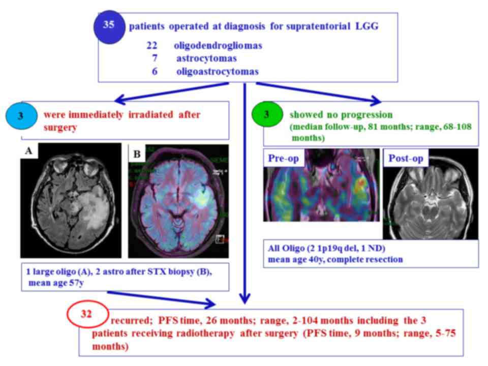 | Figure 1.Patient selection. A total of 32/35
patients who underwent surgical resection for supratentorial LGGs
exhibited tumor recurrence [PFS 26 months (range 2–104 months)]
including the 3 patients receiving radiotherapy after surgery [PFS
9 months (range 5–75 months)], whereas 3 patients exhibited no
tumor progression after median FU 81 months (range 68–108 months).
All 3 patients with no tumor progression were diagnosed with
oligodendroglioma and based on MRI and metabolic exams, these
patients underwent complete resection. The pre-op image is a fusion
image of the PET-Met with MRI, while the post-op image is the MRI
following surgery. A total of 3 patients (mean age, 57 years)
underwent irradiation therapy immediately after surgery. One was a
large oligodendroglioma after patial resection in dominant left
hemisphere (A) and 2 astrocytomas after biopsy with a
hypermetabolic spot as illustrated in (B). LGG, low-grade glioma;
MRI, magnetic resonance imaging; STX, stereotactic brain tumor
biopsy; PFS, progress-free survival; oligo, oligodendroglioma;
astro, astrocytoma; PET, positron emission tomography; met, C-11
methionine. |
Tumor recurrence occurred in the 32 remaining
patients with supratentorial LGGs (PFS, 23 months; range, 2–104)
and included 3 patients at high-risk of tumor recurrence who
received EBRT following surgery (PFS, 9 months; range, 5–75 months;
Fig. 1). The 3 patients at high-risk
of tumor recurrence (median age, 58 years) included 2 patients with
astrocytoma who underwent stereotactic biopsies and one patient
with a large oligodendroglioma who underwent a partial surgery
(Fig. 1).
The diagnosis of tumor recurrence was based on the
following: MRI and metabolic evolution for 13; combined MRI,
metabolic and clinical evolution for 11; metabolic and clinical
evolution for 4; MRI and clinical evolution for 1; metabolic
evolution alone for 2; and clinical evolution alone for 1
patient.
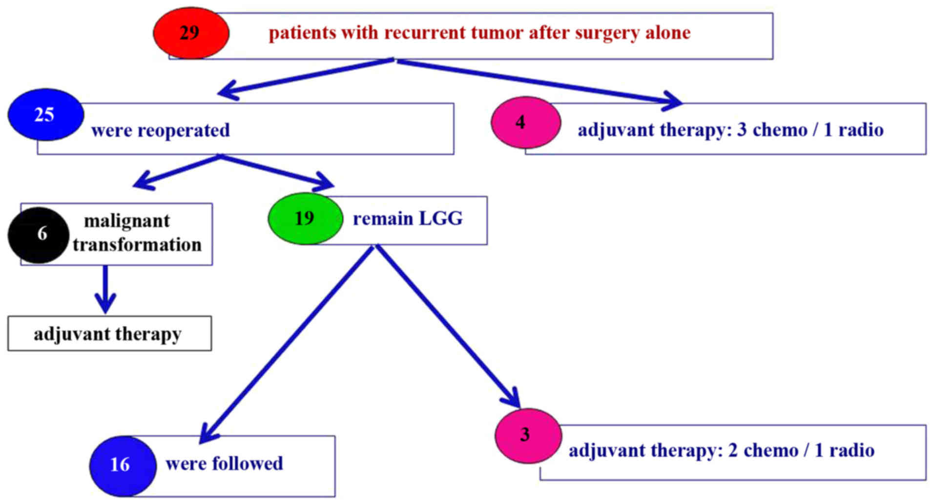
In total, 25 out of the 29 (86%) patients who
received surgery alone at diagnosis underwent reoperation at tumor
recurrence (Fig. 2). As no definite
position is available concerning reoperation at tumor recurrence
[confirmed in a recent review by Nahed et al (18)], the strategy implemented was the
proposal of a new resection once the multidisciplinary team had
concluded that there was evident tumor recurrence in a brain
region, for which the probability of post-surgical morbidity was
low. This strategy is based on several arguments, including the
following: Acquisition of histological and biological information
on the recurrent tumor (with potential impact on further
management), and the reduction of tumor load with a potential
impact on current and future clinical signs, and on the probability
and timing of future anaplastic transformation.
High-grade transformation of tumors occurred in 6/25
(24%) patients who underwent reoperation, 5 of which were
anaplastic tumors, in addition to 1 secary glioblastoma. All 6
patients received adjuvant therapy following reoperation and 3
patients were alive at the FU (FU time, 116 months; range,
114–127); however, 3 patients succumbed (OS, 36 months; range,
15–46 months).
A total of 4 patients diagnosed with tumor
recurrence received adjuvant therapy without a second surgery
(4/29, 14%). Furthermore, 3 of these patients received
chemotherapy, and 1 patient received EBRT (Fig. 2). In addition, 3/19 (16%) patients
diagnosed with recurrent LGGs following reoperation received
adjuvant therapy; 2 of these patients received chemotherapy and 1
patient received EBRT. The 16 patients who did not receive adjuvant
therapy were regularly followed via multimodal imaging (Fig. 2). The patients (n=7) who received
adjuvant therapy with (n=3) or without (n=4) a second surgery had a
median age of 38 years and a large tumor volume (median diameter,
>61 mm; range, 55–83 mm). All 5 patients who received
chemotherapy were diagnosed with oligodendroglioma, including 3
patients with chromosome 1p19q codeletion. The median PFS time
prior to adjuvant therapy was 5 months for patients who received
radiotherapy and 23 months for patients who received
chemotherapy.
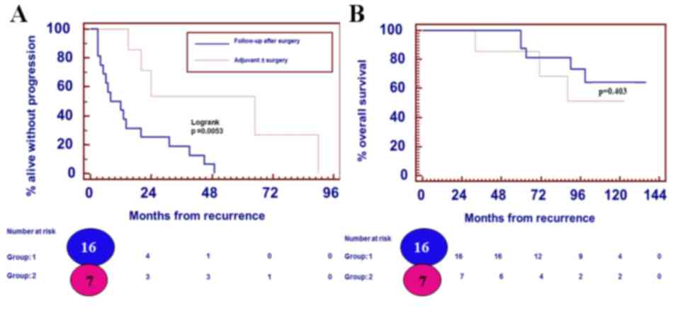
A significant difference was identified between the
PFS time for patients reoperated on with close FU (n=16) and the
PFS time for patients receiving adjuvant therapy with or without
surgery (n=7) at first recurrence, at 10 and 24 months,
respectively (P=0.005) (Fig. 3). No
significant difference was observed for OS time (P=0.403; Fig. 3).
At the time of writing the present study, 22/35 of
the included patients were alive following a median post-diagnosis
FU time of 109 months (range, 55–136 months).
Discussion
In the present study, 3 patients (3/35) exhibited no
tumor progression following the initial surgical resection. This
non-recurrent population (mean age, 40 years) was exclusively
composed of oligodendrogliomas, including 2 with 1p19q codeletion
after complete surgical resection. This indicates the importance of
maintaining a good quality of life for patients with LGGs who
experience long periods of stable disease (20).
In the present study, 3 patients who presented with
a high risk of tumor recurrence (2 patients with astrocytoma who
underwent stereotactic biopsy and 1 patient with a large
oligodendroglioma who underwent partial surgery) received EBRT
following surgery. The definition of low- vs. high-risk patients,
including the cut-off age of 40 years, varies across studies
(4,9).
To the best of our knowledge, there is no evidence demonstrating
that immediate (post-operative) EBRT is advantageous in improving
the OS time of patients with LGGs compared with deferred
radiotherapy (21,22). The PFS of patients with LGGs is
lengthened; however, the optimal timing for receiving EBRT remains
debatable (21,22). Based on 19 years of experience,
Youland et al (23) suggested
that immediate postoperative EBRT should be implemented only for
high-risk patients. It appears to be safe to delay EBRT in those
patients in which it is possible to do so (23). A recent retrospective study revealed
that upfront radiation was associated with an improvement in the
PFS (time) of patients with LGGs, but also with a significantly
decreased OS (time) and an increased rate of malignant degeneration
(24). These findings indicate the
limitations of retrospective studies with a small cohort and should
be interpreted with caution.
The long-term FU of the Radiation Therapy Oncology
Group 9802 clinical trial demonstrated a 5.5-year improvement in
the median OS following EBRT+adjuvant procarbazine, CCNU and
vincristine (PCV) chemotherapy compared with EBRT alone in patients
with high-risk LGGs (25). While an
analysis of the methods to determine the therapies best suited to
individual patients according to tumor type and molecular profile
is not currently available, EBRT+PCV remains the standard procedure
for the treatment of patients with LGGs who require postsurgical
adjuvant therapy (26). The optimal
parameter for selecting patients to receive adjuvant PCV and the
role of temozolomide (TMZ)-based chemotherapy remains unclear
(27). The European Organization for
the Research and Treatment of Cancer C2203 clinical trial
investigated the effects of TMZ-based chemotherapy vs. EBRT in
patients with LGGs. In this phase III clinical trial, patients were
randomized to receive 12 cycles of TMZ-based chemotherapy or EBRT
following stratification for genetic 1p loss. The preliminary
results identified no significant difference between patients who
received TMZ compared with patients who received EBRT (median PFS,
40 vs. 47 months) (28). Patients
with 1p-intact LGGs showed a trend towards worse PFS with TMZ. The
preliminary results of the Radiation Therapy Oncology Group 0424
clinical trial, a single-arm phase II study investigating the
effects of concurrent and adjuvant TMZ with 54 Gy of EBRT in
patients with LGGs suggested that TMZ improves survival in
high-risk patients compared with a historical control group that
received EBRT alone (29). Further
studies investigating the effects of adjuvant TMZ compared with
adjuvant PCV following EBRT are warranted. In addition, studies are
required investigating the additional benefit of EBRT and
chemotherapy agents in light of our new understanding of molecular
markers.
In the present study, tumor recurrence occurred in
32/35 patients (PFS time, 26 months; range, 2–104 months),
including 3 patients who received EBRT following surgery (PFS, 9
months; range, 5–75 months). Reoperation was performed on 25/29
(86%) patients who received surgery alone. The data on the effect
of reoperation on the OS and PFS times in patients with LGGs is
limited. The results of a recent study on 52 patients with LGGs who
underwent reoperation demonstrated that the extent of surgical
re-resection remains the most efficient predictor of OS (24). A recent systematic review by Nahed
et al (18) suggested that
there is insufficient evidence to make specific recommendations for
surgery at the time of tumor recurrence. However, reoperation
appears to be an efficacious treatment for recurrent grade II
glioma even in eloquent areas (30).
In the present study, 4 patients (4/29, 14%)
received adjuvant therapy without a second surgery (3 chemotherapy
and 1 EBRT), and only 3 received adjuvant therapy (2 chemotherapy
and 1 EBRT) following the second surgery. All 5 patients who
received chemotherapy were diagnosed with oligodendroglioma
including 3 patients with chromosome 1p19q codeletions. The
majority of patients with recurrent LGGs following the second
surgery were followed through MRI and PET (16/19 remaining LGGs at
the second surgery).
Whether EBRT with second-line chemotherapy delivered
at the time of relapse can provide the same survival advantage as
first-line chemotherapy delivered with EBRT is yet unknown.
Furthermore, long-term FU with investigations into the quality of
life and cognition of patients with LGGs is warranted for this
‘chronic’ disease.
In the present study, malignant transformation was
identified in 24% of the tumors following the second surgery (6/25
patients who did not received other intervening therapy). The
incidence of malignant tumor transformation in several clinical
series has ranged between 13 and 86%. However, in the majority of
these studies, malignant transformation has not been well reported.
In addition, a number of these patients received EBRT and/or
chemotherapy, which may have interfered with histological
interpretation and confounded the natural progression of the LGG
(31). The results of a study by
Schmidt et al (31) revealed
that 50% of patients with an initial diagnosis of infiltrative LGGs
exhibited progression in terms of grade of malignancy by the time
of reoperation.
In the present study, the PFS times of reoperated
patients with close FU (n=16) and patients receiving adjuvant
therapy with or without surgery (n=7) at first recurrence were 10
and 24 months, respectively. However, no significant difference was
determined with regard to the OS time. This could be explained by
the fact that patients who received adjuvant therapy were
considered high-risk patients with an expected shorter survival
time. In addition, adjuvant therapies, including EBRT with
sublethal doses of irradiation close to the vicinity of the
irradiated target, select a subpopulation of resistant and more
aggressive cells, as demonstrated by Wild-Bode et al
(32). It has not yet been
established whether immediate post-radiation alkylating
chemotherapy will possess the same benefit in patients previously
treated with single modality alkylating chemotherapy, since
previously treated tumors may have acquired resistance (25).
Since writing the present study, the majority of
patients have remained alive (22/35) following a median FU time of
109 months (range, 55–136 months). The outcome data from the LGG
patients who underwent multiple surgeries continues to be
assessed.
The limitations of this study were its retrospective
nature and the lack of a uniform FU evaluation (such as volumetric
analysis, perfusion and metabolic assessments) across a small
cohort, and the fact that molecular profiles were not available for
the entire cohort during the study period.
Based on retrospective studies suggesting that
patients who undergo early, extensive and maximal tumor resections
have an improved survival, the use of radical surgery is frequently
supported (12–15,30).
Prospective studies investigating the role of surgery are lacking,
and the presumed benefit from extensive resection may be largely
due to the patient selection process. A biopsy is performed to
pathologically diagnose patients with LGGs in circumstances where
the tumor location makes surgery difficult or impossible (16).
Gozé et al (6)
reported that patients with a fast velocity of diametric expansion
may be considered as ‘high-risk’, with an increased chance of early
malignant transformation, particularly in those cases where a
single biopsy has been performed due to the risk of undergrading.
As there are common outcomes between LGGs and malignant gliomas,
treatment modalities should be selected accordingly, and a 3-month
FU period is preferred (33).
In conclusion, reoperation for the majority of
patients with recurrent LGGs is preferred at the Hôpital Erasme,
and is primarily motivated by the high level of malignant
transformation observed. In addition, a second surgery aids in the
accurate molecular characterization of LLGs. As a better
understanding of the molecular biology of all types of brain tumor
is gained, molecular signatures, longitudinal evaluation, and
improved preclinical modeling and imaging technology may aid in the
improvement of therapeutic strategies (34). The results of the present study may
change the multidisciplinary approach into a more aggressive
approach using adjuvant therapy with or without surgery for the
treatment of a selected subpopulation of patients with LGGs at the
instance of first recurrence.
As emphasized by Zadeh et al (16), the neuro-oncological community must
continue to develop improved treatments for patients with LGGs,
using further clinical trials and studies on better-targeted
therapeutics. In addition, future directions should include the
neurocognitive assessment of patients with LGGs with prolonged OS
times.
References
|
1
|
Louis DN, Ohgaki H, Wiestler OD and
Cavenee WK: WHO Classification of Tumours of the Central Nervous
System. IARC Press; Lyon, France: 2007
|
|
2
|
Louis DN, Perry A, Reifenberger G, von
Deimling A, Figarella-Branger D, Cavenee WK, Ohgaki H, Wiestler OD,
Kleihues P and Ellison DW: The 2016 World Health Organization
classification of tumors of the central nervous system: A summary.
Acta Neuropathol. 131:803–820. 2016. View Article : Google Scholar : PubMed/NCBI
|
|
3
|
Chang EF, Smith JS, Chang SM, Lamborn KR,
Prados MD, Butowski N, Barbaro NM, Parsa AT, Berger MS and
McDermott MM: Preoperative prognostic classification system for
hemispheric low-grade gliomas in adults. J Neurosurg. 109:817–824.
2008. View Article : Google Scholar : PubMed/NCBI
|
|
4
|
Daniels TB, Brown PD, Felten SJ, Wu W,
Buckner JC, Arusell RM, Curran WJ, Abrams RA, Schiff D and Shaw EG:
Validation of EORTC prognostic factors for adults with low-grade
glioma: A report using intergroup 86-72-51. Int J Radiat Oncol Biol
Phys. 81:218–224. 2011. View Article : Google Scholar : PubMed/NCBI
|
|
5
|
Gorlia T, Wu W, Wang M, Baumert BG, Mehta
M, Buckner JC, Shaw E, Brown P, Stupp R, Galanis E, et al: New
validated prognostic models and prognostic calculators in patients
with low-grade gliomas diagnosed by central pathology review: A
pooled analysis of EORTC/RTOG/NCCTG phase III clinical trials.
Neuro Oncol. 15:1568–1579. 2013. View Article : Google Scholar : PubMed/NCBI
|
|
6
|
Gozé C, Blonski M, Le Maistre G, Bauchet
L, Dezamis E, Page P, Varlet P, Capelle L, Devaux B, Taillandier L,
et al: Imaging growth and isocitrate dehydrogenase 1 mutation are
independent predictors for diffuse low-grade gliomas. Neuro Oncol.
16:1100–1109. 2014. View Article : Google Scholar : PubMed/NCBI
|
|
7
|
Koekkoek JA, Dirven L, Heimans JJ, Postma
TJ, Vos MJ, Reijneveld JC and Taphoorn MJ: Seizure reduction in a
low-grade glioma: More than a beneficial side effect of
temozolomide. J Neurol Neurosurg Psychiatry. 86:366–373. 2015.
View Article : Google Scholar : PubMed/NCBI
|
|
8
|
Pallud J, Capelle L, Taillandier L,
Fontaine D, Mandonnet E, Guillevin R, Bauchet L, Peruzzi P,
Laigle-Donadey F, Kujas M, et al: Prognostic significance of
imaging contrast enhancement for WHO grade II gliomas. Neuro Oncol.
11:176–182. 2009. View Article : Google Scholar : PubMed/NCBI
|
|
9
|
Pignatti F, van den Bent M, Curran D,
Debruyne C, Sylvester R, Therasse P, Afra D, Cornu P, Bolla M,
Vecht C, et al: Prognostic factors for survival in adult patients
with cerebral low-grade glioma. J Clin Oncol. 20:2076–2084. 2002.
View Article : Google Scholar : PubMed/NCBI
|
|
10
|
Sanson M, Marie Y, Paris S, Idbaih A,
Laffaire J, Ducray F, El Hallani S, Boisselier B, Mokhtari K,
Hoang-Xuan K and Delattre JY: Isocitrate dehydrogenase 1 codon 132
mutation is an important prognostic biomarker in gliomas. J Clin
Oncol. 27:4150–4154. 2009. View Article : Google Scholar : PubMed/NCBI
|
|
11
|
Weiler M and Wick W: Molecular predictors
of outcome in low-grade glioma. Curr Opin Neurol. 25:767–773. 2012.
View Article : Google Scholar : PubMed/NCBI
|
|
12
|
Berger MS, Deliganis AV, Dobbins J and
Keles GE: The effect of extent of resection on recurrence in
patients with low grade cerebral hemisphere gliomas. Cancer.
74:1784–1791. 1994. View Article : Google Scholar : PubMed/NCBI
|
|
13
|
Gousias K, Schramm J and Simon M: Extent
of resection and survival in supratentorial infiltrative low-grade
gliomas: Analysis of and adjustment for treatment bias. Acta
Neurochir (Wien). 156:327–337. 2014. View Article : Google Scholar : PubMed/NCBI
|
|
14
|
Jakola AS, Myrmel KS, Kloster R, Torp SH,
Lindal S, Unsgård G and Solheim O: Comparison of a strategy
favoring early surgical resection vs a strategy favoring watchful
waiting in low-grade gliomas. JAMA. 308:1881–1888. 2012. View Article : Google Scholar : PubMed/NCBI
|
|
15
|
Smith JS, Chang EF, Lamborn KR, Chang SM,
Prados MD, Cha S, Tihan T, Vandenberg S, McDermott MW and Berger
MS: Role of extent of resection in the long-term outcome of
low-grade hemispheric gliomas. J Clin Oncol. 26:1338–1345. 2008.
View Article : Google Scholar : PubMed/NCBI
|
|
16
|
Zadeh G, Khan OH, Vogelbaum M and Schiff
D: Much debated controversies of diffuse low-grade gliomas. Neuro
Oncol. 17:323–326. 2015. View Article : Google Scholar : PubMed/NCBI
|
|
17
|
Soffietti R, Baumert BG, Bello L, von
Deimling A, Duffau H, Frénay M, Grisold W, Grant R, Graus F,
Hoang-Xuan K, et al: Guidelines on management of low-grade gliomas:
Report of an EFNS-EANO task force. Eur J Neurol. 17:1124–1133.
2010. View Article : Google Scholar : PubMed/NCBI
|
|
18
|
Nahed BV, Redjal N, Brat DJ, Chi AS, Oh K,
Batchelor TT, Ryken TC, Kalkanis SN and Olson JJ: Management of
patients with recurrence of diffuse low grade glioma: A systematic
review and evidence-based clinical practice guideline. J
Neurooncol. 125:609–630. 2015. View Article : Google Scholar : PubMed/NCBI
|
|
19
|
Nihashi T, Dahabreh IJ and Terasawa T:
Diagnostic accuracy of PET for recurrent glioma diagnosis: A
meta-analysis. AJNR Am J Neuroradiol. 34(944–950): S1–S11.
2013.PubMed/NCBI
|
|
20
|
Boele FW, Douw L, Reijneveld JC, Robben R,
Taphoorn MJ, Aaronson NK, Heimans JJ and Klein M: Health-related
quality of life in stable, long-term survivors of low-grade glioma.
J Clin Oncol. 33:1023–1029. 2015. View Article : Google Scholar : PubMed/NCBI
|
|
21
|
Suneja G, Alonso-Basanta M, Lustig R, Lee
JY and Bekelman JE: Postoperative radiation therapy for low-grade
glioma: Patterns of care between 1998 and 2006. Cancer.
118:3735–3742. 2012. View Article : Google Scholar : PubMed/NCBI
|
|
22
|
van den Bent MJ, Afra D, de Witte O, Ben
Hassel M, Schraub S, Hoang-Xuan K, Malmström PO, Collette L,
Piérart M, Mirimanoff R, et al: Long-term efficacy of early versus
delayed radiotherapy for low-grade astrocytoma and
oligodendroglioma in adults: The EORTC 22845 randomised trial.
Lancet. 366:985–990. 2005. View Article : Google Scholar
|
|
23
|
Youland RS, Brown PD, Giannini C, Parney
IF, Uhm JH and Laack NN: Adult low-grade glioma: 19-year experience
at a single institution. Am J Clin Oncol. 36:612–619. 2013.
View Article : Google Scholar : PubMed/NCBI
|
|
24
|
Ramakrishna R, Hebb A, Barber J, Rostomily
R and Silbergeld D: Outcomes in reoperated low-grade gliomas.
Neurosurgery. 77:175–184. 2015. View Article : Google Scholar : PubMed/NCBI
|
|
25
|
Buckner JC, Shaw EG, Pugh SL, Chakravarti
A, Gilbert MR, Barger GR, Coons S, Ricci P, Bullard D, Brown PD, et
al: Radiation plus Procarbazine, CCNU, and vincristine in low-grade
glioma. N Engl J Med. 374:1344–1355. 2016. View Article : Google Scholar : PubMed/NCBI
|
|
26
|
van den Bent MJ: Practice changing mature
results of the RTOG study 9802: Another positive PCV trial makes
adjuvant chemotherapy part of standard of care in low-grade glioma.
Neuro Oncol. 16:1570–1574. 2014. View Article : Google Scholar : PubMed/NCBI
|
|
27
|
Laack NN, Sarkaria JN and Buckner JC:
Radiation therapy oncology group 9802: Controversy or consensus in
the treatment of newly diagnosed low-grade glioma? Semin Radiat
Oncol. 25:197–202. 2015. View Article : Google Scholar : PubMed/NCBI
|
|
28
|
Baumert B, Mason W, Ryan G, Bromberg JE,
Van Den Bent MJ, Xuan KH, Brandes AA, Kantor G, Taphoom MJ, Hassel
MB, et al: Temozolomide chemotherapy versus radiotherapy in
molecularly characterized (1p loss) low-grade glioma: A randomized
phase III intergroup study by the EORTC/NCIC-CTG/TROG/MRC-CTU
(EORTC 22033–26033). J Clin Oncol. 31:(Suppl). 2007.
|
|
29
|
Fisher BJ, Hu C, Macdonald DR, Lesser GJ,
Coons SW, Brachman DG, Ryu S, Werner-Wasik M, Bahary JP, Liu J, et
al: Phase 2 study of temozolomide-based chemoradiation therapy for
high-risk low-grade gliomas: Preliminary results of radiation
therapy oncology group 0424. Int J Radiat Oncol Biol Phys.
91:497–504. 2015. View Article : Google Scholar : PubMed/NCBI
|
|
30
|
Martino J, Taillandier L, Moritz-Gasser S,
Gatignol P and Duffau H: Re-operation is a safe and effective
therapeutic strategy in recurrent WHO grade II gliomas within
eloquent areas. Acta Neurochir (Wien). 151:427–436. 2009.
View Article : Google Scholar : PubMed/NCBI
|
|
31
|
Schmidt MH, Berger MS, Lamborn KR, Aldape
K, McDermott MW, Prados MD and Chang SM: Repeated operations for
infiltrative low-grade gliomas without intervening therapy. J
Neurosurg. 98:1165–1169. 2003. View Article : Google Scholar : PubMed/NCBI
|
|
32
|
Wild-Bode C, Weller M, Rimner A, Dichgans
J and Wick W: Sublethal irradiation promotes migration and
invasiveness of glioma cells: Implications for radiotherapy of
human glioblastoma. Cancer Res. 61:2744–2750. 2001.PubMed/NCBI
|
|
33
|
Pallud J, Blonski M, Mandonnet E, Audureau
E, Fontaine D, Sanai N, Bauchet L, Peruzzi P, Frénay M, Colin P, et
al: Velocity of tumor spontaneous expansion predicts long-term
outcomes for diffuse low-grade gliomas. Neuro Oncol. 15:595–606.
2013. View Article : Google Scholar : PubMed/NCBI
|
|
34
|
Huse JT, Wallace M, Aldape KD, Berger MS,
Bettegowda C, Brat DJ, Cahill DP, Cloughesy T, Haas-Kogan DA, Marra
M, et al: Where are we now? And where are we going? A report from
the Accelerate Brain Cancer Cure (ABC2) low-grade glioma research
workshop. Neuro Oncol. 16:173–178. 2014. View Article : Google Scholar : PubMed/NCBI
|