Introduction
Cancer is currently the second leading cause of
human mortalities worldwide and is expected to surpass heart
diseases as the leading cause of human mortality in the next few
years. Esophageal cancer is the seventh leading cause of human
cancer-associated mortality worldwide (1). Esophageal squamous cell carcinoma (ESCC)
is the most major histological type of esophageal cancer. In China,
the annual incidence and mortality rates of esophageal carcinoma
rank fifth and fourth, respectively (2). The majority of patients with ESCC are
diagnosed at an advanced stage of the disease, despite the
development of rapid advances in surgical techniques, radiotherapy
and chemotherapy, and the 5-year survival rate remains poor
(between 10 and 30%) (3). ESCC is
hypothesized to develop due to risk factors including the
accumulation of genetic mutations, tobacco, alcohol consumption,
consuming hot food or water frequently, a low intake of fresh fruit
and vegetables, obesity and poor diet, local environmental
conditions and lifestyle (4). There
are a limited number of specific biomarkers available for
diagnostic use and targeted therapies against ESCC. Therefore,
identifying biomarkers which may be used for early detection,
prognostic stratification and novel therapeutic interventions are
required to effectively treat patients with ESCC.
Melanoma-associated antigens (MAGEs) are a group of
well-characterized members of the cancer/testis antigen (CTA)
family, which are expressed in a number of types of malignant
tumors, including hepatocellular carcinoma, breast cancer, renal
carcinoma and non-small cell lung cancer (5–8). MAGE-A is
a subfamily of MAGEs and MAGE-A is highly homologous with the MAGE
gene on chromosome X in CTAs (9). In
addition, MAGE-A genes have been identified to be expressed at
increased levels in a number of types of malignant tumor, but are
rarely expressed in healthy tissues (10). The biological functions of MAGE genes
remain unknown, therefore MAGE-A may be identified as a target for
cancer immunotherapy. MAGE-A9 has been studied in a number of types
of cancer, including hepatocellular carcinoma, breast cancer, renal
carcinoma, non-small cell lung cancer and laryngeal squamous cell
carcinoma (11–14). MAGE-A9 has been associated with tumor
recurrence and progression in bladder cancer (15). The results of the present study
revealed that MAGE-A9 was involved in the progress of malignant
tumors, but the association between MAGE-A9 expression and the
clinicopathological outcome in ESCC remains unknown.
In the present study, the expression level of
MAGE-A9 protein and mRNA in ESCC and normal tissues was determined
using immunohistochemistry (IHC) and the quantitative polymerase
chain reaction (qPCR). Furthermore, the association between MAGE-A9
expression and the clinicopathological features observed in
patients with ESCC, and the prognostic significance, were
analyzed.
Materials and methods
Patient specimens
A total of 103 paraffin-embedded ESCC tissues and 30
healthy tissue samples were collected from the archives of the
Department of Pathology at The First Affiliated Hospital of
Zhengzhou University (Zhengzhou, China), between August 2011 and
January 2012. All patients were staged in accordance with the
tumor-node-metastasis classification system (Union for
International Cancer Control 2009) (16). Prior to surgery, none of the patients
had received neoadjuvant chemotherapy, radiation therapy or
immunotherapy. Clinical data, including sex, age, smoking history,
alcohol, tumor size, pathological grade, lymph node metastasis and
the degree of infiltration, were collected. The present study was
approved by the Ethics Committee of The First Affiliated Hospital
of Zhengzhou University (Zhengzhou, China).
Tissue microarray (TMA) construction
and IHC analysis
A total of 54 ESCC tissues and 30 healthy tissues
were collected, and TMAs were produced by the Department of
Clinical Pathology, The First Affiliated Hospital of Zhengzhou
University (Zhengzhou, China). The TMA was sectioned (thickness, 4
um) and incubated at 60°C for 3 h. Slides were subsequently
deparaffinized with xylene, rehydrated in an ethanol series,
submerged in 0.01 M citrate buffer (pH, 6.0) antigen retrieval
buffer and microwaved for antigenic retrieval. Tissues were blocked
with 3% H2O2 for 20 min at room temperature,
followed by incubation with goat serum for 20 min at room
temperature. Subsequently, samples were incubated at 4°C with human
anti-MAGEA9 immunoglobulin G antibody (catalog no. ab38493;
dilution, 1:50; Abcam, Cambridge, UK) in a dark box overnight,
followed by incubation at 37°C with a biotinylated anti-rabbit
secondary antibody (catalog no. ab150077; dilution, 1:50; Abcam)
for 20 min. As a negative control, samples were incubated with PBS
instead of a specific antibody. The sections were incubated with
biotinylated horseradish peroxidase-labeled streptavidin. The
reaction was revealed using 3,3-diaminobenzidine, and finally the
sections were stained with hematoxylin and incubated at room
temperature for 4 min. Results were determined using a light
microscope (magnification, ×40, ×200 and ×400). The proportion of
MAGE-A9-positive cells was scored as follows: 0, 0%; 1, <30%; 2,
between 30 and 70%; and 3, >70%. In addition, the intensity of
MAGE-A9 staining was scored as follows: 0, negative staining; 1,
yellow staining; 2, light brown staining; and 3, brown staining.
The two scores were multiplied and used as the final staining
score. The degree of cytoplasmic MAGE-A9 staining was quantified
using a two-level grading system and staining scores were defined
as follows: 0–1, no expression; 2–4, low expression; and 5–9, high
expression.
Reverse transcription (RT)-qPCR
analysis
A total of 20 ESCC tissue samples and 20
corresponding healthy tissues were collected. Total RNA was
extracted from tissues using TRIzol reagent (Thermo Fisher
Scientific, Inc., Waltham, MA, USA), according to the manufacturers
protocol, for performing RT-PCR and qPCR analysis. Total RNA (4 µg)
was reverse-transcribed into cDNA using RevertAid First Strand cDNA
Synthesis (Thermo Fisher Scientific, Inc.), according to the
manufacturers protocol. qPCR was performed using the Quantitative
SYBR Green PCR kit (Thermo Fisher Scientific, Inc.) on an ABI PRISM
7500 Fast system (Applied Biosystems; Thermo Fisher Scientific,
Inc.). The primers used were as follows: MAGE-A9 forward primer,
5-CAC TGT ATG TCA TCT CTG-3; reverse primer, 5-ACT ACT GTCATT CAT
TAA CT-3; GADPH, used as a reference gene, forward primer, 5-GTC
ATC ATG TCT CTC GAG-3; reverse primer, 5-AAC TCA GCC ACC TTC AA-3.
qPCR amplification was conducted using the SYBR Green Master Mix
(Takara Bio, Inc., Otsu, Japan). The reaction mixtures were
analyzed in an ABI PRISM 7500 Fast Real-Time PCR system. The
thermocycling conditions were as follows: 95°C for 30 sec followed
by 40 cycles of 95°C for 5 sec and 60°C for 30 sec. All samples
were analyzed for 3 parallel samples in triplicate. The relative
expression of the target gene was calculated using
2−ΔΔCq values (17).
Statistical analysis
All statistical analyses were conducted using
PrismDemo (version 5.0; GraphPad Software, Inc., La Jolla, CA, USA)
and SPSS (version 17.0; SPSS, Inc., Chicago, IL, USA). The MAGE-A9
mRNA expression in ESCC tissues, compared with matched healthy
tissues, was analyzed using the Wilcoxon signed rank non-parametric
test. The significance of MAGE-A9 protein expression on
clinicopathological parameters of ESCC was determined using the
χ2 test. The Kaplan-Meier estimator survival method was
used to evaluate the associations between MAGE-A9 expression and
the outcome for patients with ESCC. Coxs proportional hazards
regression models was used to determine factors that were
independently associated with overall survival rates. For all
tests, P<0.05 was considered to indicate a statistically
significant difference.
Results
Determination of MAGE-A9 protein
expression in ESCC using IHC
IHC was used to determine the expression levels of
MAGE-A9. Increased MAGE-A9 expression was determined in 57/103
(55.34%) ESCC tissues and 4/30 (13.3%) non-cancerous healthy
tissues. A significant difference was determined in the increased
expression levels of MAGE-A9 protein between ESCC tissues and
healthy tissues (P<0.05). MAGE-A9 positive staining was
predominantly localized in the cytoplasm of cancer cells. The
typical ESCC staining for MAGE-A9 expression in ESCC is presented
in Fig. 1. The association between
MAGE-A9 expression and the clinicopathological characteristics of
103 patients with ESCC is presented in Table I. Increased MAGE-A9 expression levels
were associated with the pathological grade (P=0.008), tumor size
(P=0.027) and lymph node metastasis (P=0.009). In comparison, no
significant associated was discovered between MAGE-A9 expression
and other clinical characteristics, including sex, age, smoking
status, alcohol consumption, Tumor-Node-Metastasis stage (16) and histopathological grade (Table I).
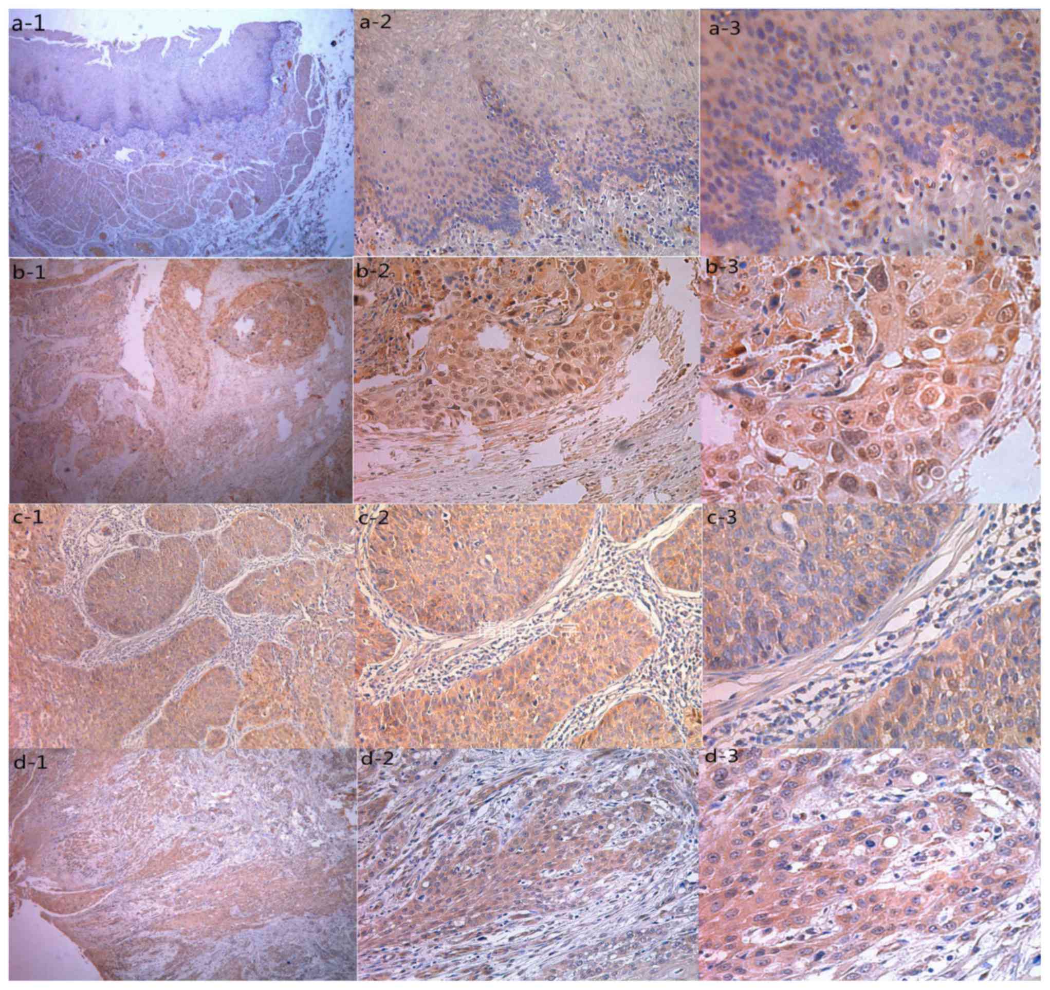 | Figure 1.Representative pattern of MAGE-A9
protein expression in ESCC and normal tissues in a tissue
microarray. (A) Negative IHC staining of MAGE-A9 of non-cancerous
tissue (magnifications: 1, ×40; 2, ×200; 3, ×400). (B) IHC staining
of MAGE-A9 of well-differentiated ESCC tissue (magnifications: 1,
×40; 2, ×200; 3, ×400). (C) High IHC staining of MAGE-A9 of
moderately-poorly differentiated ESCC tissue (magnifications: 1,
×40; 2, ×200; 3, ×400). (D) IHC staining of MAGE-A9 of
well-differentiated ESCC sample (magnifications: 1, ×40; 2, ×200;
3, ×400). MAGE-A9, melanoma-associated antigen-9; ESCC, esophageal
squamous cell carcinoma; IHC, immunohistochemistry. |
 | Table I.Association between increased MAGE-A9
expression and the clinicopathological characteristics in
esophageal squamous cell carcinoma. |
Table I.
Association between increased MAGE-A9
expression and the clinicopathological characteristics in
esophageal squamous cell carcinoma.
|
|
| MAGE-A9 |
|
|
|
|---|
|
|
|
|
|
|
|
|---|
| Group | n | + | % | χ2 | P-value |
|---|
| Total | 103 | 57 | 55.3 |
|
|
| Sex |
|
|
|
|
|
| Male | 75 | 45 | 60.0 | 2.424 | 0.119 |
|
Female | 28 | 12 | 42.9 |
|
|
| Age, years |
|
|
|
|
|
| ≤60 | 42 | 24 | 57.1 | 0.093 | 0.917 |
|
>60 | 61 | 33 | 54.1 |
|
|
| Smoking |
|
|
|
|
|
| No | 51 | 25 | 49.0 | 1.633 | 0.201 |
| Yes | 52 | 32 | 61.5 |
|
|
| Alcohol |
|
|
|
|
|
| No | 81 | 44 | 54.3 | 0.159 | 0.690 |
| Yes | 22 | 13 | 59.1 |
|
|
| Tumor size, cm |
|
|
|
|
|
| ≤3 | 48 | 21 | 43.8 | 4.886 | 0.027a |
|
>3 | 55 | 36 | 65.5 |
|
|
| pT |
|
|
|
|
|
| T1 | 12 | 4 | 33.3 | 2.717 | 0.257 |
| T2 | 37 | 21 | 56.8 |
|
|
| T3 | 54 | 32 | 59.3 |
|
|
| Lymph node
metastasis |
|
|
|
|
|
| No | 69 | 32 | 46.4 | 6.794 | 0.009a |
| Yes | 34 | 25 | 73.5 |
|
|
| Pathological
grade |
|
|
|
|
|
| I | 10 | 4 | 40.0 | 9.606 | 0.008a |
| II | 70 | 23 | 32.9 |
|
|
| III | 23 | 16 | 69.9 |
|
|
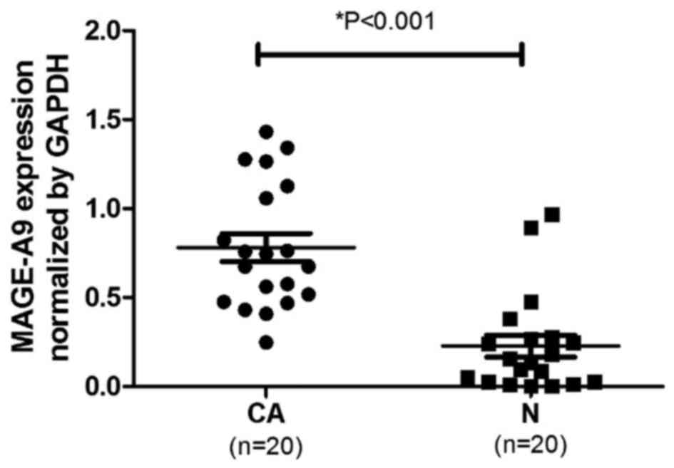
Analysis of MAGE-A9 mRNA expression in
ESCC using qPCR
Total RNA was extracted from the ESCC tissues and
corresponding non-cancerous tissues, and qPCR was used to evaluate
MAGE-A9 mRNA expression. As presented in Fig. 2, the mean ± standard error of MAGE-A9
mRNA in ESCC tissues (0.722±0.075) was significantly increased
compared with that of the corresponding non-cancerous tissues
(0.223±0.067), when normalized to GAPDH (P<0.05; Fig. 2).
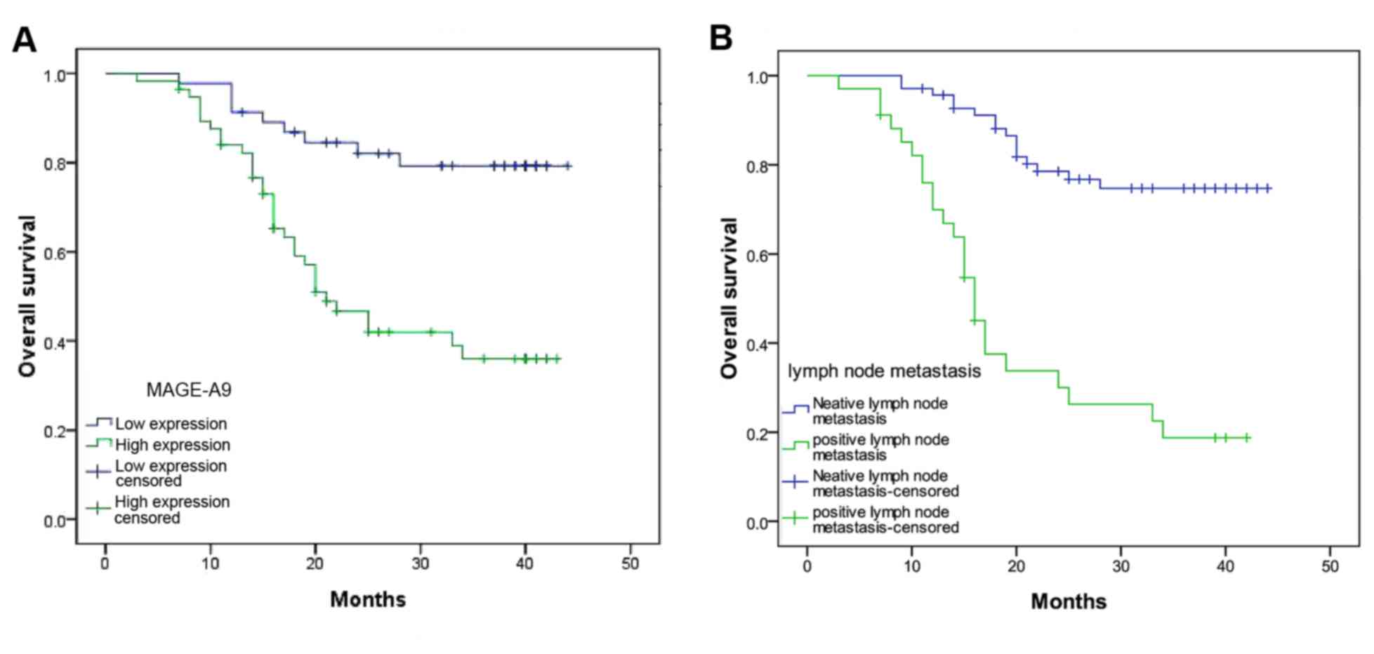
Survival analysis
Univariate analysis demonstrated that the overall
survival of 103 patients with ESCC was associated with lymph node
metastasis (P=0.001), tumor size (P=0.041) and MAGE-A9 expression
(P=0.001)(Table II). In addition,
multivariate analysis using Coxs regression model suggested that
lymph node metastasis (P=0.001) and MAGE-A9 expression (P=0.006)
may be independent prognostic factors for the overall survival
rate. The Kaplan-Meier estimator survival curves demonstrated that
patients with ESCC exhibiting lymph node metastasis and increased
MAGE-A9 presented a markedly poorer survival rate (Fig. 3).
| Table II.Univariate and multivariate analysis
of the association of prognostic factors in esophageal squamous
cell carcinoma with overall survival. |
Table II.
Univariate and multivariate analysis
of the association of prognostic factors in esophageal squamous
cell carcinoma with overall survival.
|
| Univariate
analysis | Multivariate
analysis |
|---|
|
|
|
|
|---|
| Characteristic | RR | P-value | 95% Cl | RR | P-value | 95% Cl |
|---|
| Sex |
|
|
|
|
|
|
| Male
vs. female | 1.794 | 0.138 | 0.828–3.887 |
|
|
|
| Age, years |
|
|
|
|
|
|
| ≤60 vs.
>60 | 0.821 | 0.531 | 0.443–1.522 |
|
|
|
| Smoking |
|
|
|
|
|
|
| No vs.
yes | 1.237 | 0.497 | 0.670–2.287 |
|
|
|
| Alcohol |
|
|
|
|
|
|
| No vs.
yes | 1.446 | 0.296 | 0.724–2.887 |
|
|
|
| Tumor size, cm |
|
|
|
|
|
|
| ≤3 vs.
>3 | 2.058 | 0.041a | 1.029–4.114 | 1.322 | 0.449 | 0.642–2.721 |
| pT |
|
|
|
|
|
|
| T1 and
T2 vs. T3 | 1.493 | 0.102 | 0.924–2.414 |
|
|
|
| Pathological
grade |
|
|
|
|
|
|
| II and
II vs. III | 1.344 | 0.267 | 0.797–2.264 |
|
|
|
| Lymph node
metastasis |
|
|
|
|
|
|
| No vs.
yes | 5.76 | 0.001a | 3.043–10.905 | 4.256 | 0.001a | 2.161–8.380 |
| MAGE-A9 |
|
|
|
|
|
|
| High
vs. low | 4.067 | 0.001a | 1.932–8.560 | 2.93 | 0.006a | 1.367–6.277 |
Discussion
MAGE proteins have been identified to exhibit a
prognostic value in a number of tumor tissues (18–22);
however, the normal physiological role of MAGE-A remains unknown
and their contribution to the development of cancer is poorly
understood. There is evidence that MAGE proteins are involved in
the regulation of apoptosis; for example, MAGE-A3 has been
identified to inhibit the activation of caspase 12 in vitro
and caspase 12 in turn is capable of inducing apoptosis (23,24). In
addition, MAGE-A protein has been identified to inhibit the
function of p53 through direct and indirect mechanisms (25). These previous studies have indicated
that MAGE-As may serve an important role in human cancers, but the
association between MAGE-A9 and ESCC, and whether MAGE-A9 may be
used as a targeted for diagnosis and therapy against ESCC, remains
unknown.
In the present study, MAGE-A9 mRNA expression in
ESCC tissues was determined using RT-qPCR. The results revealed an
increased expression of MAGE-A9 in ESCC tissues, compared with that
in healthy tissues. In addition, IHC analysis was conducted to
evaluate MAGE-A9 protein expression in ESCC TMA specimens. This
analysis identified increased MAGE-A9 expression in the cytoplasm
and mesenchyme of ESCC tissues, compared with that in healthy
tissues. Specific parameters, including the pathological grade,
lymph node metastasis and tumor size, were associated with MAGE-A9
protein expression. Univariate analysis identified that MAGE-A9
protein expression, lymph node metastasis and tumor size were
associated with the overall survival rate of patients with ESCC.
Multivariate analysis using Coxs regression model validated that
MAGE-A9 protein expression and lymph node metastasis may serve as
independent prognostic factors for overall survival. Increased
MAGE-A9 expression was associated with a poor prognosis in patients
with ESCC. The results of the present study were consistent with
previous studies carried out in hepatocellular carcinoma and
non-small cell lung cancer (7,11).
Additionally, the Kaplan-Meier estimator analysis demonstrated that
patients with ESCC with increased MAGE-A9 expression exhibited a
markedly unfavorable outcome.
In the present study, expression levels of the
protein and gene of MAGE-A9 were investigated. For MAGE-A9-positive
tumors, decreased MAGE-A9 expression cannot be excluded, since gene
activity may be influenced by other factors. Notably, it was
identified that p53 interacts with MAGE-A9 and the underlying
molecular mechanism which may be investigated using gene knockdown
or gene knockout of MAGE-A9. Additional analysis of the mechanism
of MAGE-A9 expression in ESCC cells is required.
The present study is the first, to the best of our
knowledge, to demonstrate an increased expression of MAGE-A9 in
ESCC tissues, which exhibited an association with the poor survival
of patients with ESCC. MAGE-A9 may provide a therapeutic strategy
for ESCC treatment.
References
|
1
|
Siegel RL, Miller KD and Jemal A: Cancer
statistics, 2015. CA Cancer J Clin. 65:5–29. 2015. View Article : Google Scholar : PubMed/NCBI
|
|
2
|
He J and Chen WQ: Chinese cancer registry
annual report. Military Medical Science Press; Beijing: 2012. pp.
P56–58. 2012, (In Chinese).
|
|
3
|
Bosset JF, Gignoux M, Triboulet JP, Tiret
E, Mantion G, Elias D, Lozach P, Ollier JC, Pavy JJ, Mercier M and
Sahmoud T: Chemoradiotherapy followed by surgery compared with
surgery alone in squamous-cell cancer of the esophagus. N Engl J
Med. 337:161–167. 1997. View Article : Google Scholar : PubMed/NCBI
|
|
4
|
Zhang Y: Epidemiology of esophageal
cancer. World J Gastroenterol. 19:5598–6006. 2013. View Article : Google Scholar : PubMed/NCBI
|
|
5
|
Roch N, Kutup A, Vashist Y, Yekebas E,
Kalinin V and Izbicki JR: Coexpression of MAGE-A peptides and HLA
class I molecules in hepatocellular carcinoma. Anticancer Res.
30:1617–1623. 2010.PubMed/NCBI
|
|
6
|
Lian Y, Sang M, Ding C, Zhou X, Fan X, Xu
Y, Lü W and Shan B: Expressions of MAGE-A10 and MAGE-A11 in breast
cancers and their prognostic significance: A retrospective clinical
study. J Cancer Res Clin Oncol. 138:519–527. 2012. View Article : Google Scholar : PubMed/NCBI
|
|
7
|
Hatiboglu G, Pritsch M, Macher-Goeppinger
S, Zöller M, Huber J, Haferkamp A, Pahernik S, Wagener N and
Hohenfellner M: Prognostic value of melanoma-associated antigen A9
in renal cell carcinoma. Scand J Urol. 47:311–322. 2012. View Article : Google Scholar : PubMed/NCBI
|
|
8
|
Melloni G, Ferreri AJ, Russo V, Gattinoni
L, Arrigoni G, Ceresoli GL, Zannini P and Traversari C: Prognostic
significance of cancer-testis gene expression in resected non-small
cell lung cancer patients. Oncol Rep. 12:145–151. 2004.PubMed/NCBI
|
|
9
|
Chen YT, Chiu R, Lee P, Beneck D, Jin B
and Old LJ: Chromosome X-encoded cancer/testis antigens show
distinctive expression patterns in developing gonads and in
testicular seminoma. Hum Reprod. 26:3232–3243. 2011. View Article : Google Scholar : PubMed/NCBI
|
|
10
|
Meek DW and Marcar L: MAGE-A antigens as
targets in tumour therapy. Cancer Lett. 324:126–132. 2012.
View Article : Google Scholar : PubMed/NCBI
|
|
11
|
Gu X, Fu M, Ge Z, Zhan F, Ding Y, Ni H,
Zhang W, Zhu Y, Tang X, Xiong L, et al: High expression of MAGE-A9
correlates with unfavorable survival in hepatocellular carcinoma.
Sci Rep. 4:66252014. View Article : Google Scholar : PubMed/NCBI
|
|
12
|
Xu X, Tang X, Lu M, Tang Q, Zhang H, Zhu
H, Xu N, Zhang D, Xiong L, Mao Y and Zhu J: Overexpression of
MAGE-A9 predicts unfavorable outcome in breast cancer. Exp Mol
Pathol. 97:579–584. 2014. View Article : Google Scholar : PubMed/NCBI
|
|
13
|
Zhang S, Zhai X, Wang G, Feng J, Zhu H, Xu
L, Mao G and Huang J: High expression of MAGE-A9 in tumor and
stromal cells of non-small cell lung cancer was correlated with
patient poor survival. Int J Clin Exp Pathol. 8:541–550.
2015.PubMed/NCBI
|
|
14
|
Han L, Jiang B, Wu H, Zhang S and Lu X:
Expression and prognostic value of MAGE-A9 in laryngeal squamous
cell carcinoma. Int J Clin Exp Pathol. 7:6734–6742. 2014.PubMed/NCBI
|
|
15
|
Bergeron A, Picard V, LaRue H, Harel F,
Hovington H, Lacombe L and Fradet Y: High frequency of MAGE-A4 and
MAGE-A9 expression in high-risk bladder cancer. Int J Cancer.
125:1365–1371. 2009. View Article : Google Scholar : PubMed/NCBI
|
|
16
|
Sobin L, Gospodarowicz M and Wittekind C:
TNM classification of malignant tumors. 7th. New Jersey;
Wiley-Blackwell; 2009
|
|
17
|
Schmittgen TD and Livak KJ: Analyzing
real-time PCR data by the comparative C(T) method. Nat Protoc.
3:1101–1108. 2008. View Article : Google Scholar : PubMed/NCBI
|
|
18
|
Jungbluth AA, Busam KJ, Kolb D, Iversen K,
Coplan K, Chen YT, Spagnoli GC and Old LJ: Expression of
MAGE-antigens in normal tissues and cancer. Int J Cancer.
85:460–465. 2000. View Article : Google Scholar : PubMed/NCBI
|
|
19
|
Svobodová S, Browning J, MacGregor D,
Pollara G, Scolyer RA, Murali R, Thompson JF, Deb S, Azad A, Davis
ID and Cebon JS: Cancer-testis antigen expression in primary
cutaneous melanoma has independent prognostic value comparable to
that of Breslow thickness, ulceration and mitotic rate. Eur J
Cancer. 47:460–469. 2011. View Article : Google Scholar : PubMed/NCBI
|
|
20
|
Ogata K, Aihara R, Mochiki E, Ogawa A,
Yanai M, Toyomasu Y, Ando H, Ohno T, Asao T and Kuwano H: Clinical
significance of melanoma antigen-encoding gene-1 (MAGE-1)
expression and its correlation with poor prognosis in
differentiated advanced gastric cancer. Ann Surg Oncol.
18:1195–1203. 2011. View Article : Google Scholar : PubMed/NCBI
|
|
21
|
Jeon CH, Shin IH, Park JB and Chae HD:
Prognostic significance of MAGE in peritoneal washes in gastric
carcinoma patients without peritoneal metastasis: Results of a
5-year follow-up study. J Clin Gastroenterol. 44:682–686. 2010.
View Article : Google Scholar : PubMed/NCBI
|
|
22
|
Pastorcic-Grgic M, Sarcevic B, Dosen D,
Juretic A, Spagnoli GC and Grgic M: Prognostic value of MAGE-A and
NY-ESO-1 expression in pharyngeal cancer. Head Neck. 32:1178–1184.
2010. View Article : Google Scholar : PubMed/NCBI
|
|
23
|
Sang M, Wang L, Ding C, Zhou X, Wang B,
Wang L, Lian Y and Shan B: Melanoma-associated antigen genes-an
update. Cancer Lett. 302:85–90. 2011. View Article : Google Scholar : PubMed/NCBI
|
|
24
|
Morishima N, Nakanishi K, Takenouchi H,
Shibata T and Yasuhiko Y: An endoplasmic reticulum stress-specific
caspase cascade in apoptosis cytochrome c-independent activation of
caspase-9 by caspase-12. J Biol Chem. 277:34287–34294. 2002.
View Article : Google Scholar : PubMed/NCBI
|
|
25
|
Marcar L, MacLaine NJ, Hupp TR and Meek
DW: Mage-A cancer/testis antigens inhibit p53 function by blocking
its interaction with chromatin. Cancer Res. 70:10362–10370. 2010.
View Article : Google Scholar : PubMed/NCBI
|