Introduction
Malignant mesothelioma (MM) is a rare but rapidly
fatal cancer that can develop from mesothelial cells in the pleura,
peritoneum, pericardium and tunica vaginalis (1,2). The
latency period of MM is exceptionally long and patients diagnosed
with MM generally have poor prognoses (3), with an average life expectancy of less
than a year, dependent on the tissue in which it originates
(4,5).
It has been reported that asbestos is the main carcinogen
associated with MM (6,7); however, the molecular mechanism
responsible for the pathogenesis of MM has not been elucidated.
Standard therapies for MM include chemotherapy,
surgery and radiotherapy, but more effective strategies are
required to improve the result of conventional therapeutic methods
or cure this cancer. Immunotherapy may be a useful treatment
(7,8);
however, despite research progress, immunotherapy using
tumor-associated antigens has been barely attempted in MM. Tumor
antigens are classified into several categories, including
differentiation antigens (9), mutated
gene products (10), overexpressed
oncogenes (11) and cancer/testis
(CT) antigens (12,13). Of these, CT antigens are considered a
promising tumor antigen group, since these proteins are expressed
in the normal testis and tumor tissues (14,15).
Various methods have been devised to identify this
group of tumor antigens. Serological analysis of recombinant tumor
cDNA expression library (SEREX) is one such method, which has been
shown to be powerful and effective (15). This technique screens cDNA expression
libraries using the sera of cancer patients, and identifies cancer
antigens recognized by autologous serum IgG (16). At present, >2,500 tumor antigens,
including melanoma-associated antigen (MAGEA) (13), synaptonemal complex protein 1
(17), NY-ESO-1 (18), NY-SAR-35 (19) and KP-CoT-23 (20) have been identified using SEREX.
Certain antigens, such as MAGE-A3 and NY-ESO-1, have been evaluated
for immunogenicity and for their immunotherapeutic efficacies
(21). In addition, CT antigens are
expected to be utilized as biomarkers for diagnosis and prognosis
(21,22).
In the present study, SEREX methodology was applied
to define the spectrum of immunogenic proteins in MM, with focus on
the CT-like antigens KP-MM-8 and KP-MM-5, which demonstrated high
seroreactivity in patients with MM.
Materials and methods
Human sera and cell lines
Human sera from 8 healthy donors and 8 MM patients
were obtained between February 2008 and December 2013, from the
Department of Pathology and Environmental Health Center for
Asbestos, Pusan National University Hospital (Yangsan, Korea)
subsequent to diagnosis and staging. Human MM NCI-H226, NCI-H2452,
NCI-H28 and MSTO-211H cell lines, human lung cancer A-427 and A549
cell lines, and the human melanoma cancer A375 cell line were
obtained from the American Type Culture Collection (Manassas, VA,
USA). Human colon cancer SNU-C1, SNU-C2A, SNU-C4 and SNU-C5 cell
lines, human ovarian cancer SNU-8 and SNU-840 cell lines were
obtained from the Korean Collection for Type Cultures (Jeongeup,
Korea). All cell lines were maintained in RPMI-1640 medium (Gibco;
Thermo Fisher Scientific, Inc., Waltham, MA, USA) supplemented with
10% fetal bovine serum medium (Gibco; Thermo Fisher Scientific,
Inc.), 2 mM L-glutamine, 100 units/ml penicillin and 100 µg/ml
streptomycin. Cells were cultured at 37°C in a humidified 5%
CO2 atmosphere. The present study was performed with
approval from the Ethical Committee of Pusan National University
Yangsan Hospital (Yangsan, Korea).
Total RNA extraction from cell
lines
Total RNA of human malignant mesothelioma NCI-H226,
NCI-H2452, NCI-H28 and MSTO-211H cell lines, human lung cancer
A-427 and A549 cell lines, human colon cancer SNU-C1, SNU-C2A,
SNU-C4 and SNU-C5 cell lines and human ovarian cancer SNU-8 and
SNU-840 cell lines were isolated using a RNA isolation kit (RNeasy
Maxi kit; Qiagen, Hilden, Germany). Amounts of RNA isolated were
assessed at 260 nm using a spectrophotometer (Ultrospec 2000;
Pharmacia Biotech; GE Healthcare, Chicago, IL, USA). Total RNAs
from: Human colon cancer HCT-15, SK-CO-1, SW-1116 and KM12/L4A cell
lines; human lung cancer SK-LC-14, SK-LC-19, SK-LC-5, NCI-H23 and
NCI-H740 cell lines; human melanoma cancer A375/C5, A375/P,
A375/SM, SK-MEL-19, SK-MEL-128, SK-MEL-21 and SK-MEL-37 cell lines;
human ovarian cancer OV-CAR-3, and SK-OV-3 cell lines; human breast
cancer SK-OV-1, BT-474, MDA-MB-453, MCF-7, MDA-MB-231 and SK-BR-5
cell lines; human renal cancer SK-RC-18 and SK-RC-59 cell lines;
human leukemia CEM/S and K-562 cell lines; human rhabdomyosarcoma
A-204 cell lines; human thyroid cancer KAT-18 cell lines; and human
liver cancer SK-HEP-1, SNU-354, SNU-423, SNU-449 and SNU-475 cell
lines were obtained from the Ludwig Institute for Cancer Research
(Memorial Sloan-Kettering Cancer Center, New York, NY, USA). Normal
tissue total RNA was purchased from Clontech Laboratories, Inc.
(Mountain View, CA, USA, cat. nos. 636742 and 636743).
Preparation of the cDNA library and
sera
Poly(A)+ RNA from normal testis was purchased from
Clontech Laboratories Inc. mRNA of the MSTO-211H cell line was
extracted using the PolyATtract mRNA Isolation System (Promega
Corp., Madison, WI, USA). Poly(A)+ RNA (5 µg) was used to construct
a cDNA library in ZAP Express vector (Stratagene; Agilent
Technologies, Inc., Santa Clara, CA, USA), according to the
manufacturer's instructions. The cDNA library contained
approximately one million recombinants and was used for
immunoscreening without amplification.
Sera from 8 healthy individuals and 8 patients with
MM were diluted with 0.2% skimmed milk to 1:200. To remove serum
antibodies that react with Escherichia
coli/bacteriophage-associated antigens, sera were absorbed with
E. coli/bacteriophage lysates, as previously described
(20).
Immunoscreening of the cDNA
library
Immunoscreening of the cDNA library was performed as
described by Song et al (20).
Briefly, E. coli XL1 blue MRF cells (Stratagene; Agilent
Technologies, Inc.) were transfected with the recombinant phages,
plated at a density of ~5,000 pfu/150 mm plate (NZCYM-IPTG agar),
and incubated for 8 h at 37°C. The cells were then transferred to
nitrocellulose filters (PROTRAN BA 85; 0.45 µm; Schleicher &
Schuell, Keene, NH, USA). The filters were incubated with patient
sera, which had been preabsorbed with E. coli-phage lysate,
at a dilution of 1:200, at room temperature overnight. The
serum-reactive clones were detected using alkaline
phosphatase-conjugated secondary antibody (Abcam, Cambridge, UK;
cat. no., ab6859; dilution, 1:2,000) incubated for 1 h at room
temperature and then visualized by incubation with
5-bromo-4-chloro-3-indolylphosphate/nitroblue tetrazolium.
Subsequent to screening, isolated positive clones were removed from
plates and preserved in suspension medium buffer with 25 µl
chloroform.
Positive phages were mixed with a helper phage to
co-infect XL-1 Blue MRF cells, and then rescued into pBluescript
phagemid forms by in vivo excision. Excised phagemids were
transformed into host bacteria (XLOLR) to multiply for plasmid
extraction and stock. The size of the cDNA inserted was determined
by double restriction enzyme digestion with EcoRI and
XhoI. The cDNA was sequenced commercially (Macrogen, Inc.,
Seoul, Korea).
Reverse transcription-polymerase chain
reaction (RT-PCR)
The cDNA preparations used as templates for RT-PCR
reactions were prepared using 1 µg total RNA isolated from all cell
lines and the Superscript First Strand Synthesis kit (Invitrogen;
Thermo Fisher Scientific, Inc., Waltham, MA, USA). Primers were as
follows: Amyotrophic lateral sclerosis 2 (juvenile) chromosome
region, candidate 11 (ALS2CR11) forward, 5′-TCTGCAGGCTGGAGGTGGAA-3′
and reverse, 5′-CCTTTCCCGGGGTTGTTGCT-3′. The conditions used for
RT-PCR were initial activation for 5 min at 94°C, 35 amplification
cycles of denaturation for 30 sec at 94°C, and annealing for 30 sec
at 63°C, extension for 1 min at 72°C, followed by a final
elongation for 10 min at 72°C. A total of 20 µl of PCR product were
loaded onto a 1.2% agarose gel and electrophoresis was performed at
room temperature at 100 V 30 min using 1X Tris-acetate-EDTA buffer.
The gels were stained with ethidium bromide solution diluted with
distilled water to a ratio of 2:104 for 10 min, and
washed with H2O. Data were analyzed by Gel Doc™ + Imager
with Image Lab™ software v.5.2.1 (Bio-Rad, Richmond, CA, USA).
Seroreactivity by plaque assay
Selected immunoreactive clones were tested for
reactivity against sera from individual MM patients and healthy
donors using a plaque assay. λ-ZAP clone without an insert was
co-plated and included in each assay as a negative control. KP-MM-5
was tested for reactivity against 10-fold serially diluted serum
(1:103 to 1:106) using the same plaque
assay.
Results
Identification of MM antigens by
SEREX
To identify tumor antigens in MM, immunoscreening of
a testis cDNA library and MSTO-211H cDNA library was performed
using the sera of patients with MM. In total, 31 immunogenic clones
were identified in this manner, and 16 of the 31 genes were
verified as tumor antigen candidates and termed KP-MM-1 to KP-MM-16
(Table I). Of the 16, 3 were
previously identified by SEREX in other tumor types, and 13
antigens did not match entries in the cancer immunome database, and
were therefore considered new antigens.
 | Table I.Malignant mesothelioma antigens
identified by SEREX. |
Table I.
Malignant mesothelioma antigens
identified by SEREX.
| Antigen | Gene name | UniGene
clustera | Library source | Serum
sourceb | Redundancy, n | Previously identified
by SEREXc |
|---|
| KP-MM-1 | IFT74 | Hs.145402 | Testis | M4 | 1 | No |
| KP-MM-2 | TMF1 | Hs.267632 | Testis | M4 | 2 | Yes |
| KP-MM-3 | BRAP | Hs.530940 | Testis | M4 | 1 | Yes |
| KP-MM-4 | RPLP0 | Hs.546285 | MSTO-211H | M8 | 1 | Yes |
| KP-MM-5 | CDC25B | Hs.153752 | Testis | M8 | 2 | No |
| KP-MM-6 | ZNF429 | Hs.572567 | Testis | M49 | 1 | No |
| KP-MM-7 | ZNF320 | Hs.369632 | Testis | M49 | 1 | No |
| KP-MM-8 | ALS2CR11 | Hs.335788 | Testis | M8 | 1 | No |
| KP-MM-9 | DNAJC2 | Hs.558476 | Testis | M8 | 1 | No |
| KP-MM-10 | NUB1 | Hs.647082 | Testis | M10 | 1 | No |
| KP-MM-11 | CCDC11 | Hs.658630 | Testis | M1 | 2 | No |
| KP-MM-12 | ZNF93 | Hs.723768 | Testis | M49 | 1 | No |
| KP-MM-13 | ZNF234 | Hs.235992 | Testis | M49 | 1 | No |
| KP-MM-14 | ZNF595 | Hs.709469 | Testis | M49 | 1 | No |
| KP-MM-15 | CCDC40 | Hs.202542 | Testis | M49 | 1 | No |
| KP-MM-16 | ZNF85 | Hs.37138 | Testis | M49 | 1 | No |
KP-MM-8 as a CT-like antigen
Preliminary in silico mRNA expression profile
analysis and characterization of the gene products identified in
the present study were undertaken based on the tissue distributions
of expressed sequence tags and serial analysis of gene expression
tags in the Cancer Genome Anatomy Database (http://cgap.nci.nih.gov/) and the information
contained in the GeneCards database (http://www.genecards.org/). One antigen, KP-MM-8, was
identified as a CT-like antigen. KP-MM-8 was identified as ALS2CR11
by a nucleotide BLAST search.
Conventional RT-PCR was performed using mRNA from
normal tissues to examine the expression of ALS2CR11 (Fig. 1A). RT-PCR showed that ALS2CR11 was
strongly expressed only in the normal testis, and weakly in the
spleen, prostate, ovary, heart and skeletal muscle. However, the
expression in various cancer cell lines was relatively high. The
expression of this antigen was positive in 3 of 4 MM cell lines,
consisting of the NCI-H226, NCI-H2452 and MSTO-211H cell lines
(Fig. 1B). In addition, it was
expressed in various cancer cell lines, including lung cancer (5/7)
(Fig. 1C), melanoma (5/7) (Fig. 1D) and liver cancer (5/5) cell lines,
but was not expressed in colon cancer, renal cancer or leukemia
cell lines (Table II). This result
suggests that ALS2CR11 may be a putative CT like antigen.
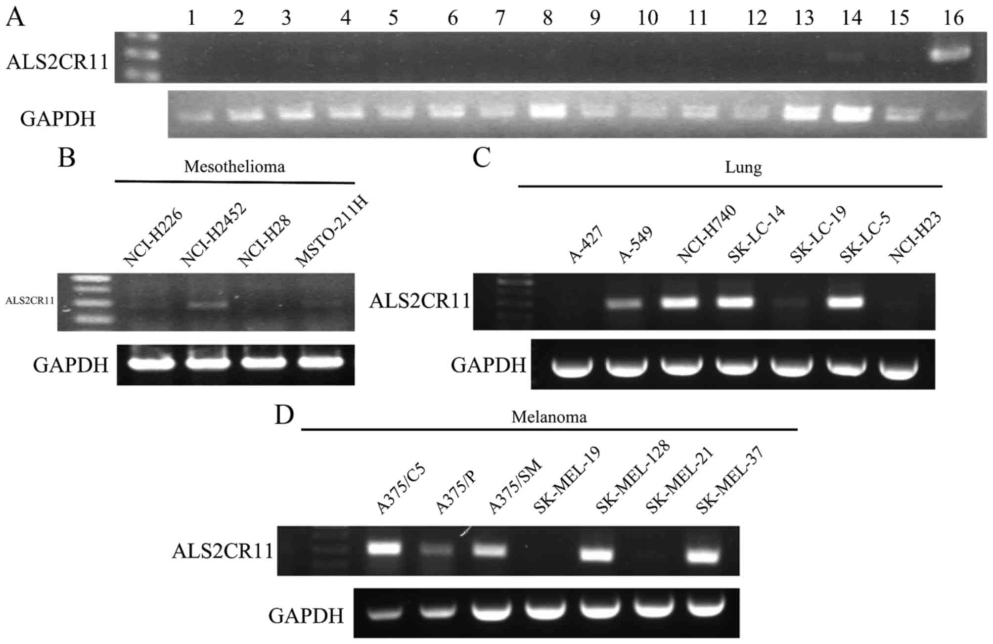 | Figure 1.Reverse transcription-polymerase chain
reaction analysis of ALS2CR11 expression in (A) normal tissues, as
follows: 1, spleen; 2, thymus; 3, prostate; 4, ovary; 5, small
intestine; 6, colon; 7, leukocyte; 8, heart; 9, brain; 10,
placenta; 11, lung; 12, pancreas; 13, liver; 14, skeletal muscle;
15, kidney; and 16, testis. ALSC2CR11 expression is also shown in
(B) mesothelioma cancer, (C) lung cancer and (D) melanoma cell
lines. cDNA templates were normalized using GAPDH as shown in the
bottom panel. ALS2CR11, amyotrophic lateral sclerosis 2 (juvenile)
chromosome region, candidate 11. |
| Table II.Summary of ALS2CR11 mRNA expression
as determined by reverse transcription-polymerase chain
reaction. |
Table II.
Summary of ALS2CR11 mRNA expression
as determined by reverse transcription-polymerase chain
reaction.
| Cancer cell
line | Frequency, n |
|---|
| Mesothelioma | 3/4 |
| Colon cancer | 0/8 |
| Lung cancer | 5/7 |
| Melanoma | 5/7 |
| Ovarian cancer | 2/4 |
| Breast cancer | 1/6 |
| Renal cancer | 0/2 |
| Leukemia | 0/2 |
|
Rhabdomyosarcoma | 1/1 |
| Thyroid cancer | 1/1 |
| Liver cancer | 5/5 |
Seroreactivity of isolated
mesothelioma antigens
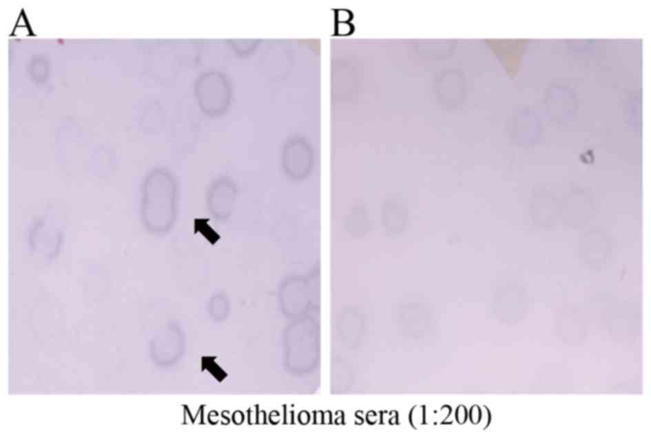
Seroreactivity testing of 7 phage clones, consisting
of KP-MM-2, KP-MM-5, KP-MM-6, KP-MM-7, KP-MM-8, KP-MM-9 and
KP-MM-12, was performed using the sera of 8 MM patients and 8
healthy donors by phage plaque assay (Fig. 2). The CT-like antigen KP-MM-8 was only
positive in one MM serum (M8), and negative in all other 7 MM sera
and to all normal sera. In total, 2 of the 8 MM patients had
antibodies reactive to KP-MM-6-containing phagemids, whereas the 8
normal sera showed no reactivity against KP-MM-6 (Table III). Notably, all MM patients and
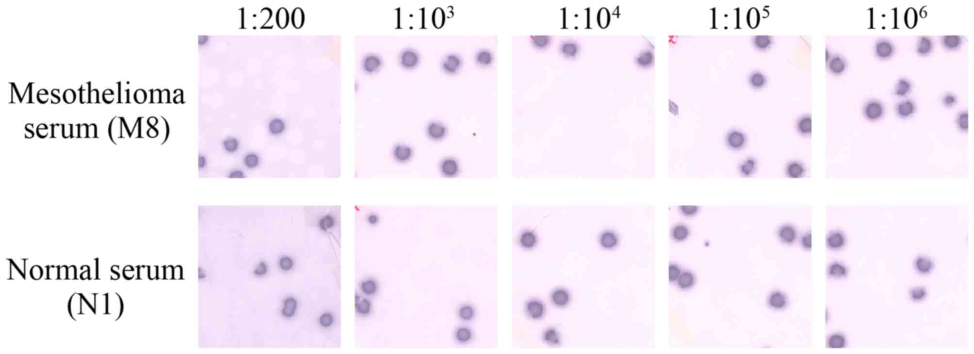
normal individuals showed high reactivity to the KP-MM-5 antigen.
Representative phage plaque assay for KP-MM-5 from the sera of M8
and normal individual N1, showed high-titer reactivity
(1:106) (Fig. 3). The
presence of consistently high-titer antibodies against KP-MM-5
suggested it to be an immunogenic tumor antigen in MM patients.
However, normal sera also showed extremely strong reactivity for
KP-MM-5; this observation requires further investigation.
| Table III.Detection of selected antigens
against sera obtained from malignant mesothelioma patients and
normal individuals. |
Table III.
Detection of selected antigens
against sera obtained from malignant mesothelioma patients and
normal individuals.
|
| No. of cases |
|---|
|
|
|
|---|
| KP-MM-antigen | Mesothelioma | Normal |
|---|
| KP-MM-2 | 3/8 | 0/8 |
| KP-MM-5 | 8/8 | 8/8 |
| KP-MM-6 | 2/8 | 0/8 |
| KP-MM-7 | 1/8 | 0/8 |
| KP-MM-8 | 1/8 | 0/8 |
| KP-MM-9 | 2/8 | 2/8 |
| KP-MM-12 | 1/8 | 0/8 |
Discussion
Previously, it was reported that several major CT
antigens are expressed in MM (23);
however, it is theorized that numerous tumor antigens remain
unidentified, and that even identified tumor antigens require
further functional study. To identify new CT antigens, MM was
studied using the SEREX technique, and as a result, 31 sequences
were isolated and 16 antigens were identified as independent
tumor-specific antigens.
Notably, 6 of the 16 identified genes (KP-MM-6,
KP-MM-7, KP-MM-12, KP-MM-13, KP-MM-14 and KP-MM-19) belonged to the
zinc finger gene family. Associations between the ZNF gene and
cancer properties are not well understood, although it has been
reported that certain ZNF genes have potential as biological
markers of cancer (24). Thus,
additional investigation of the functional roles of ZNF gene during
the pathogeneses of cancers, including MM, is required.
KP-MM-8 was identified in a testis library that
reacted to mesothelioma serum, and BLAST search identified this
gene as ALS2CR11, a member of the ALS2CR multigene family, which is
associated with amyotrophic lateral sclerosis (25). Nevertheless, the present study
identifies the ALS2CR11 gene as putative CT antigen, and little is
known about the contribution made by this gene to
carcinogenesis.
In the present study, ALS2CR11 was found to have
CT-like antigen features, particularly a restricted mRNA expression
pattern. Considering the mRNA expression profile of ALS2CR11 in the
GeneCards database and the present RT-PCR results, ALS2CR11 appears
to be expressed in various cancer cell lines and in few normal
tissues, which is a noteworthy feature of CT antigens.
However, seroreactivity testing showed KP-MM-8
reacted with only 1 MM serum, although this result was based on a
limited number of patient sera samples. Additional investigation is
required with more samples to determine whether ALS2CR11 functions
as a CT antigen, and to identify the molecular basis for its
contribution to tumorigenesis.
KP-MM-5 was identified in a testis library that
reacted with mesothelioma serum. This clone was identified as cell
division cycle (CDC) 25B, and CDC25 phosphatases activate
cyclin-dependent kinases (CDKs) and targets of the checkpoint
kinase (CHK) 1/CHK2-mediated checkpoint pathway (26). Numerous studies have been conducted on
members of the CDC25 protein phosphatase family, and it has been
reported that CDC25B plays an important role in tumorigenesis
(27,28). Specifically, CDC25B activates M phase
promoting factor and promotes G2/M transition, and the
overexpression of CDC25B contributes to excessive cell division
(29).
Presence of high-titer reactive antibodies against
KP-MM-5 suggests that KP-MM-5 is an immunogenic tumor antigen in MM
patients. However, normal sera showed strong reactivity for
KP-MM-5. This result requires additional investigation, and
suggests that numerous aspects of the actions of CDC25B have yet to
be identified.
Acknowledgements
The study was supported by a grant from Pusan
National University Yangsan Hospital Environmental Health Center
for Asbestos (Korea Ministry of Environment, grant no.
076-1900-1939-302-320-01).
References
|
1
|
Raptopoulos V: Peritoneal mesothelioma.
Crit Rev Diagn Imaging. 24:293–328. 1985.PubMed/NCBI
|
|
2
|
Cunha P, Luz Z, Seves I, Sousa C, Skiappa,
Ribeiro L, Marques C and Oliveira M: Malignant peritoneal
mesothelioma-diagnostic and therapeutic difficulties. Acta Med
Port. 15:383–386. 2002.(In Portuguese). PubMed/NCBI
|
|
3
|
Ahmed I, Tipu Ahmed S and Ishtiaq S:
Malignant mesothelioma. Pak J Med Sci. 29:1433–1438.
2013.PubMed/NCBI
|
|
4
|
Røe OD and Stella GM: Malignant pleural
mesothelioma: History, controversy and future of a manmade
epidemic. Eur Respir Rev. 24:115–131. 2015. View Article : Google Scholar : PubMed/NCBI
|
|
5
|
Mirabelli D, Roberti S, Gangemi M, Rosato
R, Ricceri F, Merler E, Gennaro V, Mangone L, Gorini G, Pascucci C,
et al: Survival of peritoneal malignant mesothelioma in Italy: A
population-based study. Int J Cancer. 124:194–200. 2009. View Article : Google Scholar : PubMed/NCBI
|
|
6
|
Rudd RM: Malignant mesothelioma. Br Med
Bull. 93:105–123. 2010. View Article : Google Scholar : PubMed/NCBI
|
|
7
|
Robinson BW and Lake RA: Advances in
malignant mesothelioma. N Engl J Med. 353:1591–1603. 2005.
View Article : Google Scholar : PubMed/NCBI
|
|
8
|
Kondola S, Manners D and Nowak AK:
Malignant pleural mesothelioma: An update on diagnosis and
treatment options. Ther Adv Respir Dis. 10:275–288. 2016.
View Article : Google Scholar : PubMed/NCBI
|
|
9
|
Coulie PG, Brichard V, Van Pel A, Wölfel
T, Schneider J, Traversari C, Mattei S, De Plaen E, Lurquin C,
Szikora JP, et al: A new gene coding for a differentiation antigen
recognized by autologous cytolytic T lymphocytes on HLA-A2
melanomas. J Exp Med. 180:35–42. 1994. View Article : Google Scholar : PubMed/NCBI
|
|
10
|
Labrecque S, Naor N, Thomson D and
Matlashewski G: Analysis of the anti-p53 antibody response in
cancer patients. Cancer Res. 53:3468–3471. 1993.PubMed/NCBI
|
|
11
|
Disis ML, Calenoff E, McLaughlin G, Murphy
AE, Chen W, Groner B, Jeschke M, Lydon N, McGlynn E, Livingston RB,
et al: Existent T-cell and antibody immunity to HER-2/neu protein
in patients with breast cancer. Cancer Res. 54:16–20.
1994.PubMed/NCBI
|
|
12
|
Boon T, Coulie PG and Van den Eynde B:
Tumor antigens recognized by T cells. Immunol Today. 18:267–268.
1997. View Article : Google Scholar : PubMed/NCBI
|
|
13
|
Chen YT, Güre AO, Tsang S, Stockert E,
Jäger E, Knuth A and Old LJ: Identification of multiple
cancer/testis antigens by allogeneic antibody screening of a
melanoma cell line library. Proc Natl Acad Sci USA. 95:6919–6923.
1998. View Article : Google Scholar : PubMed/NCBI
|
|
14
|
van der Bruggen P, Traversari C, Chomez P,
Lurquin C, De Plaen E, Van den Eynde BJ, Knuth A and Boon T: A gene
encoding an antigen recognized by cytolytic T lymphocytes on a
human melanoma. J Immunol. 178:2617–2621. 2007.PubMed/NCBI
|
|
15
|
Caballero OL and Chen YT: Cancer/testis
(CT) antigens: Potential targets for immunotherapy. Cancer Sci.
100:2014–2021. 2009. View Article : Google Scholar : PubMed/NCBI
|
|
16
|
Sahin U, Türeci O, Schmitt H, Cochlovius
B, Johannes T, Schmits R, Stenner F, Luo G, Schobert I and
Pfreundschuh M: Human neoplasms elicit multiple specific immune
responses in the autologous host. Proc Natl Acad Sci USA.
92:11810–11813. 1995. View Article : Google Scholar : PubMed/NCBI
|
|
17
|
Türeci O, Sahin U, Zwick C, Koslowski M,
Seitz G and Pfreundschuh M: Identification of a meiosis-specific
protein as a member of the class of cancer/testis antigens. Proc
Natl Acad Sci USA. 95:5211–5216. 1998. View Article : Google Scholar : PubMed/NCBI
|
|
18
|
Chen YT, Scanlan MJ, Sahin U, Türeci O,
Gure AO, Tsang S, Williamson B, Stockert E, Pfreundschuh M and Old
LJ: A testicular antigen aberrantly expressed in human cancers
detected by autologous antibody screening. Proc Natl Acad Sci USA.
94:1914–1918. 1997. View Article : Google Scholar : PubMed/NCBI
|
|
19
|
Lee SY, Obata Y, Yoshida M, Stockert E,
Williamson B, Jungbluth AA, Chen YT, Old LJ and Scanlan MJ:
Immunomic analysis of human sarcoma. Proc Natl Acad Sci USA.
100:2651–2656. 2003. View Article : Google Scholar : PubMed/NCBI
|
|
20
|
Song MH, Ha JM, Shin DH, Lee CH, Old L and
Lee SY: KP-CoT-23 (CCDC83) is a novel immunogenic cancer/testis
antigen in colon cancer. Int J Oncol. 41:1820–1826. 2012.
View Article : Google Scholar : PubMed/NCBI
|
|
21
|
Suri A, Jagadish N, Saini S and Gupta N:
Targeting cancer testis antigens for biomarkers and immunotherapy
in colorectal cancer: Current status and challenges. World J
Gastrointest Oncol. 7:492–502. 2015.PubMed/NCBI
|
|
22
|
Grah JJ, Katalinic D, Juretic A, Santek F
and Samarzija M: Clinical significance of immunohistochemical
expression of cancer/testis tumor-associated antigens (MAGE-A1,
MAGE-A3/4, NY-ESO-1) in patients with non-small cell lung cancer.
Tumori. 100:60–68. 2014.PubMed/NCBI
|
|
23
|
Sigalotti L, Coral S, Altomonte M, Natali
L, Gaudino G, Cacciotti P, Libener R, Colizzi F, Vianale G, Martini
F, et al: Cancer testis antigens expression in mesothelioma: Role
of DNA methylation and bioimmunotherapeutic implications. Br J
Cancer. 86:979–982. 2002. View Article : Google Scholar : PubMed/NCBI
|
|
24
|
Gaykalova DA, Vatapalli R, Wei Y, Tsai HL,
Wang H, Zhang C, Hennessey PT, Guo T, Tan M, Li R, et al: Outlier
analysis defines zinc finger gene family DNA methylation in tumors
and saliva of head and neck cancer patients. PLoS One.
10:e01421482015. View Article : Google Scholar : PubMed/NCBI
|
|
25
|
Hentati A, Bejaoui K, Pericak-Vance MA,
Hentati F, Speer MC, Hung WY, Figlewicz DA, Haines J, Rimmler J,
Ben Hamida C, et al: Linkage of recessive familial amyotrophic
lateral sclerosis to chromosome 2q33-q35. Nat Genet. 7:425–428.
1994. View Article : Google Scholar : PubMed/NCBI
|
|
26
|
Kiyokawa H and Ray D: In vivo roles of
CDC25 phosphatases: Biological insight into the anti-cancer
therapeutic targets. Anticancer Agents Med Chem. 8:832–836. 2008.
View Article : Google Scholar : PubMed/NCBI
|
|
27
|
Galaktionov K, Lee AK, Eckstein J, Draetta
G, Meckler J, Loda M and Beach D: CDC25 phosphatases as potential
human oncogenes. Science. 269:1575–1577. 1995. View Article : Google Scholar : PubMed/NCBI
|
|
28
|
Kristjánsdóttir K and Rudolph J: Cdc25
phosphatases and cancer. Chem Biol. 11:1043–1051. 2004. View Article : Google Scholar : PubMed/NCBI
|
|
29
|
Kim J, Singh AK, Takata Y, Lin K, Shen J,
Lu Y, Kerenyi MA, Orkin SH and Chen T: LSD1 is essential for oocyte
meiotic progression by regulating CDC25B expression in mice. Nat
Commun. 6:101162015. View Article : Google Scholar : PubMed/NCBI
|