Introduction
It is reported that oral squamous cell carcinoma
(OSCC), one of the most common oral malignancies of the squamous
epithelium of the oral cavity, is the sixth most common type of
cancer globally (1–3). It has been reported that developing
countries have the highest incidence rates of OSCC and it is
expected that the incidence will continue to increase (4); furthermore, OSCC commonly occurs in
middle-aged and elderly males because of tobacco and alcohol use
(5). OSCC commonly occurs in the
tissues of the oral cavity, including the gingiva, tongue, lip,
hard palate, buccal mucosa and mouth floor (4,6). OSCC
exhibits a marked propensity for invasive growth and metastasis,
leading to damage of the original tissues or that of distant organs
(2,4).
The predominant treatment strategy for OSCC is radical surgery and
postoperative chemoradiation (4).
Marked improvements have been made in clinical diagnosis and
management of OSCC; however, high recurrence rates and low 5-year
survival rates have remained constant for several decades (7).
Increasingly, previous studies have revealed that
chemotherapy using natural agents with low toxicity is a promising
approach for treating various types of cancer (8–10). In
addition, traditional Chinese medicine (TCM) has been used in the
treatment of various incurable diseases for centuries, and the
reliability and effectiveness of TCMs have been demonstrated by
long-term clinical use in China (11,12).
Homonoia riparia Lour (Euphorbiaceae), is a known source of
TCM with antipyretic and detoxification functions, and is commonly
used as an effective agent for treating infection, hepatitis,
hemorrhoids, ulcers, tumor and ambustion (13–15).
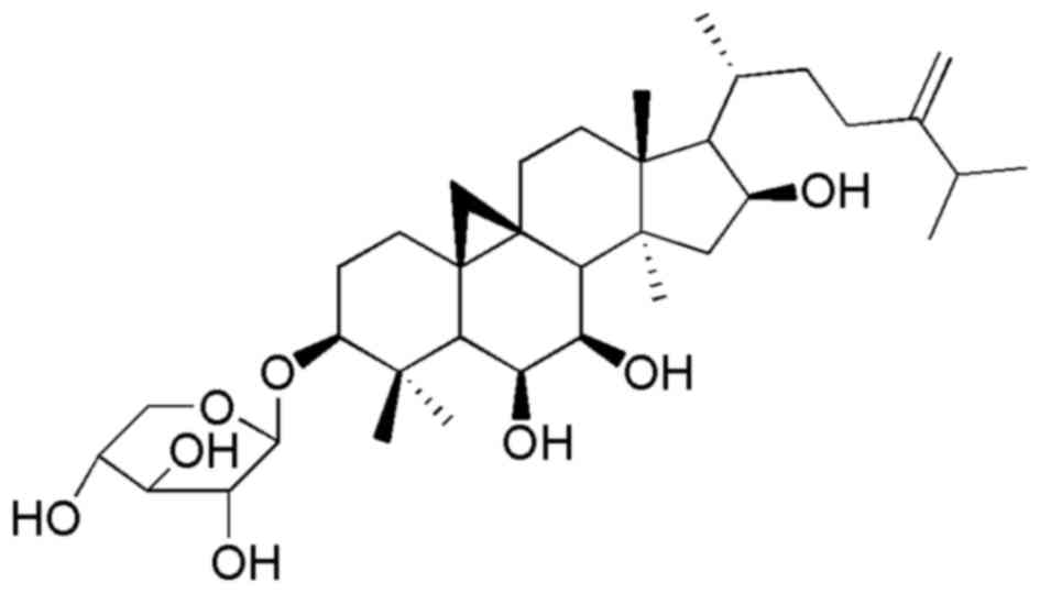
Riparsaponin (RSP; Fig. 1) is an
active constituent isolated from H. riparia that possesses
potential for treating gouty arthritis (16). In the present study, the antitumor
effects of RSP on human oral carcinoma cells via the induction of
apoptosis were systematically investigated, which has reference
value for the clinical use of RSP in the treatment of OSCC.
Materials and methods
Plant materials
The roots of H. riparia were purchased from
the Juhuacun Market of Traditional Chinese Herbs (Kunming, China)
in October 2014, and were identified by the Department of Chinese
Herbal Medicine in the Daqing Oilfield General Hospital (Daqing,
China). A voucher specimen of this plant was deposited in the
department of Stomatology laboratory, Daqing Oilfield General
Hospital (reference no. S-hptcm-20141007).
Chemicals and reagents
Dulbecco's modified Eagle's medium (DMEM), fetal
bovine serum (FBS), and anti-epithelial cadherin (E-CAD) (1:2,000;
cat. no. 13-1700) and -c-MET (1:2,000; cat. no. 44-888G) antibodies
were purchased from Invitrogen; Thermo Fisher Scientific, Inc.
(Waltham, MA, USA). MTT, DAPI and dimethyl sulfoxide (DMSO) were
purchased from Sigma-Aldrich; Merck KGaA (Darmstadt, Germany).
Antibodies against cleaved caspase-3/9 (C-caspase-3/9) (1:2,000;
cat. nos. 9664 and 7237, respectively), matrix metalloproteinase
(MMP)-2 (1:2,000; cat. no. 87809) and MMP-9 (1:2,000; cat. no.
13667) were purchased from Cell Signaling Technology, Inc.
(Danvers, MA, USA). Antibodies against B-cell lymphoma 2 (Bcl-2)
(1:1,000; cat. no. AB112), Bcl-2-associated X protein (Bax)
(1:1,000; cat. no. AB026), Bcl-2-associated death promoter (Bad)
(1:1,000; cat. no. AB008) and β-actin (1:1,000; cat. no. AB128),
goat anti-rabbit horseradish peroxidase (HRP)-conjugated secondary
antibodies (1:1,000; cat. no. A0208), the enhanced
chemiluminescence reagent and cell lysis buffer for Western and IP
kit were purchased from Beyotime Institute of Biotechnology
(Haimen, China). All other chemicals used in the present study were
of analytical reagent grade.
Cell culture
Human oral carcinoma cell lines (Cal-27, SCC-9 and
Detroit 562) were purchased from the American Type Culture
Collection (Manassas, VA, USA), and the human embryonic lung
fibroblast cell line MRC-5 was purchased from Cellcook Biotech.
Co., Ltd. (Guangzhou, China). Cells were cultured in DMEM
supplemented with 10% FBS, 100 U/ml penicillin and 100 µg/ml
streptomycin at 37°C in a humidified incubator containing 5%
CO2.
Preparation of RSP
Dried roots of H. riparia were powdered and
extracted with 60% ethanol under reflux. Then, the extracts were
sequentially extracted with petroleum ether, ethyl acetate and
n-butanol to produce petroleum ether fraction, ethyl acetate
fraction (AE), n-butanol fraction and water fraction. The AE was
subjected to column chromatography (CC) over a silica gel (200–300
mesh), eluting with ethyl acetate-acetone (20:1, 15:1, 10:1, 5:1,
3:1 and 1:1) to yield four subfractions I–IV on the basis of the
results of thin layer chromatography (TLC) analysis: The
aforementioned CC elution samples (3 µl) were analyzed using silica
gel G thin layer plate, and the chromatography was run for 8 cm
using an ethyl acetate-acetone mobile phase (10:1, 5:1 and 2:1) and
the TLC spots were visualized using 10% sulfuric acid alcohol
chromogenic agent. Fraction III was repeatedly subjected to CC over
a silica gel (200–300 mesh), eluting with ethyl acetate-acetone
(15:1, 10:1, 5:1, 3:1 and 1:1) to yield four subfractions
III1-III4. RSP was crystallized from
III2.
The isolated RSP was identified by
1H-nuclear magnetic resonance (NMR) and
13C-NMR, and compared with results of previous studies
(13,16). The 1H-NMR and
13C-NMR spectral data of this compound are as follows:
1H-NMR (500 MHz, CDCl3) δ (ppm): 0.32, 0.92
(2 H, m, H-19), 3.11 (1 H, m, H-3), 4.71 (2 H, brs, H-31), 0.84, (3
H, d, J 8.1 Hz, 21-CH3), 0.90 (3 H, s,
28-CH3), 0.93 (3 H, s, 26-CH3), 0.95 (3 H, s,
27-CH3), 1.02 (3 H, s, 29-CH3), 1.09 (3 H, s,
30-CH3), 1.17 (3 H, s, 18-CH3);
13C-NMR (125 MHz, DMSO-d6) δ (ppm): 30.97
(C-1), 29.25 (C-2), 88.97 (C-3), 42.19 (C-4), 48.91 (C-5), 71.90
(C-6), 71.04 (C-7), 46.03 (C-8), 25.14 (C-9), 18.87 (C-10), 26.99
(C-11), 33.07 (C-12), 45.81 (C-13), 45.00 (C-14), 51.03 (C-15),
71.13 (C-16), 58.31 (C-17), 21.61 (C-18), 32.04 (C-19), 28.10
(C-20), 18.04 (C-21), 37.03 (C-22), 32.82 (C-23), 160.07 (C-24),
34.21 (C-25), 22.52 (C-26), 24.01 (C-27), 20.13 (C-28), 24.82
(C-29), 16.88 (C-30), 109.27 (C-31), 105.01 (C-1′), 73.26 (C-2′),
78.38 (C-3′), 67.05 (C-4′), 68.93 (C-5′).
Determination of cell viability
Cell viability was determined using the MTT assay.
Briefly, cells (1×10⁵ cells/ml) were plated in 96-well
plates for 24 h. Subsequently, cells were treated with RSP at
various concentrations (0, 5, 10, 20, 40, 80, 150 and 200 µg/ml)
for 24 h. Subsequently, an MTT assay was used to determine the cell
viability (n=4), and the optical density value was determined at
570 nm on a microplate reader (Bio-Rad Laboratories, Inc.,
Hercules, CA, USA).
Nuclear staining with DAPI
A DAPI staining assay was carried out for the
apoptosis assays. Briefly, cells were exposed to RSP for 24 h at
37°C, and then cells were stained with DAPI at room temperature for
5 min. Alterations in the cell nuclei were determined using
fluorescence microscopy (Olympus Corporation, Tokyo, Japan) at
magnification, ×200.
Western blot analysis
Following treatment with RSP (0, 20, 40 and 80
µg/ml) for 24 h at 37°C, cells were harvested and total proteins
were extracted by using the Cell lysis buffer for Western and IP
kit according to manufacturer's protocol. The concentration of
protein was determined using a bicinchoninic acid protein assay
kit. Equal amounts of protein (30 µg) were separated by SDS/PAGE
(12% gel), then transferred onto a PVDF membrane and probed with
various primary antibodies overnight at 4°C, followed by incubation
with HRP-conjugated secondary antibodies for 1 h at room
temperature. Finally, the protein bands were visualized using
chemiluminescence detection with the enhanced chemiluminescence
reagent. Densitometric determination of the blots was carried out
using Quantity One software (version 4.0; Bio-Rad Laboratories). To
normalize protein loading, antibodies against β-actin were used,
and the protein level was expressed as a value relative to that of
β-actin.
Statistical analysis
All data are expressed as the mean ± standard
deviation, and the significance of the differences between groups
was determined using one-way analysis of variance with SPSS for
Windows software (version 19.0; IBM Corp., Armonk, NY, USA).
P<0.05 was considered to indicate a statistically significant
difference.
Results
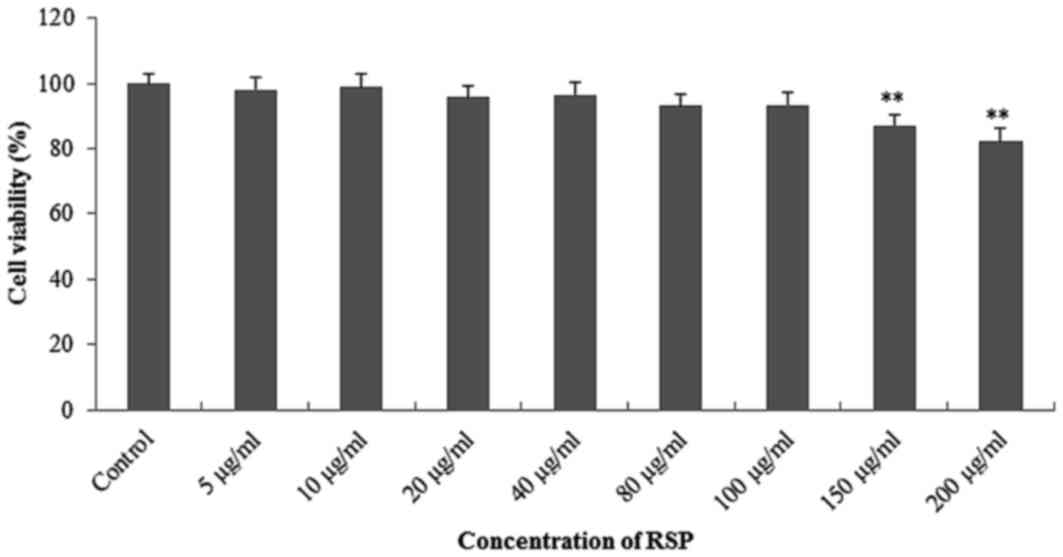
Inhibitory effects of RSP on MRC-5
cell and oral cancer cells in vitro
For investigating the toxicological effect of RSP,
the human embryonic lung fibroblast cell line MRC-5 was used. No
significant cytotoxic effect of RSP on MRC-5 cells was observed at
concentrations <100 µg/ml compared with the control (Fig. 2).
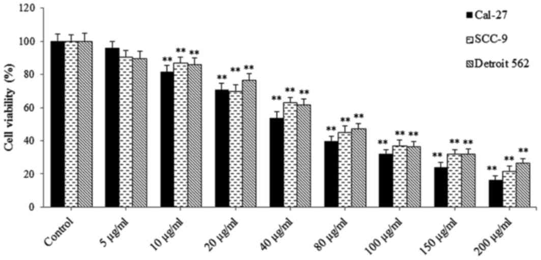
Furthermore, the cytotoxic effects of RSP on three
human oral carcinoma cell lines (Cal-27, SCC-9 and Detroit 562
cells) were evaluated. RSP exhibited a marked anti-proliferative
effect on the three oral cancer cell lines, which was
concentration-dependent between 10 and 200 µg/ml (Fig. 3).
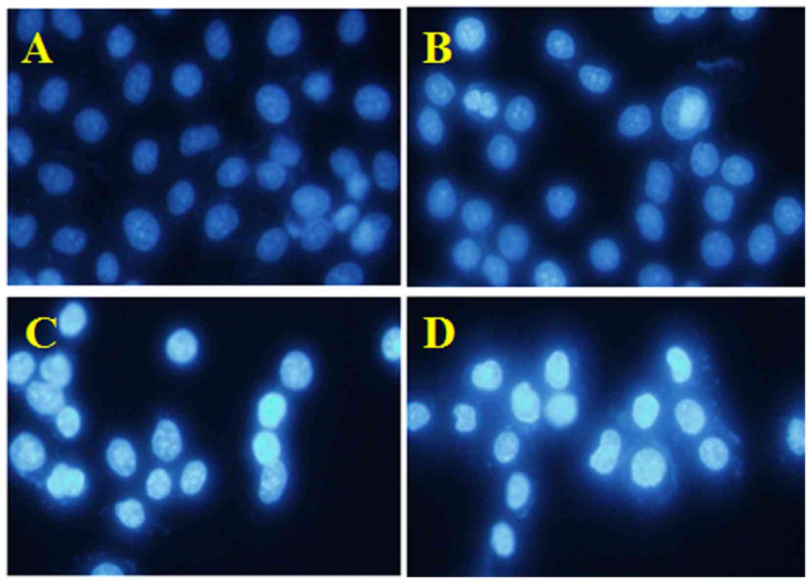
Pro-apoptotic effect of RSP on SCC-9
cells
Results of the cytotoxicity assay indicated that RSP
exerts significant antitumor potential on human oral carcinoma
cells. Furthermore, in order to determine whether the antitumor
effect of RSP was due to apoptosis, DAPI, a widely used
cell-permeant DNA dye, was used to stain SSC-9 cells for observing
the nuclear morphological changes (Fig.
4). The results indicated that the cell nuclei in the control
group were round and normal with faint staining (cells were alive).
By contrast, following treatment with RSP at concentrations of 20,
40 and 80 µg/ml, characteristic features of apoptosis were
observed, including typical nuclear condensation, increased
brightness and nuclear fragmentation. These results revealed that
RSP induced SSC-9 cell death due to apoptosis.
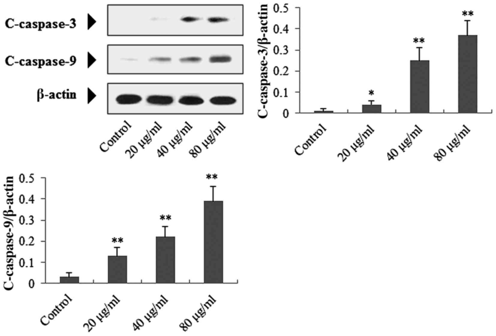
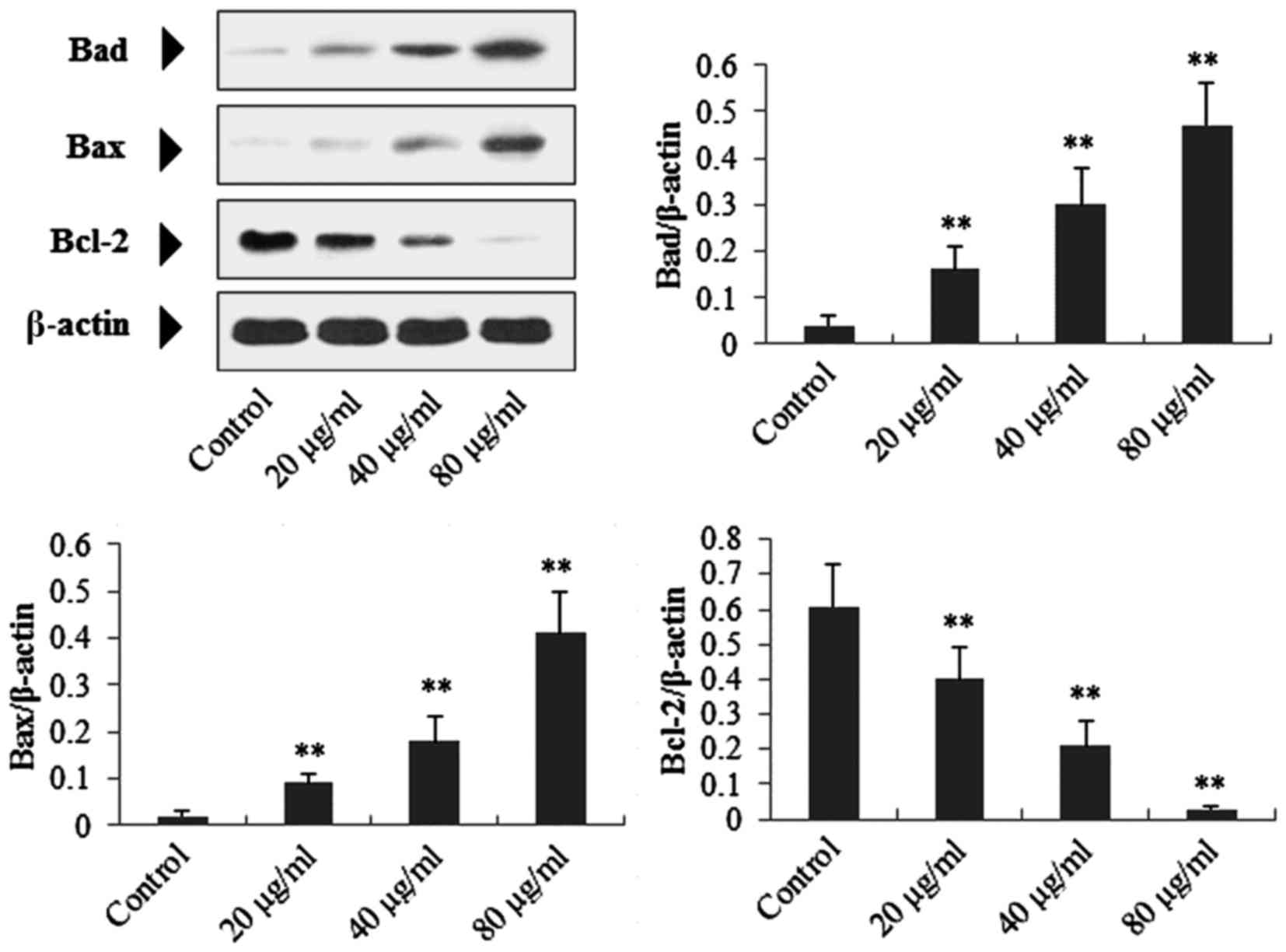
RSP upregulates C-caspase-3,
C-caspase-9, Bad and Bax, and downregulates Bcl-2 in SCC-9
cells
To investigate the potential apoptotic mechanism
induced by RSP in SSC-9 cells, the expression levels of associated
mitochondria-mediated intrinsic apoptosis proteins were determined
using western blotting. As presented in Figs. 5 and 6,
following treatment with RSP (20, 40 and 80 µg/ml), expression of
C-caspase-3 (P<0.05, P<0.01 and P<0.01, respectively),
C-caspase-9 (P<0.01), Bad (P<0.01) and Bax (P<0.01) in
SSC-9 cells were significantly increased compared with the control
group, whereas Bcl-2 expression (P<0.01) was decreased.
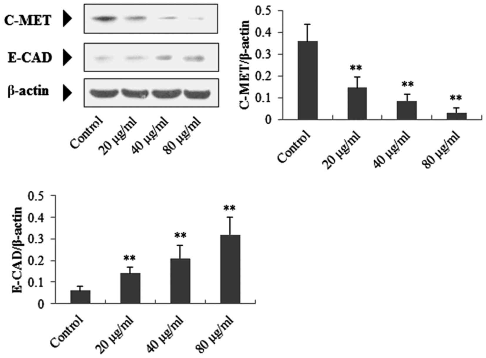
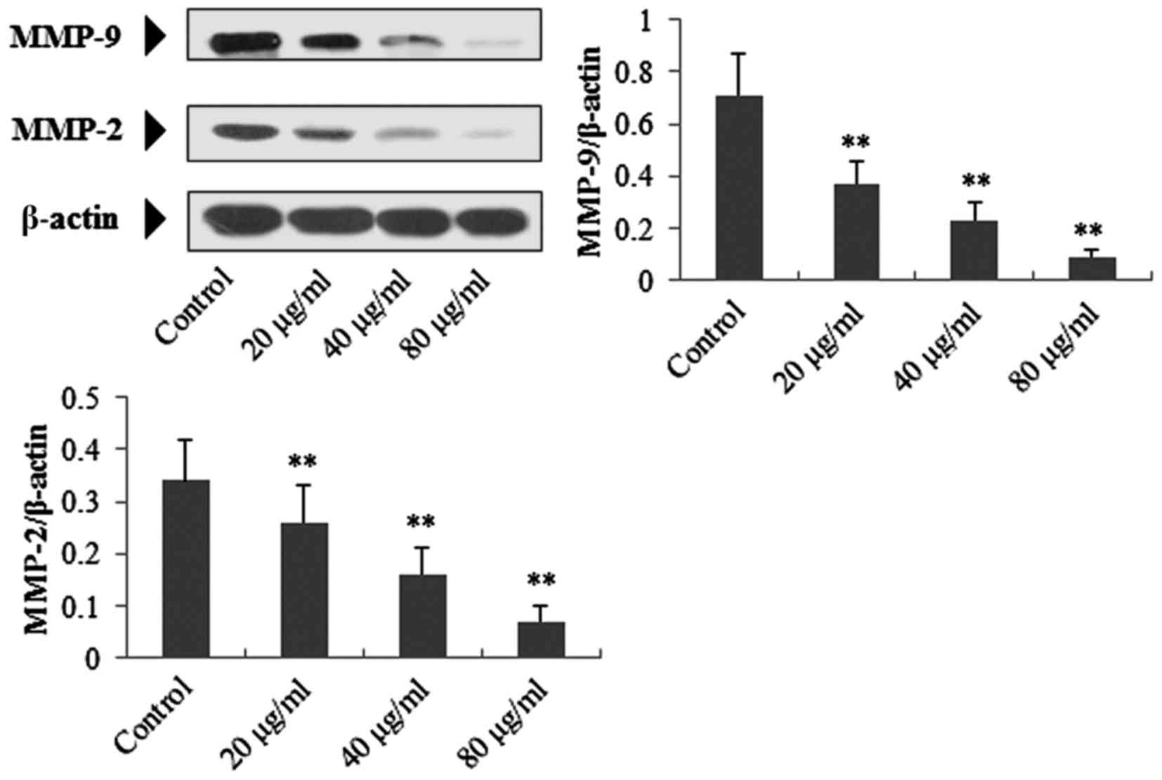
RSP downregulates c-MET, MMP-2 and
MMP-9, and upregulates E-CAD in SCC-9 cells
As presented in Figs.
7 and 8, following treatment with
RSP (20, 40 and 80 µg/ml), expression levels of c-MET (P<0.01),
MMP-2 (P<0.01) and MMP-9 (P<0.01) in SSC-9 cells were
significantly decreased compared with the control group, whereas
the expression of E-CAD (P<0.01) was increased.
Regulatory effects of RSP on the
expression of C-caspase-3, Bad, Bcl-2, c-MET, E-CAD and MMP-9 in
Cal-27 cells
The regulatory effects of RSP on caspase-3, Bax,
Bcl-2, c-MET, E-CAD and MMP-9 in Cal-27 cells were determined.
Similar to SCC-9 cells, it was identified that RSP significantly
downregulated protein expression of Bcl-2 (P<0.01), c-MET
(P<0.01) and MMP-9 (P<0.01), whereas protein expression of
C-caspase-3 (P<0.01), Bax (P<0.01) and E-CAD (P<0.01) was
upregulated (Fig. 9). These results
confirmed the antitumor effects of RSP on OSCC.
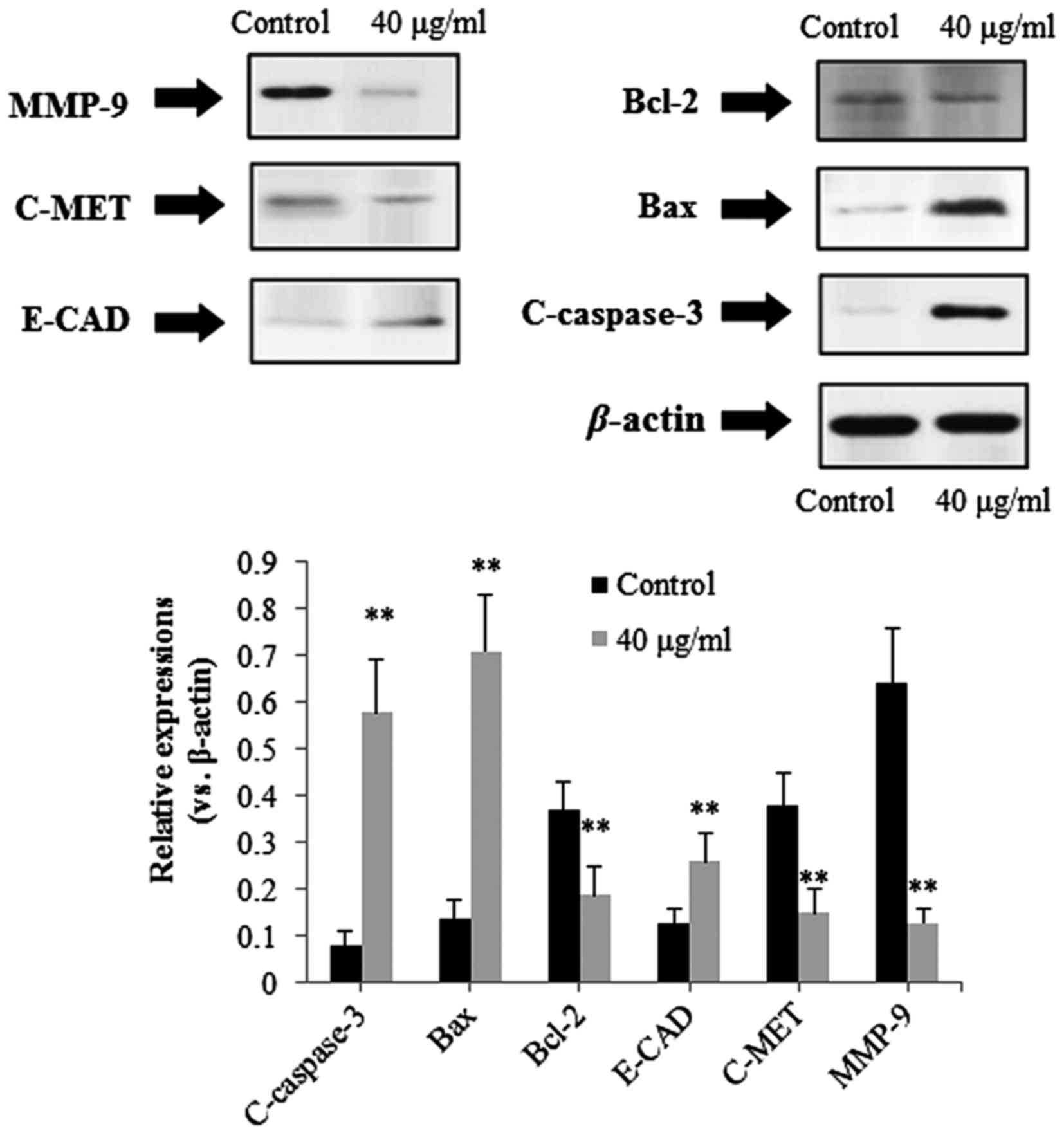 | Figure 9.Effects of RSP on caspase-3, Bax,
Bcl-2, c-MET, E-CAD and MMP-9 in Cal-27 cells. Cells were treated
with RSP (40 µg/ml) for 24 h. Total cell proteins were extracted
and subjected to western blot assay using antibodies against
caspase-3, Bax, Bcl-2, c-MET, E-CAD and MMP-9, with β-actin used as
an internal control. Results are expressed as the mean ± standard
deviation (n=4). **P<0.01 vs. control. RSP, riparsaponin; Bcl-2,
B-cell lymphoma 2; Bax, Bcl-2-associated X protein; E-CAD,
epithelial cadherin; MMP-9, matrix metalloproteinase 9. |
Discussion
To the best of our knowledge, the present study is
the first to investigate the antitumor effect of RSP on human oral
carcinoma cells. Results demonstrated that RSP exhibited a notable
antitumor effect against human oral carcinoma cells by inducing the
intrinsic apoptosis pathway.
It has been suggested previously that uncontrolled
cell proliferation and deregulation of apoptosis may be the primary
reason for the occurrence of tumors (17). In addition, evidence is accumulating
that apoptosis, a programmed physiological cell suicide process, is
considered to be an ideal strategy for inhibiting the development
and progression of cancer (18,19).
Apoptosis is characterized by distinct morphological changes
involving the nucleus (such as pyknosis, chromatin condensation,
karyorhexis and nuclear fragmentation), the plasma membrane
(phosphatidylserine exposure) and the entire cell progressively
shrinks and eventually breaks into a number of ‘apoptotic bodies’.
Furthermore, apoptosis is regulated by a series of biochemical
events and eventually results in cell death (19,20). For
apoptosis, there are two major signaling pathways including the
extrinsic and the intrinsic pathway. The extrinsic apoptosis
pathway is primarily mediated by ligand-bound death receptors of
the tumor necrosis factor receptor family, and the intrinsic
pathway, called the mitochondria-mediated apoptosis pathway, is
activated primarily by mitochondria and the Bcl-2 family. However,
the two apoptosis pathways ultimately activate members of the
caspase family (19). The present
study focused primarily on the mitochondria-mediated apoptosis
induced by RSP in oral cancer cells. Caspase-9, first activated by
cytochrome c in the cytoplasm, is the initiating caspase
protein of the caspase cascade reaction, and the activated
caspase-9 subsequently activates the executioner caspase-3.
Activated caspase-3 is commonly considered to be a biomarker for
cells undergoing apoptosis (19,21).
Proteins in the Bcl-2 family, including Bax, Bad and Bcl-2, have
been demonstrated as important members in controlling cytochrome
c release in the intrinsic apoptosis pathway (22). Bax and Bad, pro-apoptotic Bcl-2 family
members, promote the release of cytochrome c into the
cytosol (19,21). In contrast, Bcl-2 is the
anti-apoptotic protein that prevents the release of cytochrome
c by preserving mitochondrial integrity (19,20). The
results of the present study revealed that RSP upregulates
C-caspase-3, C-caspase-9, Bad and Bax, and downregulates Bcl-2,
leading to mitochondria-mediated apoptosis.
Increasingly, previous research has indicated that
metastasis is a primary reason for the recurrence and mortality of
patients with OSCC. The process of metastasis is regulated by a
variety of genes, such as E-CAD, c-MET, MMP-2 and MMP-9. E-CAD, a
membranous glycoprotein, exerts important effects in the
maintenance of cell-cell adhesion, epithelial tissue polarity
preservation and structural integrity (23–25).
Epidemiological studies have identified that the expression of
E-CAD is commonly downregulated in OSCC, thus decreased E-CAD
levels may be considered an indicator of unfavorable prognosis in
OSCC (24). c-MET, a transmembrane
tyrosine kinase receptor is commonly recognized as a crucial factor
for mediating the oncogenic activities of hepatocyte growth factor.
Upregulation of c-MET has been reported to contribute to the
progression and dissemination of various malignancies (26). MMPs, a family of zinc-dependent
proteinases, are able to degrade the majority of the extracellular
matrix components (27,28). In the development of malignancies,
activated MMPs may promote the degradation of extracellular matrix,
movement of tumor cells and tumor growth, which are directly
associated with invasion, metastasis and poor prognosis of
malignancies. Previous studies have demonstrated that MMP-2 and
MMP-9 are commonly upregulated in various types of malignant
tissues (2,29,30).
Results of the present study revealed that RSP downregulates E-MET,
MMP-2 and MMP-9, whereas RSP upregulates E-CAD, indicating that RSP
exhibits inhibitory potential on the metastasis of OSCC.
The results of the present study demonstrated that
RSP exhibited significant antitumor activity against OSCC via
inducing mitochondria-mediated apoptosis.
References
|
1
|
Huang HZ and Wang C: Molecular mechanisms
of invasion-metastasis cascade in oral cancer. J Oral Maxil Surg.
20:77–82. 2011.
|
|
2
|
Kaomongkolgit R, Cheepsunthorn P, Pavasant
P and Sanchavanakit N: Iron increases MMP-9 expression through
activation of AP-1 via ERK/Akt pathway in human head and neck
squamous carcinoma cells. Oral Oncol. 44:587–594. 2008. View Article : Google Scholar : PubMed/NCBI
|
|
3
|
Siegel R, Naishadham D and Jemal A: Cancer
statistics, 2012. CA Cancer J Clin. 62:10–29. 2012. View Article : Google Scholar : PubMed/NCBI
|
|
4
|
Malik UU, Zarina S and Pennington SR: Oral
squamous cell carcinoma: Key clinical questions, biomarker
discovery, and the role of proteomics. Arch Oral Biol. 63:53–65.
2016. View Article : Google Scholar : PubMed/NCBI
|
|
5
|
Acharya S, Rai P, Hallikeri K, Anehosur V
and Kale J: Serum lipid profile in oral squamous cell carcinoma:
Alterations and association with some clinicopathological
parameters and tobacco use. Int J Oral Maxillofac Surg. 45:713–720.
2016. View Article : Google Scholar : PubMed/NCBI
|
|
6
|
Abe S, Oikawa M, Miki Y, Shimizu Y, Suzuki
T, Takahashi T and Kumamoto H: Immunohistochemical and genetic
evaluations of epidermal growth factor receptor (EGFR) in oral
squamous cell carcinoma. J Oral Maxillofac Surg Med Pathol.
28:174–181. 2016. View Article : Google Scholar
|
|
7
|
Gohulkumar M, Gurushankar K, Prasad N
Rajendra and Krishnakumar N: Enhanced cytotoxicity and
apoptosis-induced anticancer effect of silibinin-loaded
nanoparticles in oral carcinoma (KB) cells. Mater Sci Eng C Mater
Biol Appl. 41:274–282. 2014. View Article : Google Scholar : PubMed/NCBI
|
|
8
|
Chen L, Zeng R and Zhuang Y: In vitro
anti-gastric tumor activities and possible mechanisms of action of
paederosidic acid from Paederia scandens (Lour) Merrill. Trop J
Pharm Res. 14:795–800. 2015. View Article : Google Scholar
|
|
9
|
Kinghorn AD, Chin YW and Swanson SM:
Discovery of natural product anticancer agents from biodiverse
organisms. Curr Opin Drug Discov Devel. 12:189–196. 2009.PubMed/NCBI
|
|
10
|
Peng W, Hu C, Shu Z, Han T, Qin L and
Zheng C: Antitumor activity of tatariside F isolated from roots of
Fagopyrum tataricum (L.) Gaertn against H22 hepatocellular
carcinoma via up-regulation of p53. Phytomedicine. 22:730–736.
2015. View Article : Google Scholar : PubMed/NCBI
|
|
11
|
Liu P, Yang H, Long F, Hao HP, Xu X, Liu
Y, Shi XW, Zhang DD, Zheng HC, Wen QY, et al: Bioactive equivalence
of combinatorial components identified in screening of an herbal
medicine. Pharm Res. 31:1788–1800. 2014. View Article : Google Scholar : PubMed/NCBI
|
|
12
|
Wu WY, Hou JJ, Long HL, Yang WZ, Liang J
and Guo DA: TCM-based new drug discovery and development in China.
Chin J Nat Med. 12:241–250. 2014.PubMed/NCBI
|
|
13
|
Lee I, Kim J, Kim YS, Yoo NH, Kim CS, Jo
K, Kim JH, Bach TT and Kim JS: Cycloartane-type triterpenes from
the leaves of Homonoia Riparia with VEGF-induced angiogenesis
inhibitor activity. J Nat Prod. 75:1312–1318. 2012. View Article : Google Scholar : PubMed/NCBI
|
|
14
|
State Administration of Traditional
Chinese Medicine. Chinese Material Medica; Science and Technology
Press of Shanghai: Shanghai, China. 6:824–825. 1999.
|
|
15
|
Viswanadh GS, Ramaiah PA, Laatsch H and
Maskey R: Chemical constituents of the heartwood and bark of
Homonoia riparia. J Trop Med Plants. 7:267–273. 2006.
|
|
16
|
Xu F, Zhao X, Yang L, Wang X and Zhao J: A
new cycloartane-type triterpenoid saponin xanthine oxidase
inhibitor from Homonoia riparia Lour. Molecules. 19:13422–13431.
2014. View Article : Google Scholar
|
|
17
|
Qi F, Li A, Zhao L, Xu H, Inagaki Y, Wang
D, Cui X, Gao B, Kokudo N, Nakata M and Tang W: Cinobufacini, an
aqueous extract from Bufo bufo gargarizans Cantor, induces
apoptosis through a mitochondria-mediated pathway in human
hepatocellular carcinoma cells. J Ethnopharmacol. 128:654–661.
2010. View Article : Google Scholar
|
|
18
|
Kerr JF, Winterford CM and Harnon BV:
Apoptosis. Its significance in cancer and cancer therapy. Cancer.
73:2013–2026. 1994. View Article : Google Scholar
|
|
19
|
Wang X: The expanding role of mitochondria
in apoptosis. Genes Dev. 15:2922–2933. 2001.
|
|
20
|
Galluzzi L, Zamzami N, de La Motte Rouge
T, Lemaire C, Brenner C and Kroemer G: Methods for the assessment
of mitochondrial membrane permeabilization in apoptosis. Apoptosis.
12:803–813. 2007. View Article : Google Scholar
|
|
21
|
Shi YG: A structural view of
mitochondria-mediated apoptosis. Nat Struct Biol. 8:394–401. 2011.
View Article : Google Scholar
|
|
22
|
Chipuk JE, McStay GP, Bharti A, Kuwana T,
Clarke CJ, Siskind LJ, Obeid LM and Green DR: Sphingolipid
metabolism cooperates with BAK and BAX to promote the mitochondrial
pathway of apoptosis. Cell. 148:988–1000. 2012. View Article : Google Scholar
|
|
23
|
Beavon IR: The E-cadherin-catenin complex
in tumour metastasis: Structure, function and regulation. Eur J
Cancer. 36:1607–1620. 2000. View Article : Google Scholar
|
|
24
|
Martínez A, Spencer ML, Borlando J, Flores
M and Rojas IG: E-cadherin and c-Met expression in actinic cheilits
and lip squamous cell carcinoma. Rev Clin Periodoncia Implantol
Rehabil Oral. 4:122–125. 2011. View Article : Google Scholar
|
|
25
|
Yang XK, Yang YD, Tang SQ, Xu L, Yang GH,
Xu QY, Tang H and Wu JJ: Inhibitory effect of polysaccharides from
Scutellaria barbata D. Don on invasion and metastasis of 95-D cells
lines via regulation of C-MET and E-CAD expressions. Trop J Pharm
Res. 12:517–522. 2013.
|
|
26
|
Yang L, Liu M, Deng C, Gu Z and Gao Y:
Expression of transforming growth factor-β1 (TGF-β1) and E-cadherin
in glioma. Tumour Biol. 33:1477–1484. 2012. View Article : Google Scholar
|
|
27
|
Qin Y, Ye GX, Wu CJ, Wang S, Pan DB, Jiang
JY, Fu J and Xu SQ: Effect of DAPK1 gene on proliferation,
migration, and invasion of carcinoma of pancreas BxPC-3 cell line.
Int J Clin Exp Pathol. 7:7536–7544. 2014.
|
|
28
|
Sanchavanakit N, Saengtong W,
Manokawinchoke J and Pavasant P: TNF-α stimulates MMP-3 production
via PGE2 signalling through the NF-κB and p38 MAPK pathway in a
murine cementoblast cell line. Arch Oral Biol. 60:1066–1074. 2015.
View Article : Google Scholar
|
|
29
|
Duxbury MS and Whang EE: RRM2 induces
NF-kappaB-dependent MMP-9 activation and enhances cellular
invasiveness. Biochem Biophys Res Commun. 354:190–196. 2007.
View Article : Google Scholar
|
|
30
|
Silva EJ, Argyris PP, Zou XQ, Ross KF and
Herzberg MC: S100A8/A9 regulates MMP-2 expression and invasion and
migration by carcinoma cells. Int J Biochem Cell Biol. 55:279–287.
2014. View Article : Google Scholar
|