Introduction
Royal jelly (RJ) is a thick and milky liquid
secreted by hypopharyngeal, and mandibular glands of young worker
bees (Apismellifera L.), and is used to feed the larvae
(1). RJ exhibits a wide variety of
functional properties, and has been widely used in cosmetics,
healthy foods and commercial medical products in numerous
countries. Additionally, RJ possesses various properties, including
antimicrobial (2), anti-inflammatory
(3,4),
hepatoprotective (5), antisarcopenia
(6), insulin-like action (7) and antihypercholesterolemic (8) properties. In the recent decades, there
have been an increasing number of studies investigating the effects
of RJ.
Regarding breast cancer, RJ has been reported to be
able to inhibit the growth-promoting effect of bisphenol A, which
is an estrogen that enhances the proliferation of MCF-7 mammary
cancer cells (9). RJP30, a substance
obtained from RJ crude protein, which was extracted by
precipitation with 30% ammonium sulfate, was demonstrated to be
cytotoxic for HeLa human cervicouterine carcinoma cells, and
diminish cell density to 50% of the original carcinoma cell after 7
days of treatment (10). RJ exhibited
no significant effects on the formation of metastases in
spontaneous mammary carcinoma of CBA mice when it was given
intraperitoneally or subcutaneously (11). However, when RJ and tumor cells were
injected synchronously into mice, RJ was able to inhibit the
formation of metastases in the lung (11). The therapeutic properties of RJ have
been examined in various cells, including MCF-7 (mouse
macrophages), spontaneous mammary carcinoma cells and
methylcholanthrene-induced fibrosarcoma CBA mouse cells (9,11,12).
RJ exhibits estrogenic activity. It was identified
that RJ stimulated the expression of endogenous estrogen-responsive
genes [presenilin 2 and vascular endothelial growth factor (VEGF)]
by activating estrogen receptors in MCF-7 cells, and RJ could
restore the expression of VEGF in uterus of rats that received
bilateral ovariectomy (13).
Experimental results demonstrated that four compounds from RJ can
improve the transcription of a reporter gene, which contained an
estrogen-responsive element, and subcutaneously injecting immature
rats with those compounds for 23 days induced mild hypertrophy in
the luminal epithelium of the rat uterus (14). With evidence that it was an
etiological cause of breast cancer, estrogen attracted numerous
studies on its association with breast carcinogenesis (15). Samavat and Kurzer (16) evaluated the roles and metabolites of
circulating, and urinary estrogens in human breast cancer.
Endocrine therapy is regarded as one of the earliest
molecular-targeting therapies applied in breast cancer treatment
(17). However, the effect of RJ on
breast cancer has been seldom studied.
The 4T1 tumor closely mimics human breast cancer
with regards to anatomically correct site, immunogenicity and
growth characteristics (18,19). Therefore, the 4T1 tumor is a suitable
model for testing the therapy effects under experimental
conditions.
Breast cancer is associated with high morbidity and
mortality rates among women in developed and developing countries
(20). However, the effect of RJ on
mammary cancer cells remains unclear. Therefore, a detailed and
comprehensive study on the effect of RJ on breast cancer may aid in
understanding the association between RJ and breast cancer.
The present study aimed to analyze the effect of RJ
on breast cancer. The primary purpose of the current study was to
investigate the potential antioxidant ability and immune response
of RJ on 4T1-bearing BALB/c mice, and changes in the weights of
different organs.
Materials and methods
Cell culture
Mouse mammary tumor cells (4T1) were purchased from
Culture Collection of the Chinese Academy of Sciences (Shanghai,
China). All culture work was performed under strict aseptic
conditions. Cells were cultured in RPMI-1640 (HyClone; GE
Healthcare Life Sciences, Logan, UT, USA) supplemented with 10%
fetal bovine serum (Gibco; Thermo Fisher Scientific, Inc., Waltham,
MA, USA) at 37°C in a 5% CO2 humidified incubator.
Mice
Female BALB/c mice (4 weeks old, 18–22 g) were
purchased from Shanghai SLAC Laboratory Animal Co., Ltd. (Shanghai,
China), and the present study was ethically approved by the Animal
Care and Use Committee, Laboratory Animal Center of Fujian Medical
University (Fuzhou, China). The mice were housed four to a cage
under specific pathogen-free conditions at constant temperature
(25–28°C) with a 12 h light/12 h dark schedule, and were fed with
autoclaved food and water.
Tumor model
All 4T1-bearing mice were injected subcutaneously
with 4T1 cells (4×10⁵) suspended in 0.2 ml of PBS in the
mammary fat pads (21) in the right
flank of each mouse when treated for 14 days. The tumor was
detected through palpation around the induction area.
RJ and treatment regimen
Fresh RJ was obtained from a local beekeeping
association, Yuhang Apiary (Zhejiang, China), and was stored at
−20°C before prior to use. A total of 56 female BALB/c mice were
divided into seven groups with eight mice in each group. One of the
seven groups was a 4T1 control group, and was fed with water with
no RJ content during the experiment. Three groups were administered
orally with RJ solution using a prophylactic-therapeutic method
(PTRJ groups): The mice were treated with RJ 14 days prior to the
transplantations of tumor cells, and then continued to be treated
with RJ for 28 consecutive days. The other three groups were
administered with RJ using a therapeutic method (TRJ groups): after
the transplantations of tumor cells, the mice were treated with RJ
for 28 days. For each of the three groups receiving RJ treatment,
the daily doses of RJ were 0.5, 1.0 and 1.5 g/kg, respectively. RJ
solution was prepared by dissolving RJ in distilled water (22). The weight of the mice and tumor sizes
were measured periodically. All the mice were sacrificed after 24 h
of the final administration of RJ (22), subsequently blood and tissue samples
were collected, and stored for further experiments.
Weight of organs
Following the sacrifice of the mice, necropsy was
performed by removing the tumor, kidney, thymus, spleen, lung and
liver from the bodies. Then, these organs were weighed with using a
one over ten-thousand analytical balance.
Antioxidant and immunomodulatory
activities of serum
The blood samples were centrifuged for 10 min at
3,000 × g at room temperature, and the serum was aliquoted and
stored in plastic eppendorf tubes. The serum was analyzed for the
concentrations of various cytokines, including interleukin (IL)-2,
IL-4, IL-10, interferon (IFN)-α and IFN-γ, all of which are
associated with immunomodulation, using Mouse IL-2 Quantikine ELISA
kit (cat no. M2000), Mouse IL-4 Quantikine ELISA kit (cat no.
M4000B), Mouse IL-10 Quantikine ELISA kit (cat no. M1000B), Mouse
IFN-α Quantikine ELISA kit (cat no. 42120-1) and Mouse IFN-γ
Quantikine ELISA kit (cat no. MIF00), according to the
manufacturer's protocol, respectively. These ELISA kits were
purchased from R&D Systems, Inc., Minneapolis, MN, USA. The
Total Antioxidant Capacity Assay kit (cat no. S0119) and
Glutathione Reductase Assay kit (cat no. S0055) were purchased from
Beyotime Institute of Biotechnology (Haimen, China) and were used
to determine the concentrations of various antioxidant-associated
indicators, including total antioxidant capacity (T-AOC) and
glutathione reductase (GR), according to the manufacturer's
protocol.
Antioxidant ability of liver and
kidney
The effects of RJ on antioxidant markers were
assessed by measuring the activities of liver- or kidney-associated
enzymes, including superoxide dismutase (SOD), GR and T-AOC, in the
homogenate using the following assay kits: Total Antioxidant
Capacity Assay kit (cat no. S0119), Glutathione Reductase Assay kit
(cat no. S0055) and Total Superoxide Dismutase Assay kit (cat no.
S0102), according to the manufacturer's instructions. These assay
kits were obtained from Beyotime Institute of Biotechnology.
Statistical analysis
All data are expressed as the mean ± standard error
of the mean for in vitro studies. Statistical analysis was
performed using one-way analysis of variance followed with Tukey's
post hoc test using the SPSS version 17 (SPSS, Inc., Chicago, IL,
USA). P<0.05 was considered to indicate a statistically
significant difference.
Results
Effects of RJ on the weight of
organs
As demonstrated in Fig.
1, PTRJ0.5 and PTRJ1.5 significantly inhibited tumor growth of
4T1 mammary tumors in female BALB/c mice as compared with the 4T1
control, but no significant antitumor effects were observed in the
PTRJ1.0 group. In the TRJ groups, although the development of
tumors was also inhibited, the inhibitory effect was not
significant compared with the 4T1 control group. No significant
differences were identified among the 4T1 control group and RJ
groups regarding the mean weights of the lung, and kidney of the
mice.
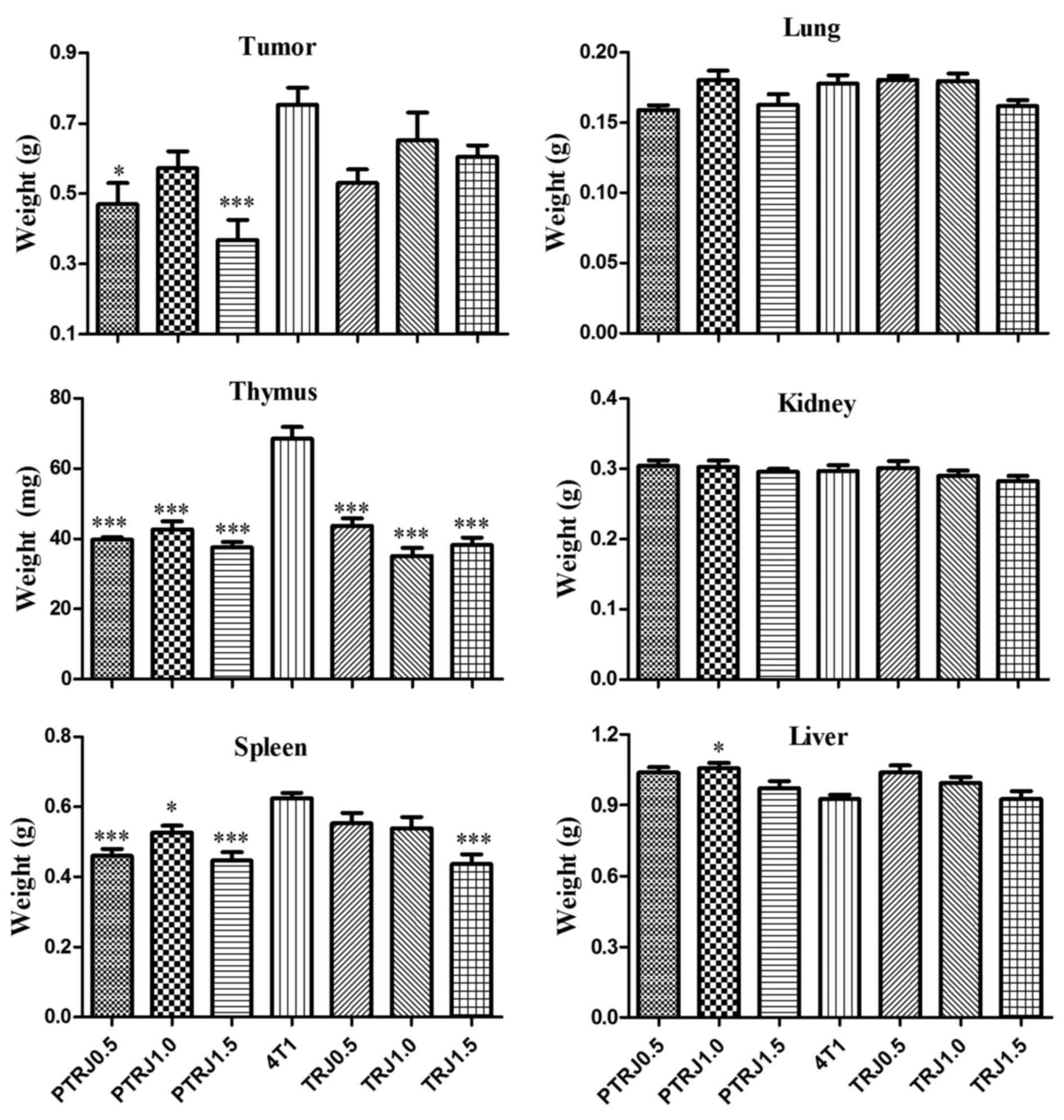 | Figure 1.Mean weight of various organs, tumor,
lung, spleen, liver, thymus and kidney, in the PTRJ, 4T1, and TRJ
groups. Data are expressed as the mean ± standard error of the
mean. *P<0.05 and ***P<0.001 vs. 4T1-bearing mice. PTRJ,
prophylactic-therapeutic royal jelly group; TRJ, therapeutic royal
jelly group. |
Compared with those in the 4T1 control group, the
weight of the thymus and spleen in all three PTRJ groups was
significantly decreased. For the TRJ groups, the weight of thymus
was only significantly reduced in the TRJ1.5 group compared with
that of 4T1 control group. Contradictory results were identified
regarding the weight of liver, all the TRJ and PTRJ groups
possessed livers that were slightly heavier compared with that of
4T1 control group, and was significantly heavier in the PTRJ1.0
group.
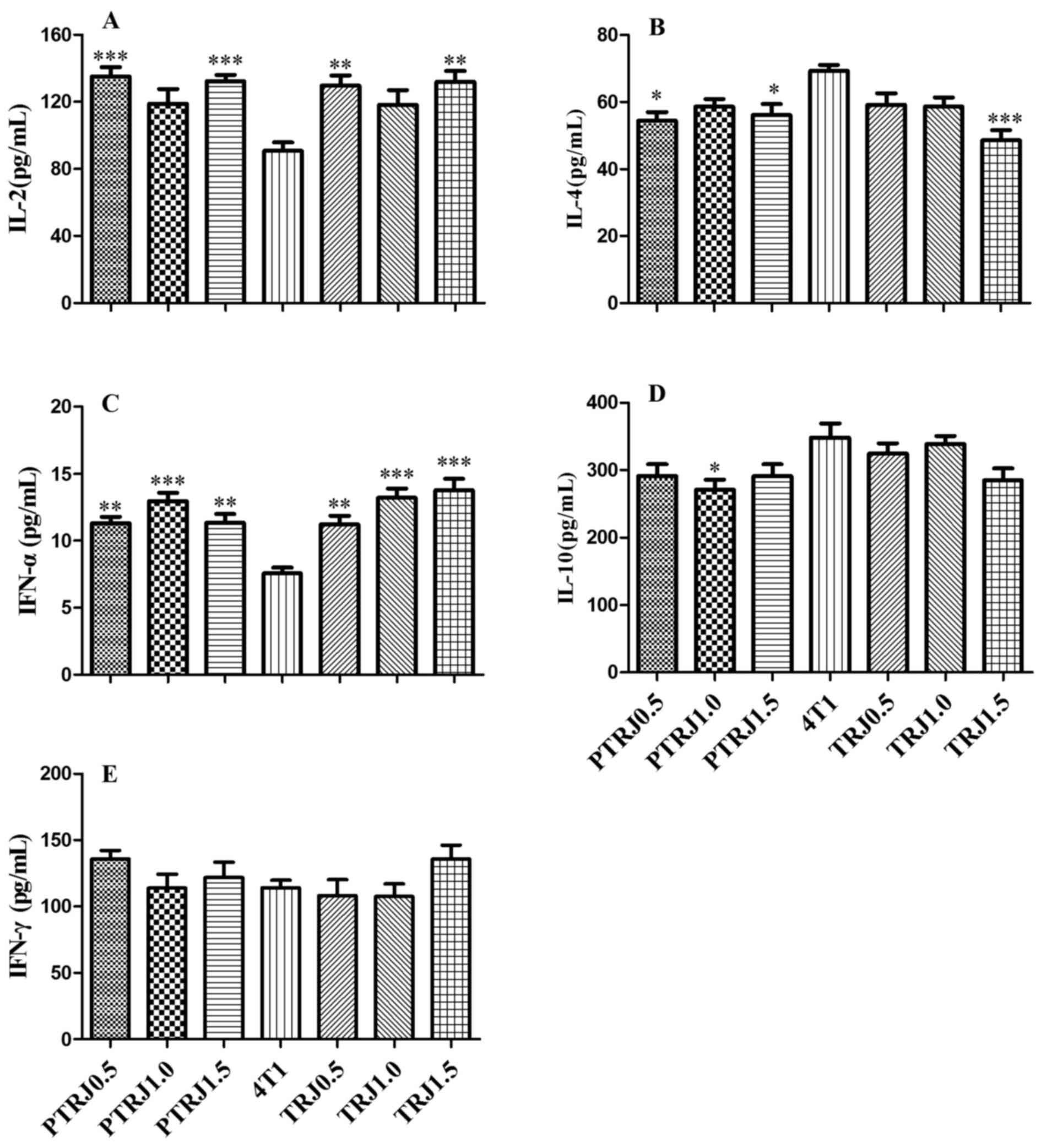
Effects of RJ on the serum
Fig. 2 presents the
cytokine concentrations of the serum collected from mice. Compared
with the 4T1 control group, PTRJ0.5, PTRJ1.5, TRJ0.5 and TRJ1.5
groups possessed significantly higher IL-2 concentrations. However,
PTRJ0.5, PTR1.0 and TRJ1.5 groups had significantly lower levels of
IL-4. The concentration of IFN-α in all RJ-treated groups was
significantly higher compared with that in 4T1 control group. The
level of IL-10 was significantly lower in the PTRJ1.0 group
compared with that in 4T1 control group. IFN-γ activity of all
RJ-treated groups had similar values with that of 4T1 control
group.
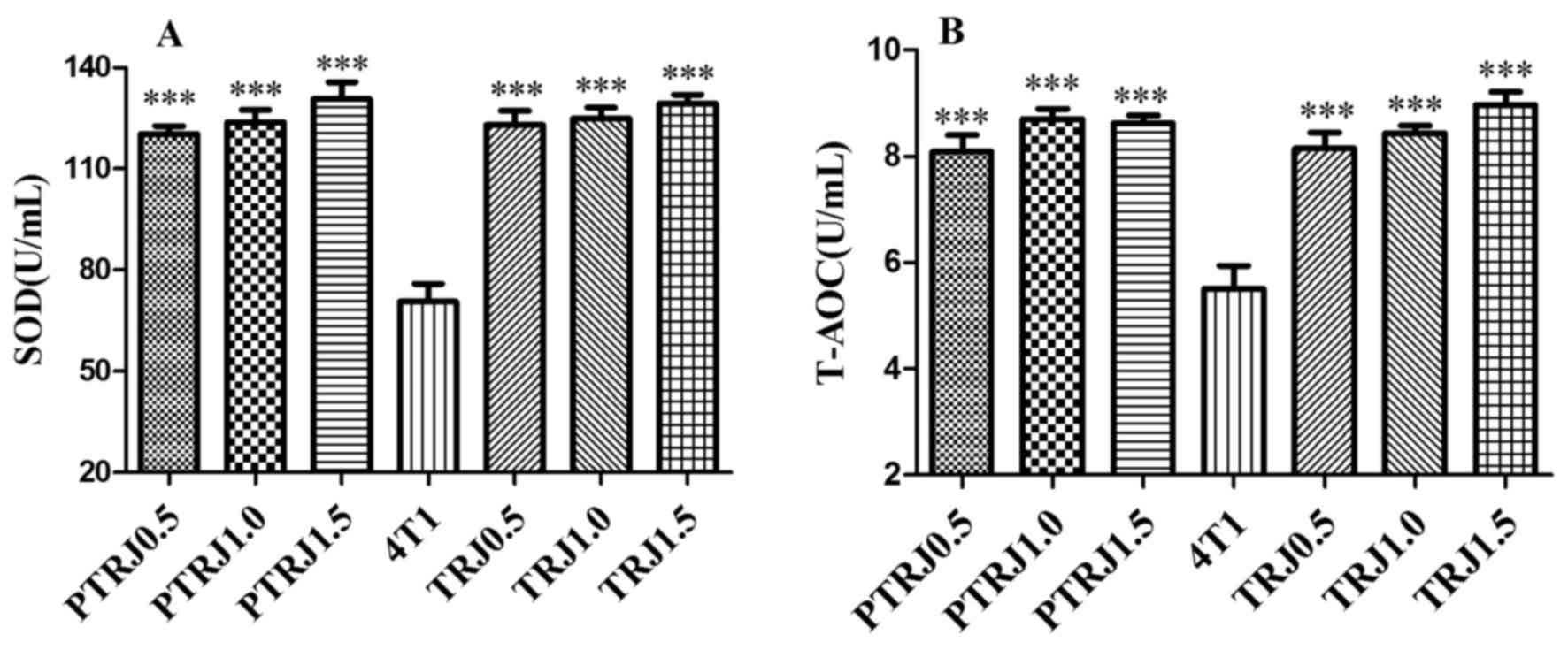
The activities of enzymatic antioxidants, including
SOD and GR in the serum are presented in Fig. 3. When compared with the 4T1 control
groups, SOD and T-AOC concentrations were significantly augmented
in all RJ-treated groups.
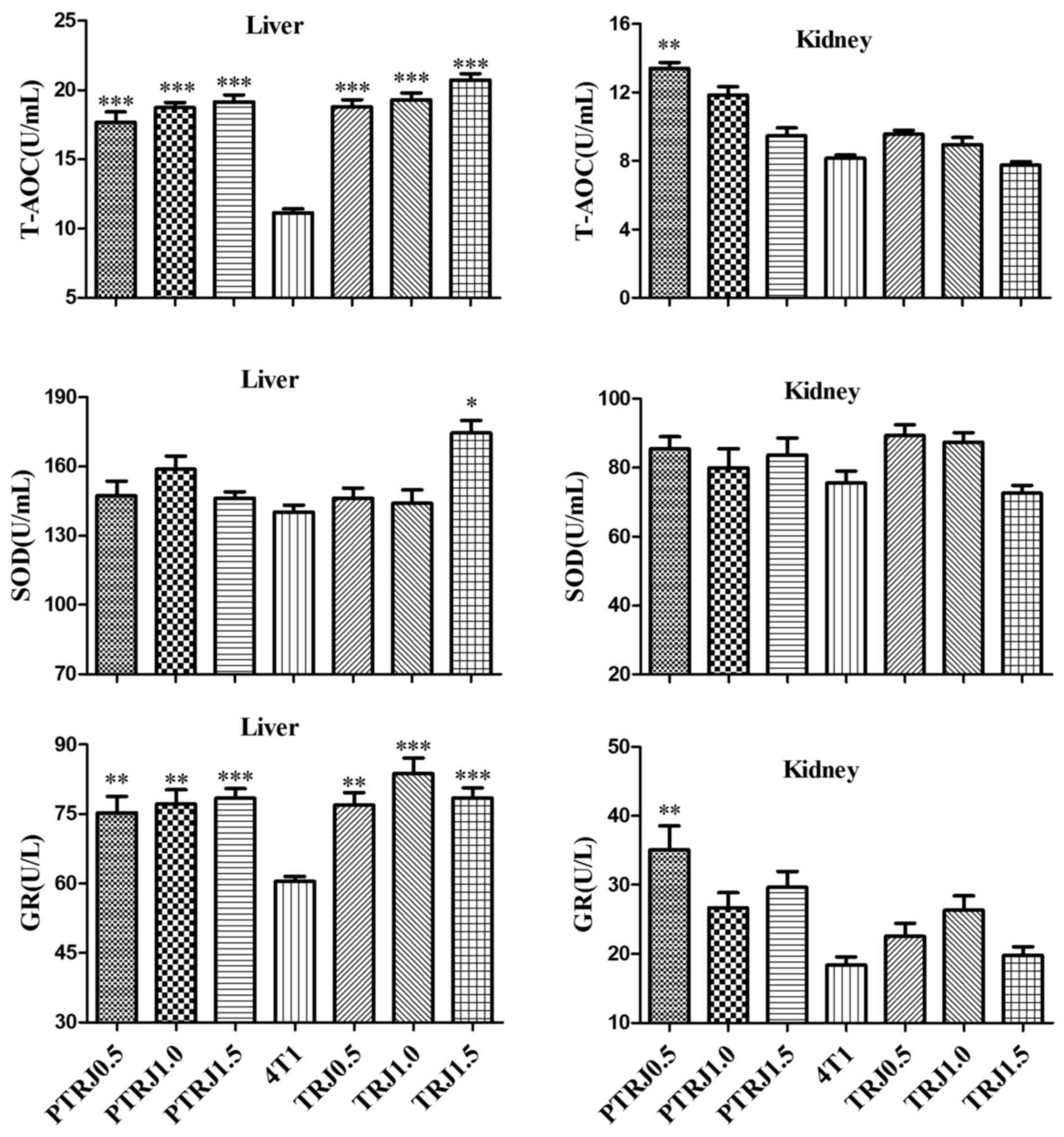
Effects of RJ on the antioxidant
ability of the liver and kidney
In the liver homogenates of RJ-treated groups, T-AOC
and GR concentrations were significantly augmented compared with
those of the 4T1 control group; while only TRJ1.5 group had an SOD
value significantly higher compared with that of the 4T1 control
group (Fig. 4). For the kidney
homogenates, there was no significant difference in SOD activity
among the groups in the experiment. The value of T-AOC and GR of
all the groups was similar to those of the 4T1 control group,
except the PTRJ0.5 group, which had significantly higher T-AOC and
GR values.
Discussion
Previous studies have reported antitumoral effects
of RJ on various types of tumor, including Ehrlich ascetic tumor
(22), Sarcoma-180 ascites (23), and fibrosarcoma transplantable tumors
(24). For the majority of tumors, RJ
significantly prolongs the average life span of murine tumor models
and the effect depends on the treatment protocol used (22,23). The
results of the present study suggested that the doses of RJ
solution applied were nontoxic to the mice, as no abnormal
fluctuation in weight or behaviors of mice was observed. It was
demonstrated that RJ significantly inhibited tumor growth in
4T1-bearing mice when orally administered in
prophylactic-therapeutic method with the doses of 0.5 and 1.5 g/kg;
however, RJ had no significant effect on growth of 4T1-bearing mice
when given to mice following tumor cell inoculation. This result
suggests that the antitumor effect of RJ primarily depends on the
time of application, and RJ has little or no estrogen agonism on
4T1-bearing mice. Furthermore, the results demonstrated that
administering RJ orally using the prophylactic-therapeutic method
may benefit controlling tumor growth. Accumulating evidence
suggested the antitumor effect of RJ may be due to
10-hydroxy-2-decenoic acid and saturated fatty acids (25,26).
No significant effects were observed on the weight
of lung and kidney following both RJ treatments. Male (27) reported that cell-mediated and humoral
immunity were affected during immune suppression, and this
phenomenon was reflected in the changes in the functions of spleen
and thymus. The thymus weight of all the groups, which were treated
with RJ, was significantly reduced compared with that of 4T1
control group. In addition, all three PTRJ groups and the TRJ1.5
group exhibited significantly reduced spleen weights compared with
the 4T1 control group. These results suggest that the weight of the
spleen and thymus are significantly reduced with RJ treatment.
Since the immune organ index is only a superficial indicator of
immune function (28), the precise
effects of RJ on immune system require further study.
It has been suggested that immunomodulation of a
host system could inhibit tumor growth; therefore, immunotherapy is
considered as an adjuvant therapy for cancer treatment (29). Certain studies have reported that
targeting tumor-associated plasmacytoid dendritic cells to restore
their IFN-α production may be an achievable therapeutic strategy to
induce antitumor immunity in breast cancer (30–32). These
studies suggested that IFN-α was important for the inhibition of
breast cancer development (30–32). In
the present study, the level of IFN-α in all six groups treated
with RJ was significantly increased, indicating that RJ serves an
important role in immunomodulation.
The immune stimulating effect of RJ has been
investigated. Low concentrations of MEL 174 (final water extract of
RJ) and MEL 147 (3–10-dihydroxydecanoic acid) stimulated rat T-cell
proliferation in vitro, and resulted in an increased
production of IL-2 of Type 1 T helper (Th1)-associated cytokines
(33). The results obtained in the
present study support the above findings. The serum concentration
of IL-2 in PTRJ and TRJ-treated mice (0.5 and 1.5 g/kg) were
significantly higher compared with that of the 4T1 control group.
For the other groups treated with RJ, the level of IL-2 was
slightly; however, not significantly increased. Thus, these results
suggest that the IL-2 cytokine is involved in tumor growth
inhibition. The levels of IL-4, a Type 2 T helper (Th2)-associated
cytokine, in the PTRJ0.5, PTRJ1.0 and TRJ1.5 groups, and IL-10 in
the PTRJ1.0 group were significantly reduced compared with that of
the 4T1 control group. Furthermore, the levels of IL-4 and IL-10
were slightly, but insignificantly decreased in all other groups
treated with RJ. These data suggest that treatment with RJ
suppressed Th2 activity and associated cytokine release in
4T1-bearing mice. The production of IL-4 may be restrained by major
royal jelly protein 3, isolated from RJ major proteins (34). No significant difference was
identified among the IFN-γ value of the groups in the experiment.
However, the level of IFN-γ in the PTRJ0.5, PTRJ1.5, and PTRJ0.5
groups was slightly higher compared that of 4T1 control group.
Together, these results suggest that the RJ immunoprotective role
acts by increasing the level of Th1-associated cytokines and
suppressing the level of Th2-associated cytokines. RJ treatment
resulted in increased Th1/Th2 cytokine ratios in the present study,
and similar results were also reported by Oka et al
(35) and Erem et al (36).
It was reported that oxidant stress was important to
carcinogenesis and the accumulation of reactive oxygen species
(37), and it highlighted the
significance of antioxidant defense enzymes for cancer therapy.
Certain reports suggested that patients with breast cancer require
improved antioxidation abilities (38,39). It
was hypothesized that the antitumor effects of RJ may be partially
due to its antioxidant properties. Regarding the observations made
in the current study, the levels of SOD and T-AOC in the serum, in
addition to the level of T-AOC, and GR in the liver were
significantly increased following RJ treatment. Furthermore, in the
liver, the level of SOD in the TRJ1.5 group was significantly
increased compared with that of the 4T1 control group. The
concentration of T-AOC and GR in the kidney was significantly
increased in the PTRJ0.5 group. Additionally, the levels of T-AOC,
GR and SOD in the kidney of the PTRJ1.0, PTRJ1.5, TRJ0.5, and
TRJ1.0 groups were augmented, but the increases were not
statistically significant. These data suggest that although the
functions of RJ on the antioxidant activities in the serum, liver
and kidney were different, overall, the antioxidant activities
increased following RJ supplementation in 4T1-bearing mice. These
results are in agreement with the literature on the antioxidant
properties of RJ (40,41). It is possible that administration of
RJ enhances antioxidant activity, since RJ contains a mixture of
peptides, flavonoids and cinnamic acid derivatives (42). Thus, the antioxidant effect of RJ may
contribute to the growth inhibition of 4T1 mammary tumors in
mice.
There are other cytokines, besides the cytokines
mentioned in the present study, which possess antitumor effects
in vivo (29,43). It is possible that the antitumor
effect of RJ on 4T1-bearing mice is associated with the action of
such cytokines, including, TNF-α, IL-1, IL-6, IL-13 (41) and IL-27 (29).
In conclusion, the antioxidant and immunomodulatory
effects of RJ on the 4T1 breast cancer mice model were investigated
in the present study. RJ treatment was demonstrated to reduce the
development of breast tumors in mice, and improve the antioxidant
activity in the serum, liver and kidney, particularly following
PTRJ treatment. These positive effects corroborate the efficacy of
RJ supplementation in diets. The antioxidant and immunomodulatory
effects of RJ likely serve an important antitumor role. Notably, it
should be taken into account that the therapeutic method of RJ may
be used ≥14 days prior to tumor inoculation.
Acknowledgements
The research was supported by the earmarked fund
provided by the Finance Department of Fujian province (grant no.
K81MLV02A) and the Modern Agro-industry Technology Research System
(grant no. CARS-45-KXJ3).
References
|
1
|
Isidorov V, Czyżewska U, Isidorova A and
Bakier S: Gas chromatographic and mass spectrometric
characterization of the organic acids extracted from some
preparations containing lyophilized royal jelly. J Chromatogr B
Analyt Technol Biomed Life Sci. 877:3776–3780. 2009. View Article : Google Scholar : PubMed/NCBI
|
|
2
|
Fujiwara S, Imai J, Fujiwara M, Yaeshima
T, Kawashima T and Kobayashi K: A potent antibacterial protein in
royal jelly. purification and determination of the primary
structure of royalisin. J Biol Chem. 265:11333–11337.
1990.PubMed/NCBI
|
|
3
|
Karaca T, Uz YH, Demirtas S, Karaboga I
and Can G: Protective effect of royal jelly in 2,4,6
trinitrobenzene sulfonic acid-induced colitis in rats. Iran J Basic
Med Sci. 18:370–379. 2015.PubMed/NCBI
|
|
4
|
Arzi A, Olapour S, Yaghooti H and
Karampour Sistani N: Effect of royal jelly on formalin
induced-inflammation in rat hind paw. Jundishapur J Nat Pharm Prod.
10:e224662015. View Article : Google Scholar : PubMed/NCBI
|
|
5
|
Yildirim S, Karadeniz A, Karakoc A,
Yildirim A, Kalkan Y and Şimşek N: Effects of royal jelly on liver
paraoxonase activity in rats treated with cisplatin. Turk J Med
Sci. 42:367–375. 2012.
|
|
6
|
Niu K, Guo H, Guo Y, Ebihara S, Asada M,
Ohrui T, Furukawa K, Ichinose M, Yanai K, Kudo Y, et al: Royal
jelly prevents the progression of sarcopenia in aged mice in vivo
and in vitro. J Gerontol A Biol Sci Med Sci. 68:1482–1492. 2013.
View Article : Google Scholar : PubMed/NCBI
|
|
7
|
Nomura M, Maruo N, Zamami Y, Takatori S,
Doi S and Kawasaki H: Effect of long-term treatment with royal
jelly on insulin resistance in otsuka long-evans tokushima fatty
(OLETF) rats. Yakugaku Zasshi. 127:1877–1882. 2007. View Article : Google Scholar : PubMed/NCBI
|
|
8
|
Kashima Y, Kanematsu S, Asai S, Kusada M,
Watanabe S, Kawashima T, Nakamura T, Shimada M, Goto T and Nagaoka
S: Identification of a novel hypocholesterolemic protein, major
royal jelly protein 1, derived from royal jelly. PLoS One.
9:e1050732014. View Article : Google Scholar : PubMed/NCBI
|
|
9
|
Nakaya M, Onda H, Sasaki K, Yukiyoshi A,
Tachibana H and Yamada K: Effect of royal jelly on bisphenol
A-induced proliferation of human breast cancer cells. Biosci
Biotechnol Biochem. 71:253–255. 2007. View Article : Google Scholar : PubMed/NCBI
|
|
10
|
Salazar-Olivo L and Paz-González V:
Screening of biological activities present in honeybee (Apis
mellifera) royal jelly. Toxicol In Vitro. 19:645–651. 2005.
View Article : Google Scholar : PubMed/NCBI
|
|
11
|
Oršolić N, Terzić S, Šver L and Bašić I:
Honey-bee products in prevention and/or therapy of murine
transplantable tumours. J Sci Food Agric. 85:363–370. 2005.
View Article : Google Scholar
|
|
12
|
Šimúth J, Bíliková K, Kováčová E, Kuzmová
Z and Schroder W: Immunochemical approach to detection of
ddulteration in honey: Physiologically active royal jelly protein
stimulating TNF-alpha release is a regular component of honey. J
Agric Food Chem. 52:2154–2158. 2004. View Article : Google Scholar : PubMed/NCBI
|
|
13
|
Mishima S, Suzuki KM, Isohama Y, Kuratsu
N, Araki Y, Inoue M and Miyata T: Royal jelly has estrogenic
effects in vitro and in vivo. J Ethnopharmacol. 101:215–220. 2005.
View Article : Google Scholar : PubMed/NCBI
|
|
14
|
Suzuki KM, Isohama Y, Maruyama H, Yamada
Y, Narita Y, Ohta S, Araki Y, Miyata T and Mishima S: Estrogenic
activities of fatty acids and a sterol isolated from royal jelly.
Evid Based Complement Alternat Med. 5:295–302. 2008. View Article : Google Scholar : PubMed/NCBI
|
|
15
|
Althuis MD, Fergenbaum JH, Garcia-Closas
M, Brinton LA, Madigan MP and Sherman ME: Etiology of hormone
receptor-defined breast cancer: A systematic review of the
literature. Cancer Epidemiol Biomarkers Prev. 13:1558–1568.
2004.PubMed/NCBI
|
|
16
|
Samavat H and Kurzer MS: Estrogen
metabolism and breast cancer. Cancer Lett. 356:231–243. 2015.
View Article : Google Scholar : PubMed/NCBI
|
|
17
|
Iwase H: Treatment strategy for metastatic
breast cancer with estrogen receptor-positive tumor. Int J Clin
Oncol. 20:249–252. 2015. View Article : Google Scholar : PubMed/NCBI
|
|
18
|
Taavoni S, Barkhordari F, Goushegir A and
Haghani H: Effect of Royal Jelly on premenstrual syndrome among
Iranian medical sciences students: A randomized, triple-blind,
placebo-controlled study. Complement Ther Med. 22:601–606. 2014.
View Article : Google Scholar : PubMed/NCBI
|
|
19
|
Pulaski BA and Ostrand-Rosenberg S:
Reduction of established spontaneous mammary carcinoma metastases
following immunotherapy with major histocompatibility complex class
II and B7. 1 cell-based tumor vaccines. Cancer Res. 58:1486–1493.
1998.PubMed/NCBI
|
|
20
|
Ferlay J, Shin HR, Bray F, Forman D,
Mathers C and Parkin DM: Estimates of worldwide burden of cancer in
2008: GLOBOCAN 2008. Int J Cancer. 127:2893–2917. 2010. View Article : Google Scholar : PubMed/NCBI
|
|
21
|
Luo KW, Ko CH, Yue GG, Lee JK, Li KK, Lee
M, Li G, Fung KP, Leung PC and Lau CB: Green tea (Camellia
sinensis) extract inhibits both the metastasis and osteolytic
components of mammary cancer 4T1 lesions in mice. J Nutr Biochem.
25:395–403. 2014. View Article : Google Scholar : PubMed/NCBI
|
|
22
|
Bincoletto C, Eberlin S, Figueiredo CA,
Luengo MB and Queiroz ML: Effects produced by royal jelly on
haematopoiesis: Relation with host resistance against ehrlich
ascites tumour challenge. Int Immunopharmacol. 5:679–688. 2005.
View Article : Google Scholar : PubMed/NCBI
|
|
23
|
Tamura T, Fujii A and Kuboyama N:
Antitumor effects of royal jelly (RJ). Nihon Yakurigaku Zasshi.
89:73–80. 1987.(In Japanese). View Article : Google Scholar : PubMed/NCBI
|
|
24
|
Shirzad M, Kordyazdi R, Shahinfard N and
Nikokar M: Does royal jelly affect tumor cells ? J HerbMed
Pharmacol. 2:45–48. 2013.
|
|
25
|
Townsend GF, Morgan JF and Hazlett B:
Activity of 10-hydroxydecenoic acid from royal jelly against
experimental leukaemia and ascitic tumours. Nature. 183:1270–1271.
1959. View Article : Google Scholar : PubMed/NCBI
|
|
26
|
Townsend GF, Morgan JF, Tolnai S, Hazlett
B, Morton HJ and Shuel RW: Studies on the in vitro antitumor
activity of fatty acids I. 10-hydroxy-2-decenoic acid from royal
jelly. Cancer Res. 20:503–510. 1960.PubMed/NCBI
|
|
27
|
Male D: Reactions against blood cells and
plateletsInL Immunology. 4th Edition. Roitt I, Brostoff J and Male
D: Mosby; London: pp. 3–23. 1996
|
|
28
|
Liu RM, Zhang XJ, Liang GY, Yang YF, Zhong
JJ and Xiao JH: Antitumor and antimetastatic activities of
chloroform extract of medicinal mushroom cordyceps taii in mouse
models. BMC Complement Altern Med. 15:2162015. View Article : Google Scholar : PubMed/NCBI
|
|
29
|
Murugaiyan G and Saha B: IL-27 in tumor
immunity and immunotherapy. Trends Mol Med. 19:108–116. 2013.
View Article : Google Scholar : PubMed/NCBI
|
|
30
|
Sisirak V, Faget J, Gobert M, Goutagny N,
Vey N, Treilleux I, Renaudineau S, Poyet G, Labidi-Galy SI,
Goddard-Leon S, et al: Impaired IFN-α production by plasmacytoid
dendritic cells favors regulatory T-cell expansion that may
contribute to breast cancer progression. Cancer Res. 72:5188–5197.
2012. View Article : Google Scholar : PubMed/NCBI
|
|
31
|
Sisirak V, Vey N, Goutagny N, Renaudineau
S, Malfroy M, Thys S, Treilleux I, Labidi-Galy SI, Bachelot T,
Dezutter-Dambuyant C, et al: Breast cancer-derived transforming
growth factor-β and tumor necrosis factor-α compromise interferon-α
production by tumor-associated plasmacytoid dendritic cells. Int J
Cancer. 133:771–778. 2013. View Article : Google Scholar : PubMed/NCBI
|
|
32
|
Escobar G, Moi D, Ranghetti A,
Ozkal-Baydin P, Squadrito ML, Kajaste-Rudnitski A, Bondanza A,
Gentner B, De Palma M, Mazzieri R and Naldini L: Genetic
engineering of hematopoiesis for targeted IFN-α delivery inhibits
breast cancer progression. Sci Transl Med. 6:217ra32014. View Article : Google Scholar : PubMed/NCBI
|
|
33
|
Gasic S, Vucevic D, Vasilijic S, Antunovic
M, Chinou I and Colic M: Evaluation of the immunomodulatory
activities of royal jelly components in vitro. Immunopharmacol
Immunotoxicol. 29:521–536. 2007. View Article : Google Scholar : PubMed/NCBI
|
|
34
|
Okamoto I, Taniguchi Y, Kunikata T, Kohno
K, Iwaki K, Ikeda M and Kurimoto M: Major royal jelly protein 3
modulates immune responses in vitro and in vivo. Life Sci.
73:2029–2045. 2003. View Article : Google Scholar : PubMed/NCBI
|
|
35
|
Oka H, Emori Y, Kobayashi N, Hayashi Y and
Nomoto K: Suppression of allergic reactions by royal jelly in
association with the restoration of macrophage function and the
improvement of Th1/Th2 cell responses. Int Immunopharmacol.
1:521–532. 2001. View Article : Google Scholar : PubMed/NCBI
|
|
36
|
Erem C, Deger O, Ovali E and Barlak Y: The
effects of royal jelly on autoimmunity in Graves' disease.
Endocrine. 30:175–183. 2006. View Article : Google Scholar : PubMed/NCBI
|
|
37
|
Cebrian A, Pharoah PD, Ahmed S, Smith PL,
Luccarini C, Luben R, Redman K, Munday H, Easton DF, Dunning AM and
Ponder BA: Tagging single-nucleotide polymorphisms in antioxidant
defense enzymes and susceptibility to breast cancer. Cancer Res.
66:1225–1233. 2006. View Article : Google Scholar : PubMed/NCBI
|
|
38
|
Şener DE, Gönenç A, Akıncı M and Torun M:
Lipid peroxidation and total antioxidant status in patients with
breast cancer. Cell Biochem Funct. 25:377–382. 2007. View Article : Google Scholar : PubMed/NCBI
|
|
39
|
Samy RP, Gopalakrishnakone P and
Ignacimuthu S: Anti-tumor promoting potential of luteolin against
7,12-dimethylbenz(a)anthracene-induced mammary tumors in rats. Chem
Biol Interact. 164:1–14. 2006. View Article : Google Scholar : PubMed/NCBI
|
|
40
|
Karadeniz A, Simsek N, Karakus E, Yildirim
S, Kara A, Can I, Kisa F, Emre H and Turkeli M: Royal jelly
modulates oxidative stress and apoptosis in liver and kidneys of
rats treated with cisplatin. Oxid Med Cell Longev. 2011:9817932011.
View Article : Google Scholar : PubMed/NCBI
|
|
41
|
Jamnik P, Goranovič D and Raspor P:
Antioxidative action of royal jelly in the yeast cell. Exp
Gerontol. 42:594–600. 2007. View Article : Google Scholar : PubMed/NCBI
|
|
42
|
Gómez-Caravaca AM, Gómez-Romero M,
Arráez-Román D, Segura-Carretero A and Fernández-Gutiérrez A:
Advances in the analysis of phenolic compounds in products derived
from bees. J Pharm Biomed Anal. 41:1220–1234. 2006. View Article : Google Scholar : PubMed/NCBI
|
|
43
|
Da Silva RJ, Da Silva MG, Vilela LC and
Fecchio D: Cytokine profile of ehrlich ascites tumor treated with
Bothrops jararaca venom. Mediators Inflamm. 11:197–201. 2002.
View Article : Google Scholar : PubMed/NCBI
|