Introduction
Ovarian cancer is the fifth leading cause of
cancer-associated mortality in women (1). The majority of patients present with
advanced-stage disease, as symptoms are rarely overt at early
disease stages. As a result, patients with ovarian cancer generally
have poor disease prognosis, with a 5-year survival rate of 45%
(2). Recent evidence suggests that
chemotherapies could increase the long-term survival of ovarian
cancer patients, acting through a multitude of different mechanisms
to cause ovarian cancer cell death. Metformin
(N,N-dimethylbiguanide) has been regarded as potential drugs for
ovarian cancer treatment (3,4). However, the mechanisms by which
metformin act son the tumorigenesis of ovarian cancer are not fully
understood.
Metformin, an oral anti-diabetic drug, is widely
used by overweight and obese people with type-2 diabetes (5). Prior studies have identified metformin
as having potential as an anticancer drug (6,7). In
addition, previous studies have demonstrated that metformin could
improve the survival of diabetic patients and decrease their
incidence of cancer (8). The detailed
mode of action of metformin in cancer cells is not yet clearly
understood; however, several reports have demonstrated that this
therapeutic agent induces apoptosis through a serine/threonine
kinase 11-mediated 5′-adenosine monophosphate-activated protein
kinase (AMPK) signaling pathway (9).
Other studies have reported that metformin inhibits proliferation
and induces cell death through the phosphoinositide 3-kinase/AKT
serine/threonine kinase/mechanistic target of rapamycin and
AMPK/acetyl coenzyme A carboxylase pathways (4).
Cisplatin (DDP) is a member of a class of
platinum-containing chemotherapy drugs used to treat a variety of
malignancies, including ovarian, lung, gastric and head and neck
cancer (3,10–12). The
predominant anti cancer mechanism of DDP is by binding to DNA,
which causes crosslinking of DNA strands and ultimately blocks
tumor cell proliferation and induces apoptosis (13). Metformin has been demonstrated to act
synergistically with DDP to decrease tumor size and inhibit
angiogenesis in mice, indicating that it could bea reasonable
candidate for combination with DDP-dependent therapy (3). However, a previous study found that
metformin antagonizes the DDP-dependent apoptotic effect by
suppressing oxidative stress and inhibiting the caspase-dependent
activation of apoptosis in certain cancer cells (14).
The present study examined the combination of
metformin and DDP as a novel treatment for ovarian cancer.
Metformin and DDP were observed to decrease cell viability and
induce apoptosis of HO-8910 cells. Further detailed analysis of
HO-8910 cells demonstrated that the anticancer effect of metformin
was mediated through the inhibition of extracellular
signal-regulated kinase 1/2 (ERK1/2) signaling and an increase in
the B-cell lymphoma 2-associated X (Bax)/B-cell lymphoma 2 (Bcl-2)
ratio and the expression of vascular endothelial growth factor
(VEGF), VEGF receptor 2 (VEGFR2), and caspase-3.
Materials and methods
Cell culture
The human ovarian HO-8910 cancer cell line was
purchased from the American Type Culture Collection (Rockville, MD,
USA), and cultured in 4,500 mg/l D-glucose Dulbecco's modified
Eagle's medium (DMEM) containing 10% fetal bovine serum (FBS)
(Thermo Fisher Scientific, Inc., Waltham, MA, USA) and 1%
penicillin-streptomycin (Beijing Solarbio Science & Technology
Co., Ltd., Beijing, China). All cells were incubated in a
humidified incubator at 37°C in 5% CO2.
Cell viability assay
Cell viability was analyzed using the Cell Counting
Kit-8 (CCK-8; Signalway Antibody LLC, College Park, MD, USA), as
previously described (15). HO-8910
cells were cultured in 96-well plates and to 80% confluence.
HO-8910 cells were treated with metformin (0.01, 0.5, 1, 5 and 10
mM) or/and DDP (5 mM). The CCK-8 solution (10 ml) at a 1:10
dilution with FBS-free DMEM (100 ml) was added to each wellat 0,
24, 48 and 72 h according to the manufacturer's protocol, followed
by a further 1-h incubation under the same incubator conditions.
Following this, the absorbance was read at 450 nm using a
microplate reader (Molecular Devices, LLC, Sunnyvale, CA, USA). The
mean optical density in the indicated groups was used to calculate
the percentage of cell viability.
Cell apoptosis assay
Flow cytometric analysis was performed to determine
the number of apoptotic cells. Following treatment with metformin
(0.5, 1 and 5 mM) and/or DDP (5 µM) for 72 h, the HO-8910 cells
were collected by trypsinization, washed twice with PBS, and
resuspended in trypsin-EDTA solution (containing 0.25% trypsin and
0.02% EDTA; Beyotime Institute of Biotechnology, Haimen, China)
binding buffer (1×10⁶ cells/ml). Next, 195 µl
fluorescein isothiocyanate (FITC)-conjugated Annexin V and 5 µl
propidium iodide (PI) were added to HO-8910 cells, followed by
incubation for 15 min at room temperature in the dark. The stained
HO-8910 cells were analyzed by flow cytometry (BD Biosciences,
Franklin Lakes, NJ, USA).
Reverse transcription-quantitative
polymerase chain reaction (RT-qPCR)
Total RNA was extracted using TRIzol reagent (Thermo
Fisher Scientific, Inc.), and single-stranded cDNA was synthesized
from 1 µg of total RNA using the Moloney Murine Leukemia Virus
Reverse Transcriptase reagent kit (Thermo Fisher Scientific, Inc.),
following the manufacturer's instructions. qPCR was performed with
the SYBR-Green PCR Master Mix on an Applied Biosystems 7500 Fast
Real-Time PCR system (both from Thermo Fisher Scientific, Inc.).
Sequences of primers used are listed in Table I. Data were normalized to GAPDH mRNA
content, using the 2−ΔΔCq method (16). The PCR cycling conditions were as
follows: 95°C for 10 min; followed by 40 cycles at 95°C for 15 sec
and 60°C for 45 sec; and a final extension step of 95°C for 15 sec,
60°C for 1 min, 95°C for 15 sec and 60°C for 15 sec. The
experiments were performed at least three times in triplicates.
 | Table I.Primers used in the present study. |
Table I.
Primers used in the present study.
| Gene | Sequences |
|---|
| VEGF (forward) |
5′-GACAGATCACAGGTACAG-3′ |
| VEGF (reverse) |
5′-GAAGCAGGTGAGAGTAAG-3′ |
| VEGFR2 (forward) |
5′-GGGAGTCTGTGGCATCTGAAG-3′ |
| VEGFR2 (reverse) |
5′-AATCTGGGCTGTGCTACCG-3′ |
| Bax (forward) |
5′-AGCTGAGCGAGTGTCTCAAG-3′ |
| Bax (reverse) |
5′-TGTCCAGCCCATGATGGTTC-3 |
| Bcl-2 (forward) |
5′-AGACCGAAGTCCGCAGAACC-3′ |
| Bcl-2 (reverse) |
5′-GAGACCACACTGCCCTGTTG-3′ |
| Caspase-3
(forward) |
5′-AACTGGACTGTGGCATTGAG-3′ |
| Caspase-3
(reverse) |
5′-ACAAAGCGACTGGATGAACC-3′ |
| GAPDH (forward) |
5′-CACCCACTCCTCCACCTTTG-3′ |
| GAPDH (reverse) |
5′-CCACCACCCTGTTGCTGTAG-3′ |
SDS-PAGE and western blot
analysis
Following metformin and/or DDP treatment for the
indicated times, cells were lysed using radioimmunoprecipitation
assay buffer containing protease inhibitor (Beyotime Institute of
Biotechnology, Inc., Shanghai, China). The protein concentration
was determined using a bicinchoninic acid assay (Thermo Fisher
Scientific, Inc.). For each lane, 30 µg of protein was separated by
12% SDS-PAGE and transferred onto a nitrocellulose membrane (EMD
Millipore, Billerica, MA, USA). Following blocking in fat-free milk
over night at 4°C, the membranes were incubated overnight at 4°C
with the following primary antibodies: Monoclonal rabbit
anti-p-ERK1/2 (1:1,000 dilution; cat no. 4376); monoclonal rabbit
anti-ERK1/2 (1:1,000 dilution; cat no. 4695) (both from Cell
Signaling Technology, Inc., Danvers, MA, USA); monoclonal rabbit
anti-VEGF (1:1,000 dilution; cat no. ab46154); monoclonal rabbit
anti-VEGFR2 (1:1,000 dilution; cat no. ab11939); monoclonal rabbit
anti-caspase-3 (1:500; cat no. ab44976) (all from Abcam, Cambridge,
MA, USA); polyclonal rsabbit anti-Bax (1:1,000 dilution; cat no.
sc-493); polyclonal rabbit anti-Bcl-2 (1:1,000 dilution; cat no.
sc-492) (both from Santa Cruz Biotechnology, Inc., Dallas, TX, USA)
or monoclonal rabbit anti-GAPDH (1:1,500 dilution; cat no. 5174;
Cell Signaling Technology, Inc.). Following 3 additional
Tris-buffered saline-Tween-20 washes, the membranes were incubated
with goat anti-rabbit horseradish peroxidase-conjugated secondary
antibodies (1:1,000 dilution; cat no. A0208; Beyotime Institute of
Biotechnology, Inc.,) for 1 h at 37°C and detected by enhanced
chemiluminescence (Pierce; Thermo Fisher Scientific). Signals were
quantified by densitometry (Quantity One software, version 4.62;
Bio-Rad Laboratories, Inc., Hercules, CA, USA).
Statistical analysis
Each experimental value was expressed as the mean ±
standard deviation. The statistical significance of the difference
between the values of control and treatment groups was determined
using either unpaired, two-tail Student's t-test or simple one-way
analysis of variance and Tukey's post-hoc test using GraphPad Prism
5 software (GraphPad Software, Inc., La Jolla, CA, USA). P<0.05
was considered to indicate a statistically significant
difference.
Results
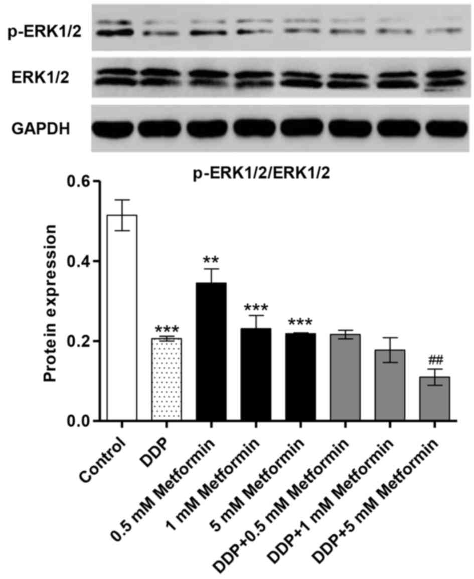
Metformin combined with DDP inhibits
ERK1/2 activation
The HO-8910 human ovarian cancer cell line was
treated with different concentrations of metformin for 6 h to
examine its effects on the ERK1/2 activity. Treatment with
metformin (0.5, 1 and 5 mM) reduced the levels of phosphorylated
(p)-ERK1/2 in a concentration-dependent manner in HO-8910 cells
(Fig. 1). Metformin combined with DDP
significantly decreased the p-ERK1/2 level to lower than in cells
treated with metformin alone. However, metformin (0.5, 1 and 5 mM)
and DDP (5 µM) alone or combination treatment had no effects on the
total expression of ERK1/2 in HO-8910 cells. These data suggest
that metformin combined with DDP may inhibit ERK1/2 activity in
ovarian cancer cells.
Metformin combined with DDP reduces
the expression of VEGF and VEGFR2 in HO-8910 cells
The expressions of VEGF and VEGFR2 in HO-8910 cells
were examined following metformin (0.5, 1 and 5 mM) and DDP (5 µM)
treatment using RT-qPCR and western blot analysis. The expression
of VEGF and VEGFR2 mRNA in HO-8910 cells was significantly
decreased by metformin treatment in a concentration-dependent
manner (Fig. 2A). Notably, metformin
combined with DDP in significantly decreased the expression of VEGF
and VEGFR2 mRNA to even lower levels than with DDP treatment alone
(Fig. 2B). Furthermore, the levels of
VEGF and VEGFR2 protein were also significantly decreased by
metformin or/and DDP treatment in HO-8910 cells (Fig. 2C-E). However, a lower concentration of
metformin (0.5 mM) had no effect on VEGF protein expression alone
or in combination with DDP in HO-8910 cells (Fig. 2D). Only higher concentrations of
metformin enhanced the inhibition of VEGFR2 protein expression
induced by DDP treatment in HO-8910 cells (Fig. 2E). These results suggest that
metformin combined with DDP effectively reduced the expression of
VEGF and VEGFR2, which may reduce angiogenesis.
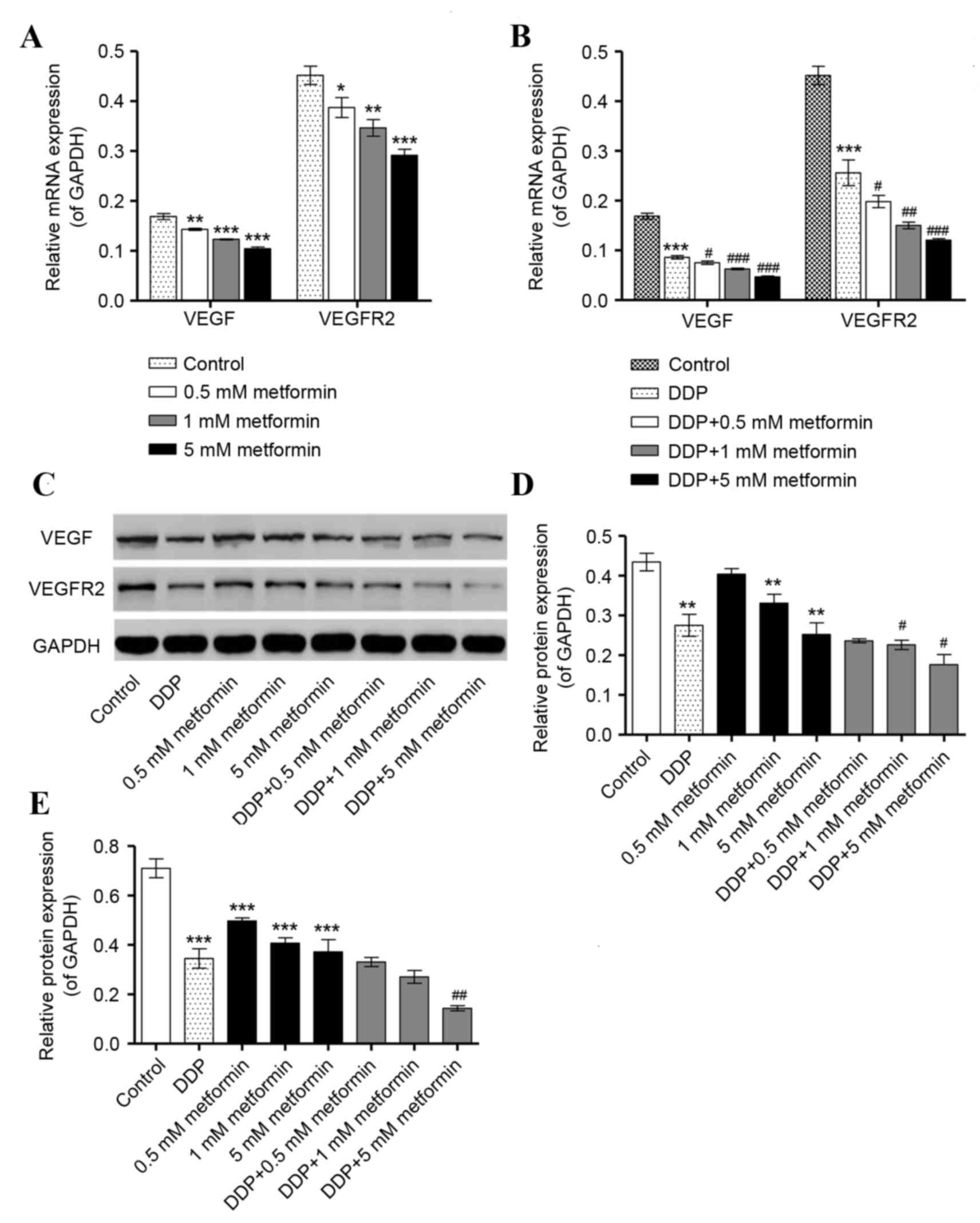 | Figure 2.Metformin and DDP inhibit the
expression of angiogenesis-associated factors in ovarian cancer
cells. HO-8910 cellswere treated with metformin (0.5, 1, and 5 mM)
or DDP (5 µM) alone or in combination (0.5, 1, or 5 mM metformin
plus 5 µM DDP) for 48 h to detect their effects on the expressions
of VEGF and VEGFR2. Reverse transcription-quantitative polymerase
chain reaction assay was used to examine the (A) VEGF and (B)
VEGFR2 mRNA expression following treatment with metformin, DDP or
both. (C) Western blot assay was used to examine the protein
expressions of (D) VEGF and (E) VEGFR2 following treatment with
metformin, DDP or both. *P<0.05, **P<0.01 and ***P<0.001
vs. control. #P<0.05, ##P<0.01 and
###P<0.001 vs. DDP. DDP, cisplatin; VEGF, vascular
endothelial growth factor; VEGFR2, VEGF receptor 2. |
Metformin combined with DDP induces
the expression of Bax, Bcl-2 and caspase-3 in HO-8910 cells
Expression of the apoptosis-associated factors, Bax,
Bcl-2 and caspase-3, were also quantified following metformin
or/and DDP treatment. Expression of Bax and caspase-3 mRNA was
elevated in a dose-dependent manner upon metformin (0.5, 1 and 5
mM) and DDP (5 µM) treatment alone, while combined metformin and
DDP treatment enhanced the increase in mRNA expression of Bax and
caspase-3 in HO-8910 cells (Fig. 3A and
B). However, expression of Bcl-2 mRNA was significantly
decreased by metformin treatment in a concentration-dependent
manner, with combined metformin and DDP treatment enhancing the
effect of treatment with either alone. Expression of Bax and
caspase-3 protein was also increased by metformin and DDP alone or
combination, whereas expression of Bcl-2 protein was decreased
(Fig. 3C-F). However, the lowest
concentration (0.5 mM) of metformin had no effect on the expression
of caspase-3 protein, either alone or in combination with DDP
(Fig. 3F). These results suggest that
metformin combined with DDP can effectively induce cell apoptosis
by increasing Bax/Bcl-2 ratio and caspase-3 expression.
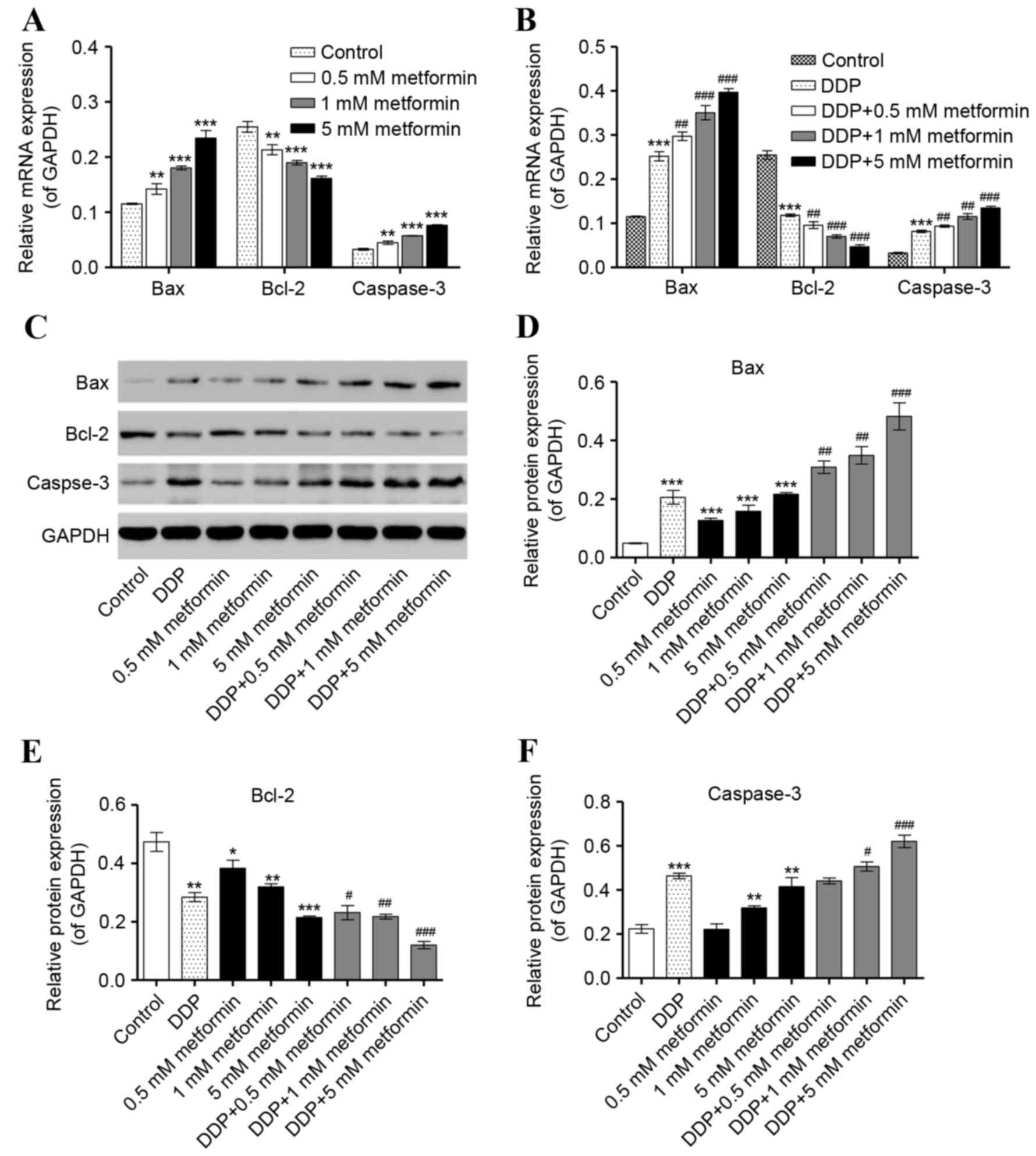 | Figure 3.Effect of metformin and DDP on the
expression of apoptosis-associated factors in ovarian cancer cells.
HO-8910 cells were treated with metformin (0.5, 1, and 5 mM), DDP
(5 µM) or the two in combination (0.5, 1, and 5 mM metformin plus 5
µM DDP) for 48 h to detect their effects on the expression of Bax,
Bcl-2 and caspase-3. Reverse transcription-quantitative polymerase
chain reaction was used to examine the mRNA expression of Bax,
Bcl-2 and caspase-3 following treatment with (A) metformin or (B)
DDP alone or DDP and metformin in combination. (C) Western blot
analysis was used to examine the protein expression of (D) Bax, (E)
Bcl-2 and (F) caspase-3 following treatment with metformin, DDP or
the two combined. *P<0.05, **P<0.01 and ***P<0.001 vs.
control. #P<0.05, ##P<0.01 and
###P<0.001 vs. DDP. DDP, cisplatin; Bcl-2, B-cell
lymphoma 2; Bax, Bcl-2 associated X. |
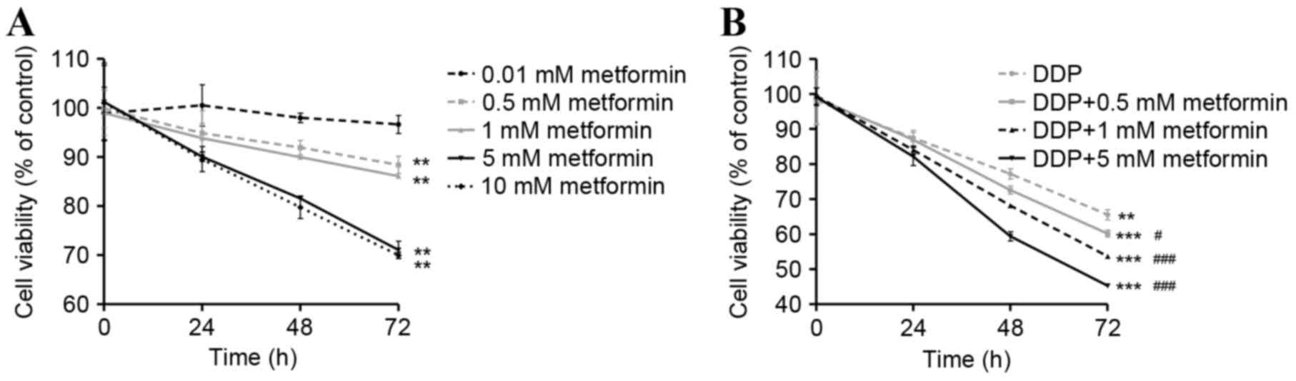
Metformin combined with DDP inhibits
the growth of HO-8910 cells
Having determined the effect of metformin and DDP on
ERK1/2 signaling, a CCK-8 cell viability and cytotoxicity assay was
performed to investigate the biological effect of the drug son the
phenotypes of HO-8910 cells. Metformin (0.01, 0.5, 1, 5 and 10 mM)
inhibited the cell viability of HO-8910 cells in a time- and
concentration-dependent manner (Fig.
4A). When combined, metformin (0.5, 1, and 5 mM) and DDP (5 µM)
inhibited cell growth in a concentration-dependent manner compared
with DDP treatment alone (Fig. 4B).
These findings suggested that metformin combined with DDP
effectively inhibited the cell viability of ovarian cancer
cells.
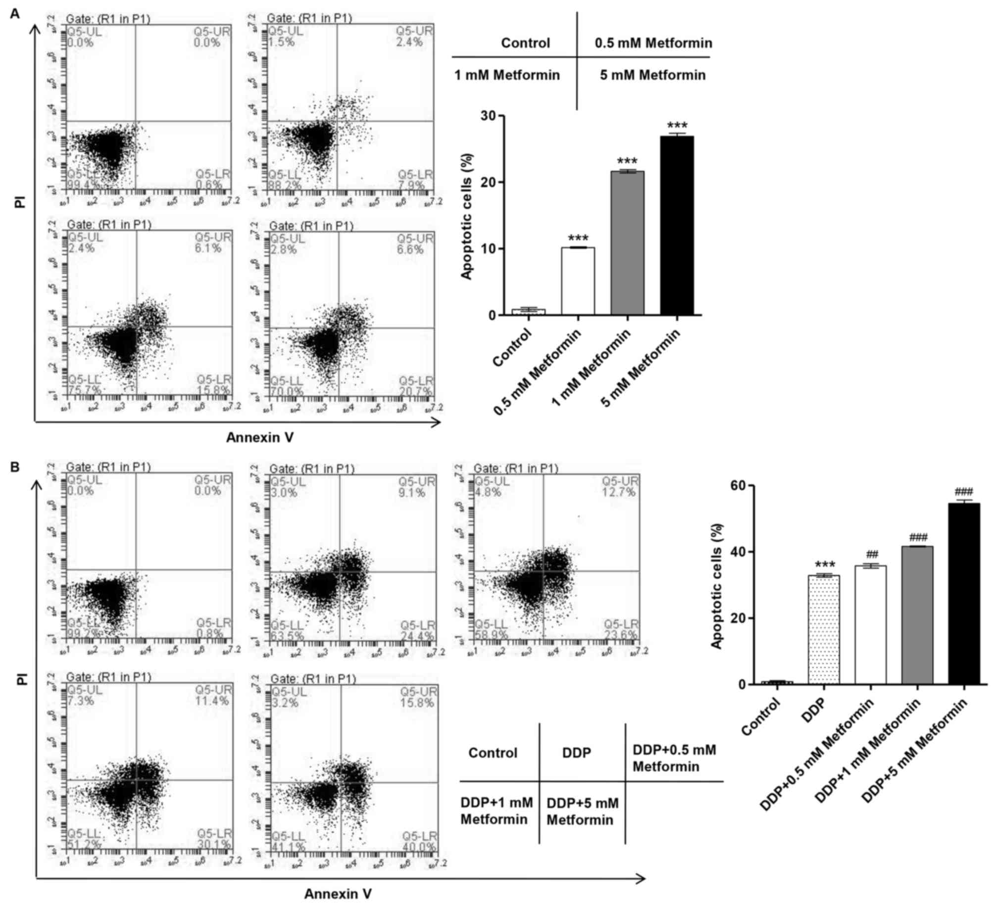
Metformin combined with DDP induces
cell apoptosis of HO-8910 cells
Due to the inhibitory effect of metformin and DDP on
ovarian cancer cell proliferation, the effects of metformin and DDP
on cell apoptosis of ovarian cancer cells were further
investigated. HO-8910 cells treated with metformin and/or DDP
treatment were stained with FITC-labeled annexinV/PI and
subsequently subjected to flow cytometry analysis. The results
revealed that metformin (0.5,1 and 5 mM) significantly increased
the number of apoptotic HO-8910 cells in a concentration-dependent
manner (Fig. 5A). Additionally,
metformin (0.5, 1, and 5 mM) treatment enhanced the apoptotic
effect of DDP (5 µM) in HO-8910 cells in a concentration-dependent
manner (Fig. 5B). These findings
suggest that metformin combined with DDP effectively induces the
apoptosis of ovarian cancer cells.
Discussion
The present study demonstrated that metformin alone
or combined with DDP inhibited the proliferation of HO-8910 ovarian
cancer cells and induced apoptosis via ERK1/2 deactivation.
Previous studies have observed that metformin inhibited tumor
growth, including in prostate and breast cancer (6,17). The ERK
pathway has a critical role in cell proliferation and survival, and
represents a conserved signaling system throughout evolution.
Activation of the ERK1/2 pathway has been documented in colorectal
(18), breast (19) and prostate cancer (20), and in several ovarian cancer cell
lines (21). Metformin treatment of
human mammary endothelial cells resulted in decreased migration and
invasion via inhibition of the ERK1/2 pathway, resulting in the
upregulation of thrombospondin-1 expression, which has been shown
to be involved in platelet aggregation, angiogenesis and
tumorigenesis (22). However,
metformin treatment had no effect on the phosphorylation of ERK in
U251 human glioma cells (23) and
treatment with DDP led to the strong activation of ERK1/2 in SKOV3
human ovarian carcinoma cells (24),
findings that are contrary to those of the present study.
VEGF is expressed by tumor cells and initiates
vasculogenesis and angiogenesis via binding to the tyrosine kinase
receptors (VEGFRs) on endothelial cells. VEGFR2 appears to mediate
the majority of the known cellular responses to VEGF. In human
breast cancer, VEGF stimulation increased the degree of ERK1/2
phosphorylation, indicating the activation of ERK1/2-associated
intra cellular pathways (25). A
previous study reported that metformin decreased vascular
permeability and expression of VEGF, suggesting that metformin
normalizes vascular permeability by regulating VEGF levels
(26). Additionally, the effects of
metformin and DDP on VEGF modulation have been also described in
ovarian cancer in vivo and in Lewis lung cancer rats
(3,27). In the present study, metformin alone
or in combination with DDP was demonstrated to significantly
decrease levels of VEGF and VEGFR2 in HO-8910 cells in a
concentration-dependent manner, implicating VEGF/VEGFR2 in the
ERK1/2-dependent anticancer effects of metformin and DDP in ovarian
cancer.
Furthermore, reduction of ERK1/2 activity causes G1
cell cycle arrest, downregulation of Bcl-2 expression levels,
increased activity of caspase-3, −6, −8, and −9, and induces
apoptotic cell death (28). In the
present study, inhibition of ERK1/2 by treatment with metformin in
ovarian cancer cells increased the expression of Bax, with the
amount of apoptosis further increased by DDP, which is in line with
previous reports (29). In the
present study, the effect of metformin and DDP on cell viability
and apoptosis was assessed using CCK-8 and flow cytometry analysis,
respectively. The results of these assays demonstrated that
metformin alone, or in combination with DDP, inhibited cell
viability and promoted apoptosis in a concentration-dependent
manner. Notably, a previous study reported that metformin treatment
inhibited the growth of metastatic nodules and enhanced
DDP-dependent cytotoxicity in ovarian cancer, resulting in the
inhibition of tumor cell growth by ~90% (3). However, Lesan et al (30) reported that metformin suppressed the
anticancer efficacy of DDP and reduced DDP-mediated apoptosis in
the MKN-45 cancer cell line. Further in vitro and in
vivo studies are therefore required to elucidate the mechanism
of action of these therapeutic agents in combination on ovarian
cancer cells, as well as other cancer types.
In conclusion, the results of the present study
support the use of metformin as an anticancer drug, either alone or
in combination with DDP, as it inhibited cell viability and
angiogenesis, and induced apoptosis. Therefore, these results
provide a strong rationale for the use of metformin as a
therapeutic drug in combination with DDP in the treatment of
patients with ovarian cancer.
References
|
1
|
Siegel RL, Miller KD and Jemal A: Cancer
statistics, 2015. CA Cancer J Clin6. 65:5–29. 2015. View Article : Google Scholar
|
|
2
|
Smith HO, Arias-Pulido H, Kuo DY, Howard
T, Qualls CR, Lee SJ, Verschraegen CF, Hathaway HJ, Joste NE and
Prossnitz ER: GPR30 predicts poor survival for ovarian cancer.
Gynecol Oncol. 114:465–471. 2009. View Article : Google Scholar : PubMed/NCBI
|
|
3
|
Rattan R, Graham RP, Maguire JL, Giri S
and Shridhar V: Metformin suppresses ovarian cancer growth and
metastasis with enhancement of cisplatin cytotoxicity in vivo.
Neoplasia. 13:483–491. 2011. View Article : Google Scholar : PubMed/NCBI
|
|
4
|
Li C, Liu VW, Chan DW, Yao KM and Ngan HY:
LY294002 and metformin cooperatively enhance the inhibition of
growth and the induction of apoptosis of ovarian cancer cells. Int
J Gynecol Cancer. 22:15–22. 2012. View Article : Google Scholar : PubMed/NCBI
|
|
5
|
Correia S, Carvalho C, Santos MS, Proença
T, Nunes E, Duarte AI, Monteiro P, Seiça R, Oliveira CR and Moreira
PI: Metformin protects the brain against the oxidative imbalance
promoted by type 2 diabetes. Med Chem. 4:358–364. 2008. View Article : Google Scholar : PubMed/NCBI
|
|
6
|
Kourelis TV and Siegel RD: Metformin and
cancer: New applications for an old drug. Med Oncol. 29:1314–1327.
2012. View Article : Google Scholar : PubMed/NCBI
|
|
7
|
Gonzalez-Angulo AM and Meric-Bernstam F:
Metformin: A therapeutic opportunity in breast cancer. Clin Cancer
Res. 16:1695–1700. 2010. View Article : Google Scholar : PubMed/NCBI
|
|
8
|
Jiralerspong S, Palla SL, Giordano SH,
Meric-Bernstam F, Liedtke C, Barnett CM, Hsu L, Hung MC, Hortobagyi
GN and Gonzalez-Angulo AM: Metformin and pathologic complete
responses to neoadjuvant chemotherapy in diabetic patients with
breast cancer. J Clin Oncol. 27:3297–3302. 2009. View Article : Google Scholar : PubMed/NCBI
|
|
9
|
Foretz M, Hébrard S, Leclerc J,
Zarrinpashneh E, Soty M, Mithieux G, Sakamoto K, Andreelli F and
Viollet B: Metformin inhibits hepatic gluconeogenesis in mice
independently of the LKB1/AMPK pathway via a decrease in hepatic
energy state. J Clin Invest. 120:2355–2369. 2010. View Article : Google Scholar : PubMed/NCBI
|
|
10
|
Scagliotti GV, Parikh P, von Pawel J,
Biesma B, Vansteenkiste J, Manegold C, Serwatowski P, Gatzemeier U,
Digumarti R, Zukin M, et al: Phase III study comparing cisplatin
plus gemcitabine with cisplatin plus pemetrexed in
chemotherapy-naive patients with advanced-stage non-small-cell lung
cancer. J Clin Oncol. 26:3543–3551. 2008. View Article : Google Scholar : PubMed/NCBI
|
|
11
|
Kang YK, Kang WK, Shin DB, Chen J, Xiong
J, Wang J, Lichinitser M, Guan Z, Khasanov R, Zheng L, et al:
Capecitabine/cisplatin versus 5-fluorouracil/cisplatin as
first-line therapy in patients with advanced gastric cancer: A
randomised phase III noninferiority trial. Ann Oncol. 20:666–673.
2009. View Article : Google Scholar : PubMed/NCBI
|
|
12
|
Lorch JH, Goloubeva O, Haddad RI, Cullen
K, Sarlis N, Tishler R, Tan M, Fasciano J, Sammartino DE and Posner
MR: TAX 324 Study Group: Induction chemotherapy with cisplatin and
fluorouracil alone or in combination with docetaxel in locally
advanced squamous-cell cancer of the head and neck: Long-term
results of the TAX 324 randomised phase 3 trial. Lancet Oncol.
12:153–159. 2011. View Article : Google Scholar : PubMed/NCBI
|
|
13
|
Kaina B: DNA damage-triggered apoptosis:
Critical role of DNA repair, double-strand breaks, cell
proliferation and signaling. Biochem Pharmacol. 66:1547–1554. 2003.
View Article : Google Scholar : PubMed/NCBI
|
|
14
|
Chang J, Jung HH, Yang JY, Lee S, Choi J,
Im GJ and Chae SW: Protective effect of metformin against
cisplatin-induced ototoxicity in an auditory cell line. J Assoc Res
Otolaryngol. 15:149–158. 2014. View Article : Google Scholar : PubMed/NCBI
|
|
15
|
Zhang YH, Wang Y, Yusufali AH, Ashby F,
Zhang D, Yin ZF, Aslanidi GV, Srivastava A, Ling CQ and Ling C:
Cytotoxic genes from traditional Chinese medicine inhibit tumor
growth both in vitro and in vivo. J Integr Med. 12:483–494. 2014.
View Article : Google Scholar : PubMed/NCBI
|
|
16
|
Livak KJ and Schmittgen TD: Analysis of
relative gene expression data using real-time quantitative PCR and
the 2(-Delta Delta C(T)) method. Methods. 25:402–408. 2001.
View Article : Google Scholar : PubMed/NCBI
|
|
17
|
Alimova IN, Liu B, Fan Z, Edgerton SM,
Dillon T, Lind SE and Thor AD: Metformin inhibits breast cancer
cell growth, colony formation and induces cell cycle arrest in
vitro. Cell Cycle. 8:909–915. 2009. View Article : Google Scholar : PubMed/NCBI
|
|
18
|
Yeh JJ, Routh ED, Rubinas T, Peacock J,
Martin TD, Shen XJ, Sandler RS, Kim HJ, Keku TO and Der CJ:
KRAS/BRAF mutation status and ERK1/2 activation as biomarkers for
MEK1/2 inhibitor therapy in colorectal cancer. Mol Cancer Ther.
8:834–843. 2009. View Article : Google Scholar : PubMed/NCBI
|
|
19
|
Sirianni R, Chimento A, De Luca A,
Casaburi I, Rizza P, Onofrio A, Lacopetta D, Puoci F, Andò S,
Maggiolini M and Pezzi V: Oleuropein and hydroxytyrosol inhibit
MCF-7 breast cancer cell proliferation interfering with ERK1/2
activation. Mol Nutr Food Res. 54:833–840. 2010. View Article : Google Scholar : PubMed/NCBI
|
|
20
|
Albrecht DS, Clubbs EA, Ferruzzi M and
Bomser JA: Epigallocatechin-3-gallate (EGCG) inhibits PC-3 prostate
cancer cell proliferation via MEK-independent ERK1/2 activation.
Chem Biol Interact. 171:89–95. 2008. View Article : Google Scholar : PubMed/NCBI
|
|
21
|
Steinmetz R, Wagoner HA, Zeng P, Hammond
JR, Hannon TS, Meyers JL and Pescovitz OH: Mechanisms regulating
the constitutive activation of the extracellular signal-regulated
kinase (ERK) signaling pathway in ovarian cancer and the effect of
ribonucleic acid interference for ERK1/2 on cancer cell
proliferation. Mol Endocrinol. 18:2570–2582. 2004. View Article : Google Scholar : PubMed/NCBI
|
|
22
|
Tan BK, Adya R, Chen J, Farhatullah S,
Heutling D, Mitchell D, Lehnert H and Randeva HS: Metformin
decreases angiogenesis via NF-kappaB and Erk1/2/Erk5 pathways by
increasing the antiangiogenic thrombospondin-1. Cardiovasc Res.
83:566–574. 2009. View Article : Google Scholar : PubMed/NCBI
|
|
23
|
Janjetovic K, Vucicevic L, Misirkic M,
Vilimanovich U, Tovilovic G, Zogovic N, Nikolic Z, Jovanovic S,
Bumbasirevic V, Trajkovic V and Harhaji-Trajkovic L: Metformin
reduces cisplatin-mediated apoptotic death of cancer cells through
AMPK-independent activation of Akt. Eur J Pharmacol. 651:41–50.
2011. View Article : Google Scholar : PubMed/NCBI
|
|
24
|
Wei SQ, Sui LH, Zheng JH, Zhang GM and Kao
YL: Role of ERK1/2 kinase in cisplatin-induced apoptosis in human
ovarian carcinoma cells. Chin Med Sci J. 19:125–129.
2004.PubMed/NCBI
|
|
25
|
Kim EH, Na HK and Surh YJ: Upregulation of
VEGF by 15-deoxy-Delta12,14-prostaglandin J2 via heme oxygenase-1
and ERK1/2 signaling in MCF-7 cells. Ann NY Acad Sci. 1090:375–384.
2006. View Article : Google Scholar : PubMed/NCBI
|
|
26
|
Elia EM, Quintana R, Carrere C, Bazzano
MV, Rey-Valzacchi G, Paz DA and Pustovrh MC: Metformin decreases
the incidence of ovarian hyperstimulation syndrome: An experimental
study. J Ovarian Res. 6:622013. View Article : Google Scholar : PubMed/NCBI
|
|
27
|
Feng P, Zhang ZL, Zhang ZH, Zhang XL,
Xiang F, Tang JH and Xiang BL: Effect of endostar combined with
cisplatin on expression of VEGF and Sema3A of lewis lung cancer
rats. Asian Pac J Trop Med. 6:57–60. 2013. View Article : Google Scholar : PubMed/NCBI
|
|
28
|
Boucher MJ, Morisset J, Vachon PH, Reed
JC, Lainé J and Rivard N: MEK/ERK signaling pathway regulates the
expression of Bcl-2, Bcl-X(L), and Mcl-1 and promotes survival of
human pancreatic cancer cells. J Cell Biochem. 79:355–369. 2000.
View Article : Google Scholar : PubMed/NCBI
|
|
29
|
Yasmeen A, Beauchamp MC, Piura E, Segal E,
Pollak M and Gotlieb WH: Induction of apoptosis by metformin in
epithelial ovarian cancer: Involvement of the Bcl-2 family
proteins. Gynecol Oncol. 121:492–498. 2011. View Article : Google Scholar : PubMed/NCBI
|
|
30
|
Lesan V, Ghaffari SH, Salaramoli J,
Heidari M, Rostami M, Alimoghaddam K and Ghavamzadeh A: Evaluation
of antagonistic effects of metformin with Cisplatin in gastric
cancer cells. Int J Hematol Oncol Stem Cell Res. 8:12–19.
2014.PubMed/NCBI
|