Introduction
Esophageal squamous cell carcinoma (ESCC) is one of
the most common malignant diseases globally, particularly in China,
where it is the fourth most common cause of cancer-associated
mortality (1–3). Currently, there are limited clinical
approaches towards the early diagnosis and treatment of ESCC,
resulting in a 10% 5-year survival rate for patients (4). Thus, clarification of the molecular
mechanisms underlying the pathogenesis of ESCC and development of
therapeutic strategies are urgently required.
MicroRNAs (miRNAs) are small non-coding RNAs
(between 18 and 22 nucleotides in length), which serve a role in
gene expression regulation primarily by repressing translation
(5,6).
Prior evidence supports the notion that miRNAs modulate
proliferation, apoptosis, metastasis and metabolism of cancer
(6,7).
Numerous miRNAs are involved in esophageal tumorigenesis (8,9). However,
the role of miRNA-1470 (miR-1470) in esophageal cancer progression
remains undefined.
Bcl-2 is an apoptosis inhibitor (10). When Bcl-2 is suppressed, tumor cells
undergo apoptosis and cease proliferation (11). Various moderators of apoptosis exert
their effects through the caspase enzyme system. Among them,
caspase-3 has been demonstrated to be the most markedly associated
with apoptosis (12,13). Furthermore, decreased caspase-3
expression has been observed in several human malignancies
including oral squamous cell carcinoma, non-small cell lung
carcinoma, breast cancer, gastric cancer, and ESCC (14–17).
Overexpressions of Bcl-2 and other apoptotic inhibitors serve the
roles in therapeutic resistance of ESCC (18,19).
Whereas decreased Bcl-2 expression promotes apoptotic responses to
anticancer drugs, increased expression of Bcl-2 leads to resistance
to chemotherapeutic drugs and radiation therapy. The inhibition of
cell apoptosis is associated with the onset and development of
cancer, as well as its sensitivity to chemotherapy (19–21).
In the present study, it was demonstrated that
miR-1470 was upregulated in ESCC tissues, and knockdown of miR-1470
significantly inhibited proliferation and migration, and promoted
senescence, of ESCC cells. Furthermore, knockdown of miR-1470
exhibited apoptosis-promoting activity, including downregulation of
the anti-apoptotic protein Bcl-2 and upregulation of the
pro-apoptotic Bcl-2-associated X protein (BAX). Taken together, the
results of the present study suggest that miR-1470 affects cancer
progression via the regulation of Bcl-2 expression and may be a
promising therapeutic target for the treatment of ESCC.
Materials and methods
Cell culture and transfection
The ESCC cell lines including KYSE30, KYSE180,
KYSE450, KYSE150 and KYSE510, provided by Dr Yutaka Shimada (Kyoto
University, Kyoto, Japan), were cultured in RPMI-1640 medium
(Invitrogen; Thermo Fisher Scientific, Inc., Waltham, MA, USA) with
10% fetal bovine serum (FBS; Hyclone; GE Healthcare Life Sciences,
Logan, UT, USA). All cells were maintained at 37°C with 5%
CO2. Homo sapiens (hsa)-miR-1470 inhibitor was
designed and provided by Shanghai GenePharma, Co., Ltd. (Shanghai,
China). Transfection was performed using Lipofectamine 2000
(Invitrogen; Thermo Fisher Scientific, Inc. Carlsbad, USA),
according to the manufacturer's protocol. Transfected cells were
incubated at 37°C with 5% CO2 for 48 h.
The sequence of the miR-1470 inhibitor was
5′-CGGGGUGCACGGGCGGAGGGC-3′; the sequence of the negative control
was 5′-CAGUACUUUUGUGUAGUACAA-3′.
Cell proliferation assay
Cells were seeded in 96-well flat-bottomed plates,
with each well containing 2×10³ cells in 100 µl cell
suspension. After incubation for 24, 48, 7 h and 9 6 h, cell
viability was determined using the Cell Counting Kit-8 assay
(CCK-8; Dojindo Molecular Technologies, Inc., Kumamoto, Japan),
according to the manufacturer's protocol using a microplate read at
450 nm. Each experiment, with 6 replicates, was repeated in
triplicate.
Cell migration assay
Cells (5×10⁵ cells in serum-free
RPMI-1640 medium) were seeded in the upper chamber. The wells in
the lower chamber were filled with RPMI-1640 medium containing 20%
FBS, and incubated at 37°C for 36 h. Cells on the upper membrane
surface were wiped off using a cotton swab, and the lower membrane
surface was fixed with methanol and stained with 0.1% crystal
violet. Each experiment was repeated in triplicate.
Reverse transcription-quantitative
polymerase chain reaction (RT-qPCR) assay
RT-qPCR was performed using the Premix Ex Taq kit
(Takara Biotechnology Co., Ltd., Dalian, China) and a 7300
Real-Time PCR system (Thermo Fisher Scientific, Inc.), according to
the manufacturer's protocol. The mRNA expression levels of genes of
interest were normalized to the endogenous expression level of
GAPDH. The following primer pairs were used for the PCR assay:
Cyclin E1 (CCNE1) forward, 5′-TGACCTAAGGGACTCCCACAA-3′; CCNE1
reverse, 5′-TGATAATGTGGAGAGGGCAGC-3′; matrix metalloproteinase 2
(MMP2) forward, 5′-GTGATGGTTCCCCTGTTCACT-3′, MMP2 reverse,
5′-CATGGGGAATGGTTGAAGGGA-3′; MMP13 forward,
5′-TCCCTTGAGATATGGAAGGATGC-3′; MMP13 reverse,
5′-TAGTTCTTCCCTTGATGGCCG-3′; MMP14 forward,
5′-TGGATGCGAGTACCCCAAGA-3′, MMP14 reverse,
5′-GTAGCCCGGTTCTACCTTCAG-3′. A Hairpin-it™ miR-1470
RT-qPCR Primer Set (Shanghai GenePharma, Co., Ltd.) was used for
the measurement of the relative quantity of hsa-miR-1470. The
expression level of miR-1470 was normalized to the endogenous
expression of U6.
Western blotting
Cells from each group were detached with trypsin,
centrifuged at 1,000 × g for 5 min at 4°C and washed twice with
pre-chilled PBS. Radioimmunoprecipitation Assay buffer (Beyotime
Institute of Biotechnology, Haimen, China) was subsequently added
to lyse cells and cells were incubated on ice for protein
extraction. Protein concentration was determined using the
Bicinchoninic Acid Protein Assay kit (Beyotime Institute of
Biotechnology). Equal amounts (10 µg per lane) of proteins were
separated by SDS-PAGE (12% gel) and transferred onto a
polyvinylidene difluoride membrane (Merck KGaA, Darmstadt,
Germany). The membrane was soaked in 10% skimmed milk (in PBS, pH
7.2, containing 0.1% Tween-20) for 2 h at 4°C and incubated with an
appropriate amount of primary antibody [working dilutions of
antibodies: Bcl-2, 1:1,000, cat. no. 10783-1-AP; BAX, 1:1,000, cat.
no. 23931-1-AP (both Proteintech Group, Inc., Chicago, IL, USA)
pro-caspase-3, 1:1,000, cat. no. sc-7148; pro-caspase-12, 1:1,000,
cat. no. sc-5627; β-actin, 1:400, cat. no. sc-47778), (Santa Cruz
Biotechnology, Inc., Dallas, TX, USA; Proteintech Group, Inc.,
Chicago, IL, USA)] at 4°C overnight. Detection was performed using
horseradish peroxidase-conjugated secondary antibodies (anti-mouse
IgG, 1:10,000; cat. no. 474-1806 or anti-rabbit IgG, 1:10,000; cat.
no. 5450-0010) for 2 h at room temperature (KPL, Inc.,
Gaithersburg, MD, USA) and Immobilon™ Western
chemiluminescent HRP substrate kit (EMD Millipore, Billerica, MA,
USA). Densitometry analysis was performed using ImageJ 1.46
software (National Institutes of Health, Bethesda, MD, USA).
Assessment of senescence
To assess senescence, β-galactosidase activity was
measured using a Senescence β-Galactosidase Staining kit (Cell
Signaling Technology, Inc., Danvers, MA, USA) and performed
according to the manufacturer's protocol. Briefly, negative control
(NC)-or miRNA-1470 inhibitor-transfected cells were washed twice
with 1X PBS, fixed in 1X fixation buffer for 5 min, washed three
times with 1X PBS and incubated with the staining mixture overnight
at 37°C. β-galactosidase-positive cells were visualized as blue
cells when examined using a light microscope (magnification, ×400).
For NC- and miRNA-1470 inhibitor-transfected cells, a total of 100
cells were counted from different fields of the plate, and the
percentage of blue cells was calculated.
Statistical analysis
All results were confirmed in at least three
independent experiments, and data from one representative
experiment are presented. Student's t-test was used to compare the
means of experimental groups. All quantitative data are presented
as the mean ± standard deviation. The analysis was performed using
GraphPad Prism (version 5.01; GraphPad Software, La Jolla, CA,
USA). For all comparisons, P<0.05 was considered to indicate a
statistically significant difference.
Results
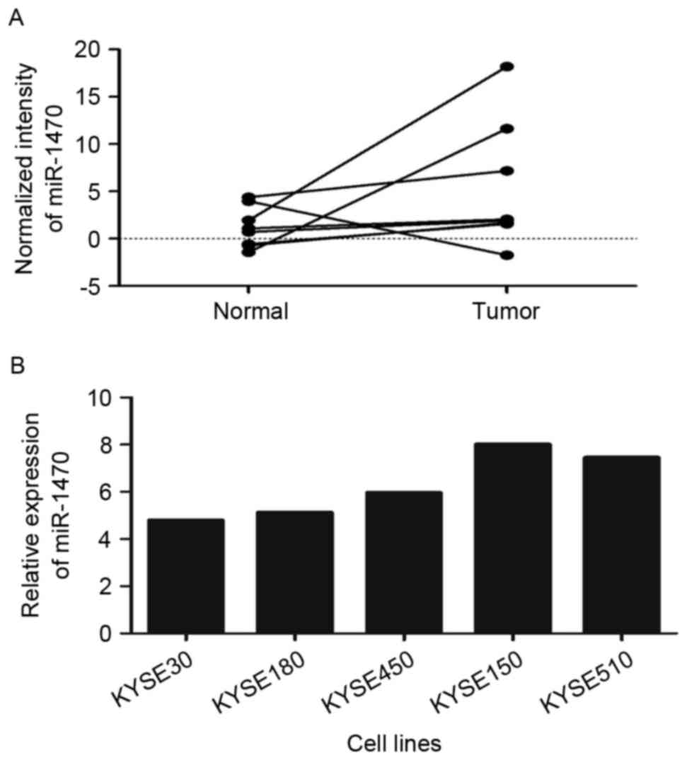
ESCC tissues overexpress miR-1470
In the present study, it was revealed that the
expression level of miR-1470 was increased in ESCC tissues compared
with their adjacent non-malignant tissues using Agilent Human miRNA
Microarrays (Fig. 1A). The expression
of miR-1470 was further evaluated in 5 ESCC cell lines (KYSE30,
KYSE180, KYSE450, KYSE150 and KYSE510) using RT-qPCR. The results
revealed that KYSE150 and KYSE510 exhibited increased expression
levels compared with other cell lines (Fig. 1B).
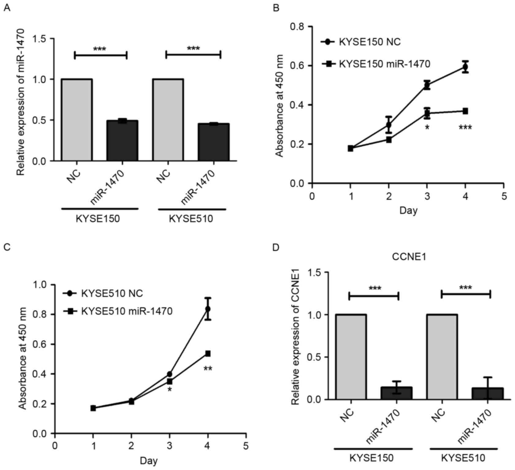
Knockdown of miR-1470 suppresses
proliferation of ESCC cells by regulating cell cycle regulatory
protein
To explore the role of miR-1470 in esophageal
carcinogenesis, the cell viability was evaluated using a CCK-8
assay. Knockdown of miR-1470 using the inhibitor significantly
inhibited proliferation of KYSE150 and KYSE510 cells (Fig. 2A-C). The mRNA level of the cell cycle
regulatory gene CCNE1 in miR-1470 inhibitor-transfected cells were
significantly decreased (Fig. 2D).
These observations indicate that miR-1470 may promote cell
proliferation via up-regulating CCNE1 in ESCC.
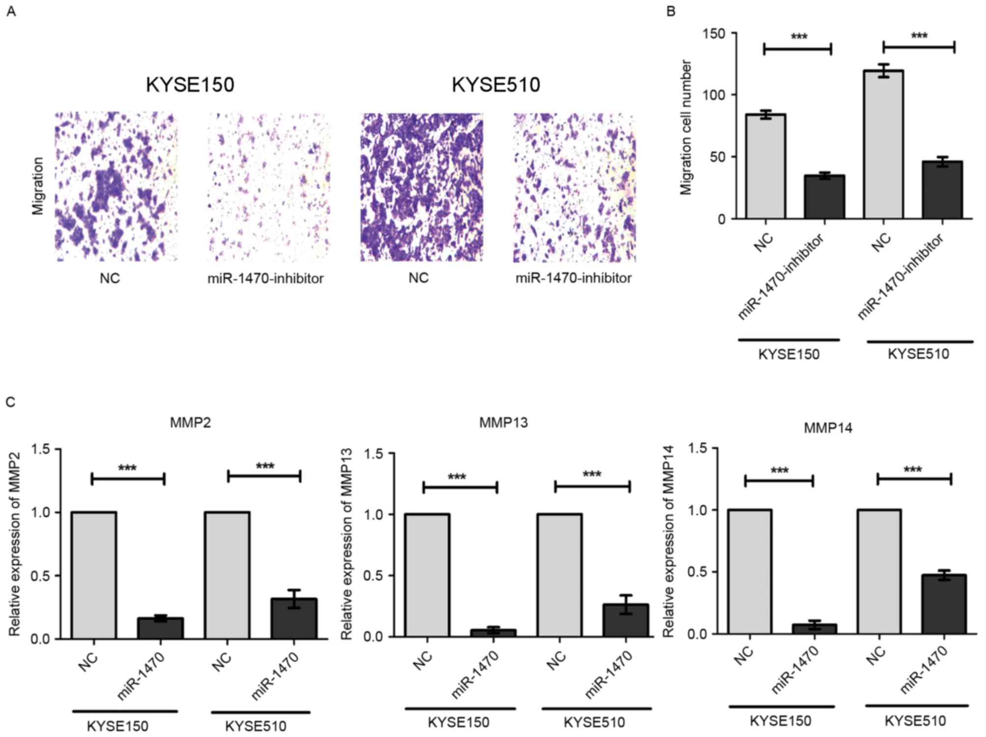
Knockdown of miR-1470 inhibits the
migration of ESCC cells by decreasing the expression of MMPs
Using a Transwell assay, it was identified that
knockdown of miR-1470 led to a decrease in the number of migratory
KYSE150 and KYSE510 cells (Fig. 3A).
Quantification and statistical analysis of the decrease in
migratory cell numbers revealed that the difference was
statistically significant (Fig. 3B).
MMPs, a family of zinc-binding proteins, have been demonstrated to
serve a role in tumor cell metastasis owing to their ability to
degrade the extracellular matrix. Using RT-qPCR, we found that
knockdown of miR-1470 significantly decreased the expression levels
of MMP2, MMP13 and MMP14. Taken together, these data suggest that
down-regulated miR-1470 may suppress the migration of ESCC cells
via inhibition of MMPs.
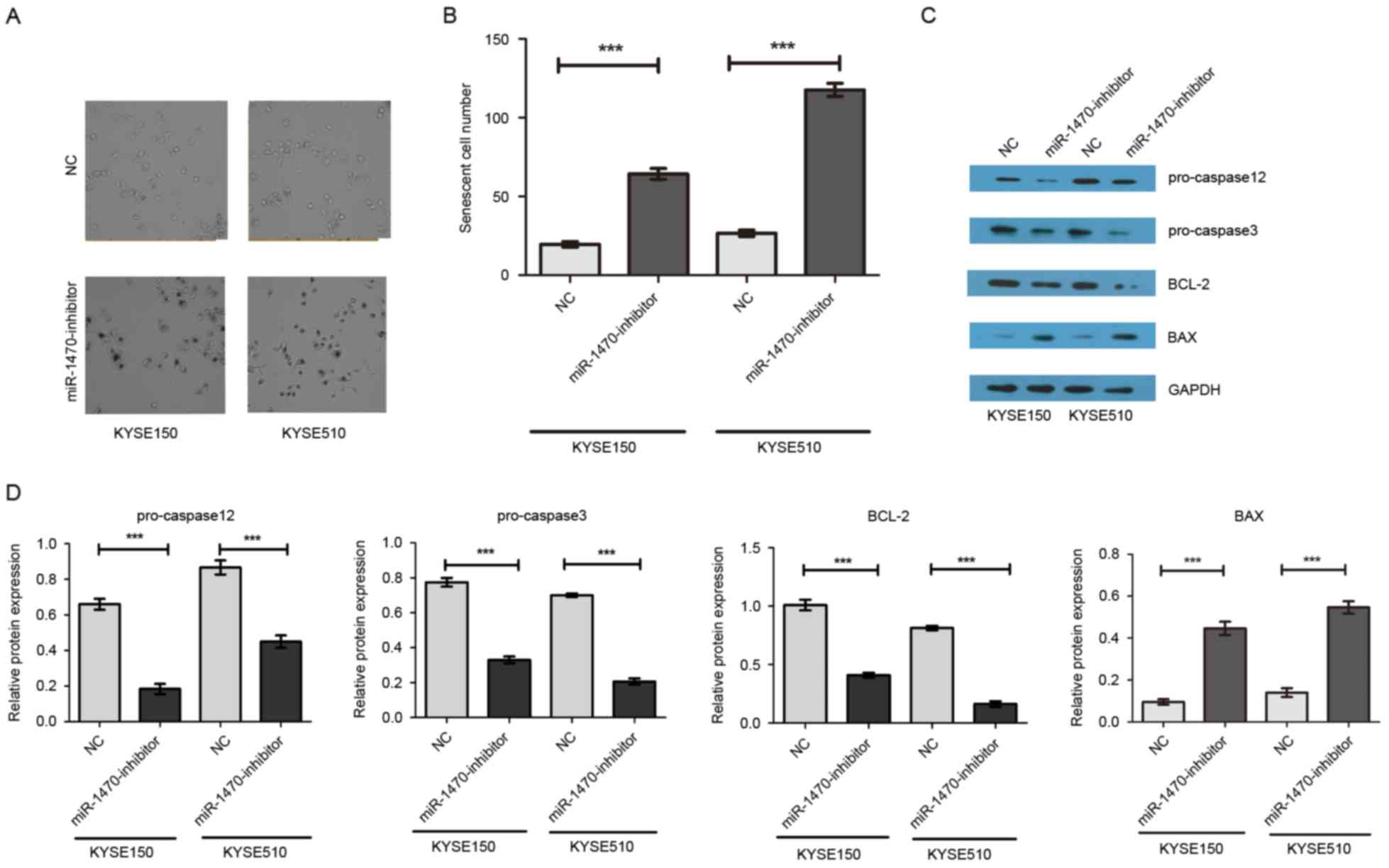
Suppression of miR-1470 induces
senescence and activates apoptosis pathway in ESCC cells
To investigate the effect of miR-1470 on cell
senescence, morphological evaluation of KYSE150 and KYSE510 cells
was performed following knockdown of miR-1470 expression. The
results revealed an elongated phenotype resembling cellular
senescence (Fig. 4A and B). As
presented in Fig. 4C and D, compared
with the control group, knockdown of miR-1470 markedly decreased
anti-apoptotic protein Bcl-2, pro-caspase-12, pro-caspase-3 and
increased pro-apoptotic protein BAX. These results suggest that
suppression of miR-1470 may induce cell apoptosis.
Discussion
Previous studies have demonstrated that miRNAs are
frequently dysregulated in numerous types of human cancer. As the
roles of miRNAs in cancer are gradually investigated, their
potential as a therapeutic target has generated interest with
regard to the development of novel strategies for treating cancer
(22–24). Previous studies have characterized the
miRNA expression profiles associated with distinct stages of ESCC,
and dysregulation of miRNAs has been demonstrated to serve a role
in esophageal carcinogenesis (25–28).
miR-375 inhibits tumor growth and metastasis in ESCC through the
suppression of insulin-like growth factor 1 receptor (27). Cui et al (29) demonstrated that targeting oncogenic
phospholipase Cε1 using miR-145 impaired tumor proliferation and
metastasis of ESCC. Although Nie et al (30) demonstrated that miR-1470 mediated
lapatinib-induced p27 up-regulation by targeting c-jun, the
expression status, role and underlying molecular oncogenic
mechanism of miR-1470 in esophageal cancer progression remain to be
established.
miR-1470 is located at 19p13.12. In the present
study, upregulation of miR-1470 in ESCC tissues was revealed using
microarray. Functional experiments further revealed that
downregulation of miR-1470 significantly inhibited the
proliferation of ESCC cells, and also decreased the level of the
cell cycle regulatory gene CCNE1. To the best of our knowledge,
prior to the present study there has been no reported association
between miR-1470 and CCNE1. The results of the present study
indicate that miR-1470 positively regulated CCNE1. The results
suggest that upregulation of miR-1470 expression in ESCC promoted
cell proliferation by accelerating cell cycle transition.
Metastasis is the primary cause of mortality in
patients with ESCC. It was identified that downregulated miR-1470
expression inhibited the migration of ESCC cells in vitro.
MMPs are proteolytic enzymes that serve a role in the
transformation and progression of tumors at all stages,
particularly during invasion and metastasis (31,32). In
the present study, downregulation of miR-1470 significantly
suppressed the expressions of MMP2, MMP13 and MMP14. The results
suggested that overexpression of miR-1470 in ESCC promoted the
migration of tumor cells via increasing the expression levels of
MMPs.
Senescence generally contributes to the elimination
of damaged cells by phagocytic cells, and to the subsequent
promotion of tissue remodeling, in a similar manner to apoptosis
(33). In the present study,
knockdown of miR-1470 significantly induced cell senescence and
activated the apoptotic signaling pathway.
In conclusion, it was demonstrated that
downregulation of miR-1470 significantly inhibited ESCC cell
proliferation and migration. The identification of miR-1470
provides insight into the pathogenesis of ESCC, and may represent a
potential therapeutic target for the treatment of ESCC.
Acknowledgements
The present study was supported by the National
Natural Science Foundation of China (grant no. 81460425 and
81760526), the Yunnan Provincial Research Foundation for Basic
Research, China (grant no. 2013FD012), the Foundation for the
Talents of Kunming University of Science and Technology (grant no.
KKSY201226099) and the Open Project Program of State Key Laboratory
of Molecular Oncology (SKL-KF-2017-11).
References
|
1
|
Zhao P, Dai M, Chen W and Li N: Cancer
trends in China. Jpn J Clin Oncol. 40:281–285. 2010. View Article : Google Scholar : PubMed/NCBI
|
|
2
|
Mariette C, Piessen G and Triboulet JP:
Therapeutic strategies in oesophageal carcinoma: Role of surgery
and other modalities. Lancet Oncol. 8:545–553. 2007. View Article : Google Scholar : PubMed/NCBI
|
|
3
|
Ferlay J, Shin HR, Bray F, Forman D,
Mathers C and Parkin DM: Estimates of worldwide burden of cancer in
2008: GLOBOCAN 2008. Int J Cancer. 127:2893–2917. 2010. View Article : Google Scholar : PubMed/NCBI
|
|
4
|
Ell C and Lorenz D: Diagnosis and
treatment of oesophageal carcinoma: Changes in every respect.
Viszeralmedizin. 31:3142015.PubMed/NCBI
|
|
5
|
Acunzo M and Croce CM: MicroRNA in Cancer
and Cachexia-A Mini-Review. J Infect Dis. 212 Suppl 1:S74–S77.
2015. View Article : Google Scholar : PubMed/NCBI
|
|
6
|
Bartel DP: MicroRNAs: Genomics,
biogenesis, mechanism, and function. Cell. 116:281–297. 2004.
View Article : Google Scholar : PubMed/NCBI
|
|
7
|
Iorio MV and Croce CM: MicroRNAs in
cancer: Small molecules with a huge impact. J Clin Oncol.
27:5848–5856. 2009. View Article : Google Scholar : PubMed/NCBI
|
|
8
|
Li S, Qin X, Li Y, Zhang X, Niu R, Zhang
H, Cui A, An W and Wang X: MiR-133a suppresses the migration and
invasion of esophageal cancer cells by targeting the EMT regulator
SOX4. Am J Transl Res. 7:1390–1403. 2015.PubMed/NCBI
|
|
9
|
Mao Y, Li L, Liu J, Wang L and Zhou Y:
MiR-495 inhibits esophageal squamous cell carcinoma progression by
targeting Akt1. Oncotarget. 7:51223–51236. 2016. View Article : Google Scholar : PubMed/NCBI
|
|
10
|
Llambi F and Green DR: Apoptosis and
oncogenesis: Give and take in the BCL-2 family. Curr Opin Genet
Dev. 21:12–20. 2011. View Article : Google Scholar : PubMed/NCBI
|
|
11
|
Fulda S, Meyer E and Debatin KM:
Inhibition of TRAIL-induced apoptosis by Bcl-2 overexpression.
Oncogene. 21:2283–2294. 2002. View Article : Google Scholar : PubMed/NCBI
|
|
12
|
Thoms HC, Dunlop MG and Stark LA:
p38-mediated inactivation of cyclin D1/cyclin-dependent kinase 4
stimulates nucleolar translocation of RelA and apoptosis in
colorectal cancer cells. Cancer Res. 67:1660–1669. 2007. View Article : Google Scholar : PubMed/NCBI
|
|
13
|
Törmänen-Näpänkangas U, Soini Y, Kahlos K,
Kinnula V and Pääkkö P: Expression of caspases-3, −6 and −8 and
their relation to apoptosis in non-small cell lung carcinoma. Int J
Cancer. 93:192–198. 2001. View
Article : Google Scholar : PubMed/NCBI
|
|
14
|
Li YH, Wang C, Meng K, Chen LB and Zhou
XJ: Influence of survivin and caspase-3 on cell apoptosis and
prognosis in gastric carcinoma. World J Gastroenterol.
10:1984–1988. 2004. View Article : Google Scholar : PubMed/NCBI
|
|
15
|
Li SX, Chai L, Cai ZG, Jin LJ, Chen Y, Wu
HR and Sun Z: Expression of survivin and caspase 3 in oral squamous
cell carcinoma and peritumoral tissue. Asian Pac J Cancer Prev.
13:5027–5031. 2012. View Article : Google Scholar : PubMed/NCBI
|
|
16
|
Nassar A, Lawson D, Cotsonis G and Cohen
C: Survivin and caspase-3 expression in breast cancer: Correlation
with prognostic parameters, proliferation, angiogenesis, and
outcome. Appl Immunohistochem Mol Morphol. 16:113–120. 2008.
View Article : Google Scholar : PubMed/NCBI
|
|
17
|
Hsia JY, Chen CY, Chen JT, Hsu CP, Shai
SE, Yang SS, Chuang CY, Wang PY and Miaw J: Prognostic significance
of caspase-3 expression in primary resected esophageal squamous
cell carcinoma. Eur J Surg Oncol. 29:44–48. 2003. View Article : Google Scholar : PubMed/NCBI
|
|
18
|
Chen J, Yang H, Wen J, Luo K, Liu Q, Huang
Y, Zheng Y, Tan Z, Huang Q and Fu J: NHE9 induces chemoradiotherapy
resistance in esophageal squamous cell carcinoma by upregulating
the Src/Akt/β-catenin pathway and Bcl-2 expression. Oncotarget.
6:12405–12420. 2015. View Article : Google Scholar : PubMed/NCBI
|
|
19
|
Hendrickson AW, Meng XW and Kaufmann SH:
Anticancer therapy: Boosting the bang of Bim. J Clin Invest.
118:3582–3584. 2008. View
Article : Google Scholar : PubMed/NCBI
|
|
20
|
Youle RJ and Strasser A: The BCL-2 protein
family: Opposing activities that mediate cell death. Nat Rev Mol
Cell Biol. 9:47–59. 2008. View
Article : Google Scholar : PubMed/NCBI
|
|
21
|
Stauber RH, Mann W and Knauer SK: Nuclear
and cytoplasmic survivin: Molecular mechanism, prognostic, and
therapeutic potential. Cancer Res. 67:5999–6002. 2007. View Article : Google Scholar : PubMed/NCBI
|
|
22
|
Zhang X, Wang C, Shan S, Liu X, Jiang Z
and Ren T: TLR4/ROS/miRNA-21 pathway underlies lipopolysaccharide
instructed primary tumor outgrowth in lung cancer patients.
Oncotarget. 7:42172–42182. 2016. View Article : Google Scholar : PubMed/NCBI
|
|
23
|
Xicola RM, Bontu S, Doyle BJ, Rawson J,
Garre P, Lee E, de la Hoya M, Bessa X, Clofent J, Bujanda L, et al:
Association of a let-7 miRNA binding region of TGFBR1 with
hereditary mismatch repair proficient colorectal cancer (MSS
HNPCC). Carcinogenesis. 37:751–758. 2016. View Article : Google Scholar : PubMed/NCBI
|
|
24
|
Bimonte S, Leongito M, Barbieri A, Del
Vecchio V, Falco M, Giudice A, Palaia R, Albino V, Di Giacomo R,
Petrillo A, et al: The therapeutic targets of miRNA in hepatic
cancer stem cells. Stem Cells Int. 2016:10652302016. View Article : Google Scholar : PubMed/NCBI
|
|
25
|
Wu BL, Xu LY, Du ZP, Liao LD, Zhang HF,
Huang Q, Fang GQ and Li EM: MiRNA profile in esophageal squamous
cell carcinoma: Downregulation of miR-143 and miR-145. World J
Gastroenterol. 17:79–88. 2011. View Article : Google Scholar : PubMed/NCBI
|
|
26
|
Chen Z, Li J, Tian L, Zhou C, Gao Y, Zhou
F, Shi S, Feng X, Sun N, Yao R, et al: MiRNA expression profile
reveals a prognostic signature for esophageal squamous cell
carcinoma. Cancer Lett. 350:34–42. 2014. View Article : Google Scholar : PubMed/NCBI
|
|
27
|
Kong KL, Kwong DL, Chan TH, Law SY, Chen
L, Li Y, Qin YR and Guan XY: MicroRNA-375 inhibits tumour growth
and metastasis in oesophageal squamous cell carcinoma through
repressing insulin-like growth factor 1 receptor. Gut. 61:33–42.
2012. View Article : Google Scholar : PubMed/NCBI
|
|
28
|
Winther M, Alsner J, Tramm T, Baeksgaard
L, Holtved E and Nordsmark M: Evaluation of miR-21 and miR-375 as
prognostic biomarkers in esophageal cancer. Acta Oncol.
54:1582–1591. 2015. View Article : Google Scholar : PubMed/NCBI
|
|
29
|
Cui XB, Li S, Li TT, Peng H, Jin TT, Zhang
SM, Liu CX, Yang L, Shen YY, Li SG, et al: Targeting oncogenic
PLCE1 by miR-145 impairs tumor proliferation and metastasis of
esophageal squamous cell carcinoma. Oncotarget. 7:1777–1795. 2016.
View Article : Google Scholar : PubMed/NCBI
|
|
30
|
Nie W, Song W, Zhang W, Wang Y, Zhu A,
Shao J and Guan X: miR-1470 mediates lapatinib induced p27
upregulation by targeting c-jun. J Cell Physiol. 230:1630–1639.
2015. View Article : Google Scholar : PubMed/NCBI
|
|
31
|
Banday MZ, Sameer AS, Mir AH, Mokhdomi TA,
Chowdri NA and Haq E: Matrix metalloproteinase (MMP)-2, −7 and −9
promoter polymorphisms in colorectal cancer in ethnic Kashmiri
population-A case-control study and a mini review. Gene. 589:81–89.
2016. View Article : Google Scholar : PubMed/NCBI
|
|
32
|
Serra R, Grande R, Gallelli L, Rende P,
Scarcello E, Buffone G, Caliò FG, Gasbarro V, Amato B and de
Franciscis S: Carotid body paragangliomas and matrix
metalloproteinases. Ann Vasc Surg. 28:1665–1670. 2014. View Article : Google Scholar : PubMed/NCBI
|
|
33
|
Muñoz-Espín D and Serrano M: Cellular
senescence: From physiology to pathology. Nat Rev Mol Cell Biol.
15:482–496. 2014. View
Article : Google Scholar : PubMed/NCBI
|