Introduction
Osteosarcoma, as a kind of malignant tumor derived
from malignant interstitial cells, has certain osteoid
characteristics, such as strong migration capacity and frequent
systemic metastasis (1). The
pathogenesis of osteosarcoma remains unclear and the osteosarcoma
of osteoblasts is a clinically common form at present, which
generally occurs in the metaphysis of long-tubular bone, but seldom
in the axial skeleton (2,3). The malignant grade of osteosarcoma is
high, the micro-lesion metastasis may be possible in the diagnosis
and lung tissue is a common metastatic site (4). Before the application of chemotherapy
drugs, amputation is generally used in the treatment of
osteosarcoma. With the rise and development of chemotherapy drugs,
cisplatin (CDDP), methotrexate and doxorubicin, have significant
treatment effects on osteosarcoma, effectively improving the
treatment level and cure rate of osteosarcoma (5,6). The
application of chemotherapy drugs is promising in the treatment of
osteosarcoma, but the cure rate of osteosarcoma is generally about
60% due to chemotherapy drug resistance (7). Chemotherapy drug resistance is generally
divided into endogenous drug resistance and acquired drug
resistance, and there are various drug resistance-related molecular
mechanisms, mainly including the increase of drug efflux, and
changes in targets of drug metabolism and mutant drug therapy
(8). Zhai et al (9) found that PAK5 and Ezrin genes are
closely related to the occurrence and development of osteosarcoma,
and patients with high expression of PAK5 and Ezrin genes are often
prone to recurrence and metastasis with a poor chemotherapy effect.
In the present study, the correlation between PAK5 and Ezrin genes
and chemotherapy resistance of osteosarcoma patients was studied by
in vivo and in vitro experimental systems, so as to
clarify the related mechanism of osteosarcoma chemotherapy
resistance from the molecular level and provide new ideas for the
clinical prevention of osteosarcoma resistance.
Materials and methods
Instruments and materials
Human osteosarcoma cell line SOSP-9607 (Shanghai
Cell Bank, Chinese Academy of Sciences, Shanghai, China), methyl
thiazolyltetrazolium (MTT) (Sigma; Merck KGaA, Darmstadt, Germany
USA), dimethylsulfoxide (DMSO; Sigma; Merck KGaA), CDDP (Shanghai
Aladdin Biochemical Technology Co., Ltd., Shanghai, China), TRIzol
reagent (Invitrogen; Thermo Fisher Scientific, Inc., Waltham, MA,
USA), reverse transcription kit (Invitrogen, Thermo Fisher
Scientific, Inc.), ELISA kit (R&D Systems, Inc., Minneapolis,
MN, USA), ECI luminescent solution (Invitrogen, Thermo Fisher
Scientific, Inc.), rabbit anti-human PAK5 polyclonal primary
antibody (dilution, 1:1,000; cat. no. 3241), goat anti-rabbit
polyclonal secondary antibody (dilution, 1:500; cat. no. 7074)
(both from Cell Signaling Technology, Inc., Boston, MA, USA),
rabbit anti-Ezrin and glyceraldehyde 3-phosphate dehydrogenase
(GAPDH; Cell Signaling Technology, Inc., Danvers, MA, USA),
inversed fluorescent microscope (Thermo Electron LED GmbH,
Langenselbold, Germany), cell culture flask (Corning Incorporated,
Corning, NY, USA), pipettor (Eppendorf, Hamburg, Germany),
Transwell chamber (Eppendorf), PCR instrument (ABI, Los Angeles,
CA, USA), UV imaging system (Biometra GmbH, Göttingen, Germany),
electronic balance (BP121S; Sartorius AG, Göttingen, Germany) were
obtained; other related equipment and reagents are described in the
relevant parts. The study was approved by the Ethics Committee of
Dezhou People's Hospital and informed consents were signed by the
patients and/or guardians.
Construction of osteosarcoma
drug-resistant cell lines
After human osteosarcoma cell line SOSP-9607 was
resurrected, they were placed and incubated in an incubator. In
vitro drug-resistance cell lines of osteosarcoma were
constructed through the induction of stepwise increasing
concentration: The cells in the exponential growth phase were
inoculated into the culture flask, and CDDP was added to induce
concentrations of 0.1, 0.2, 0.5, 1 and 2 µg/ml; the induction was
repeated for 5 times for each concentration; after cell culture for
48 h, the medium was discarded and the fresh medium was added. The
induction was repeated. After 12 months, the stable drug-resistance
cell lines of osteosarcoma were constructed in the concentration of
2 µg/ml, and frozen in liquid nitrogen for reserve.
Detection of cell growth cycle and
drug sensitive test
Detection of cell growth cycle: The drug-resistance
cell lines of osteosarcoma in the exponential growth phase were
prepared into the suspension and inoculated into the culture dish
in a concentration of 1×10⁵ cells/5 ml. The cells were
collected every 24 h for a total of 7 days. The growth curve was
drawn and the doubling time was detected. SOSP-9607 cell lines were
used as the control group. Drug sensitivity test: The suspension of
drug-resistant cell lines of osteosarcoma in the exponential growth
phase was collected and inoculated in the 96-well plate
(1×10⁵/well). The control group was set-up according to
the experimental requirements. CDDP in 8 different concentrations
was added with 10-time increase for each concentration gradient.
After cells were cultured for 72 h, 5% MTT was added for another 4
h; the culture medium was discarded and 150 µl DMSO was added. The
absorbance value at 492 nm was detected using the multi-functional
microplate reader, IC50 value of each drug was
calculated, and SOSP-9607 cell lines were used as the control group
to determine whether the drug-resistance cell line was constructed
successfully.
Detection of cell invasion
capacity
The invasion capacity of cells was determined by
Transwell assay: After drug-resistant cell line of osteosarcoma
SOSP-9607/DDP in good growing status was taken and starved for 2 h,
the cell number was adjusted into 5×10⁵/ml and added
into the Transwell chamber; and SOSP-9607 cell lines were used as
the blank control group; after staining and fixation, the number of
cells passing through the chamber was calculated under a
microscope.
Detection of PAK5 and Ezrin gene
expression levels via semi-quantitative PCR (qPCR)
The total RNA was extracted from the drug-resistant
cell lines of osteosarcoma SOSP-9607/CDDP and SOSP-9607 accurately
according to the instructions of TRIzol kit. The RNA integrity was
confirmed by agarose gel electrophoresis. The results of
electrophoresis showed that 28S, 18S and 5S bands were clear, and
the brightness of 28S band was about twice that of 18S band,
indicating that RNA is intact and can be used for subsequent
experiments. cDNA was obtained via the reverse transcription using
reverse transcription kit. The expression levels of PAK5 and Ezrin
in tumor tissues and para-tumor tissues were detected by semi-qPCR
with GAPDH as the internal reference. Reaction conditions: 95°C for
30 sec, 64°C for 25 sec, 72°C for 30 sec, a total of 35 cycles.
Primers were synthesized by Tiangen Biotech Co., Ltd. (Beijing,
China). The sequences are shown in Table
I. After the reaction, agarose gel electrophoresis was
performed, followed by observation using UV imaging system.
 | Table I.PCR primer sequence of PAK5, Ezrin and
GAPDH mRNA. |
Table I.
PCR primer sequence of PAK5, Ezrin and
GAPDH mRNA.
| Gene | Sequence |
|---|
| PAK5 | F:
5′-ATCCACCTTGACGATGCTTTAC-3′ |
|
| R:
5′-TTCAGATGTTCTAAGCCTACGG-3′ |
| Ezrin | F:
5′-TGGCCCTCGTAGCCTTGAGGAC-3′ |
|
| R:
5′-CCAGTGCTGCAGGGTCCGAGGT-3′ |
| GAPDH | F:
5′-GATGATTGGCATGGCTTT-3′ |
|
| R:
5′-CACCTTCCGTTCCAGTTT-3′ |
Detection of PAK5 and Ezrin expression
levels via western blot analysis
The proteins of drug-resistant cell lines of
osteosarcoma SOSP-9607/CDDP and SOSP-9607 were extracted, and the
supernatants were transferred to obtain the total protein after
centrifugation. The protein samples were prepared into the sample
loading system in the same concentration via protein
quantification. Sodium dodecyl sulphate-polyacrylamide gel
electrophoresis was used for sample loading. After membrane
transfer, sealing and washing, the target bands were cut-off. The
PAK5 and Ezrin primary antibodies were incubated overnight at 4°C.
The secondary antibody was incubated at room temperature for 2 h
after washing with TBST (Tris-buffered saline Tween) three times.
After washing with TBST three times, the target protein band was
obtained via development exposure, followed by scanning and result
analysis with GAPDH as the internal reference, so as to detect the
expression quantities of PAK5 and Ezrin in osteoclastoma.
Detection of PAK5 and Ezrin expression
in osteosarcoma tissues via immunohistochemistry
The paraffin sections of tumor tissue and para-tumor
tissue samples of osteosarcoma patients were soaked with absolute
ethyl alcohol, 95% ethanol, 75% ethanol and distilled water for 10
min, respectively, and washed twice with phosphate-buffered saline
(PBS) (5 min each time). After being soaked with 3%
H2O2 at room temperature for 10 min, the
samples were washed with PBS for 5 min, and the confining liquid
was dropped to seal the samples for 30 min; after that, the
redundant liquid was removed and PAK5 and Ezrin primary antibodies
were added for incubation at −4°C overnight, followed by washing
with PBS for three times (5 min each time). Then the secondary
antibody was added for incubation at room temperature for 1 h,
followed by washing with PBS three times. Then horseradish
peroxidase was added for labeling for 30 min, followed by
development via DBA solution for 5 min. At 30 min after the
reaction was terminated using distilled water, hematoxylin was
added for re-staining for 6–9 sec, followed by dehydration,
transparency, mounting and microscopic examination.
Statistical analysis
The data are presented as mean ± standard deviation
and analyzed by SPSS 19.0 (SPSS, Inc., Chicago, IL, USA) software.
t-test was used for measurement data, while Chi-square test was
used for enumeration data. Pearson's analysis was used for
correlation analysis. One-way analysis of variance (ANOVA) was used
for other data. Bonferronic method was used for pairwise comparison
under homogeneity of variance, while Welch method was used under
heterogeneity of variance. Dunnett's T3 method was used for
multiple comparisons.
Results
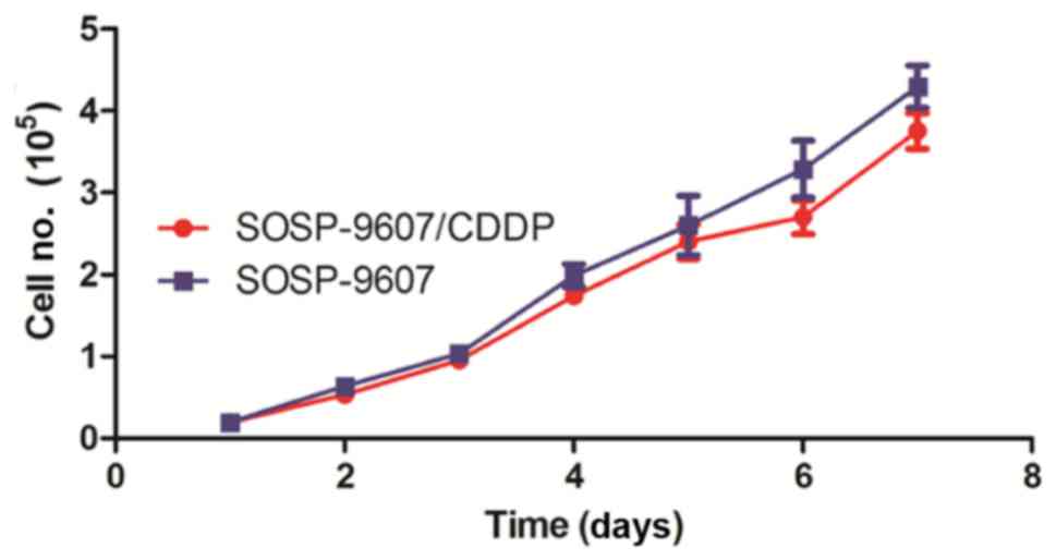
Cell growth cycle and doubling
time
The drug-resistance cell line of osteosarcoma
SOSP-9607/CDDP was constructed, and cell line SOSP-9607 was used as
the control group. The growth cycle and doubling time of cells in
each group were detected by MTT assay. The results are showed in
Fig. 1: The growth rate of SOSP-9607
was similar to that of SOSP-9607/DDP, with incubation time as the
x-axis and average cell count as the y-axis, and the difference was
not statistically significant (P>0.05). The doubling time of
SOSP-9607/CDDP was similar to that of SOSP-9607, and the difference
was not statistically significant (P>0.05).
Sensitivity of SOSP-9607/CDDP and
SOSP-9607 to drugs
CDDP in 8 different concentrations was added into
the drug-resistance cell line of osteosarcoma SOSP-9607/CDDP.
SOSP-9607 was used as the control group. The sensitivity of
SOSP-9607/CDDP to the above drug was detected via MTT assay and
IC50 value was calculated, and the results are shown in
Table II. The results showed that
IC50 values of SOSP-9607 and SOSP-9607/CDDP were
3.28±0.42 and 18.27±0.39, and the difference was statistically
significant (P<0.01).
| Table II.Sensitivity of SOSP-9607 and
SOSP-9607/CDDP to cisplatin. |
Table II.
Sensitivity of SOSP-9607 and
SOSP-9607/CDDP to cisplatin.
|
| IC50 (mean
± SD, µg/ml) |
|
|---|
|
|
|
|
|---|
| Agent | SOSP-9607 | SOSP-9607/CDDP | P-value |
|---|
| CDDP | 3.28±0.42 | 18.27±0.39 | 0.0087 |
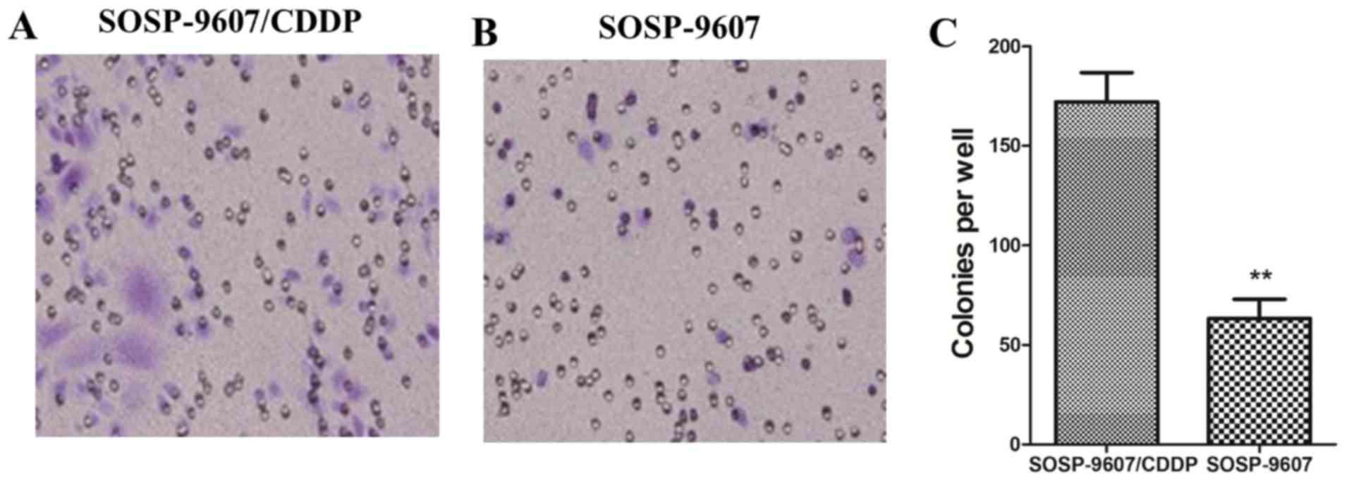
Detection of cell migration
capacity
The migration capacity of drug-resistance cell line
of osteosarcoma SOSP-9607/CDDP was detected via Transwell assay.
SOSP-9607 was used as the control group. The results are shown in
Fig. 2: The migration capacity of
SOSP-9607/CDDP was significantly increased compared with that of
SOSP-9607, and the difference was statistically significant
(P<0.01).
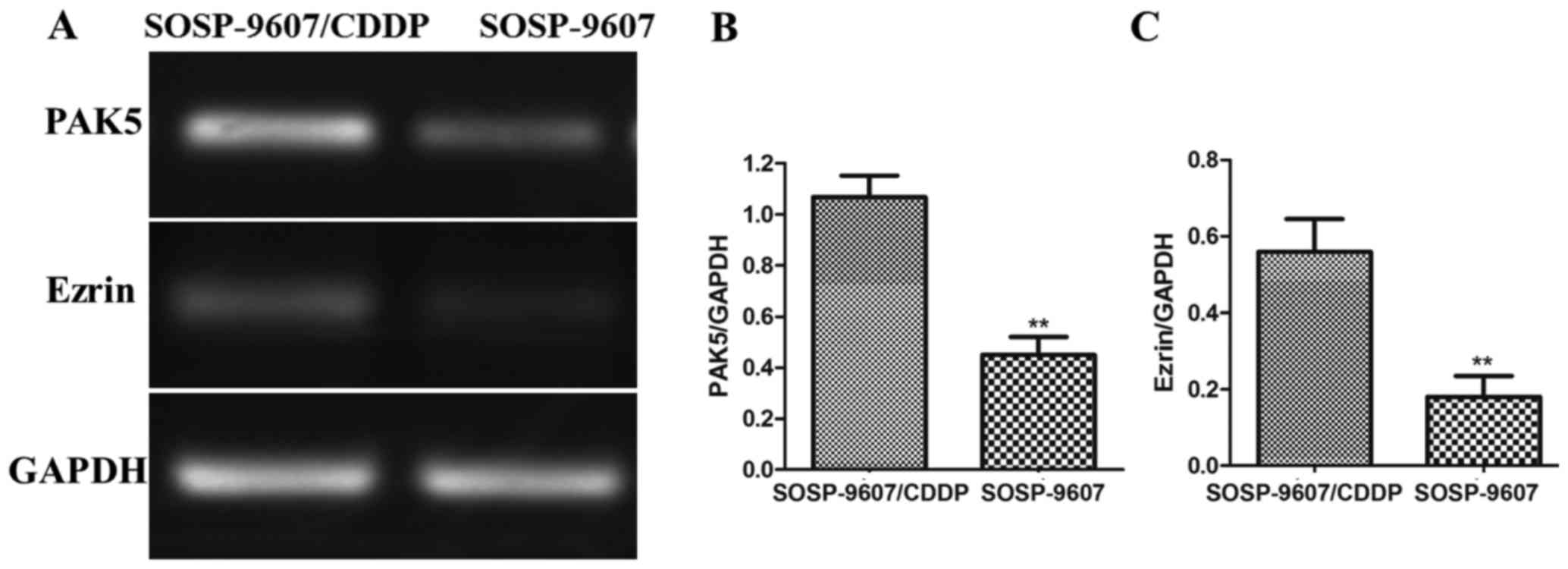
Detection of PAK5 and Ezrin
expressions via semi-qPCR
The expression levels of PAK5 and Ezrin in
SOSP-9607/CDDP and SOSP-9607 were detected by semi-qPCR. The
results are shown in Fig. 3: The
expression levels of PAK5 and Ezrin in SOSP-9607/CDDP were
significantly higher than those in SOSP-9607, and the differences
were statistically significant (P<0.01).
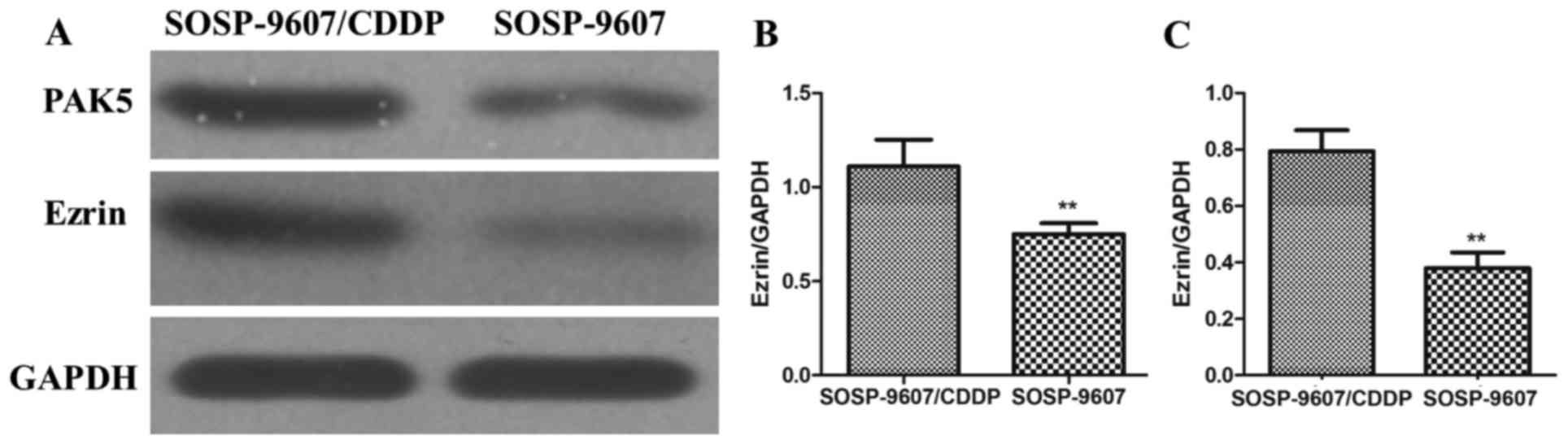
Detection of PAK5 and Ezrin expression
via western blot analysis
The expression levels of PAK5 and Ezrin in
SOSP-9607/CDDP and SOSP-9607 were detected via western blot
analysis. The results are shown in Fig.
4: The PAK5 and Ezrin protein expression levels in
SOSP-9607/CDDP were significantly higher than those in SOSP-9607,
and the differences were statistically significant (P<0.01).
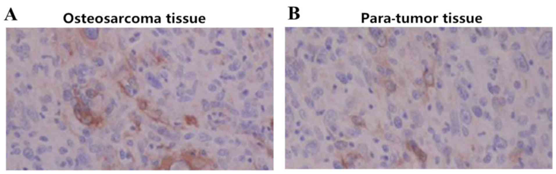
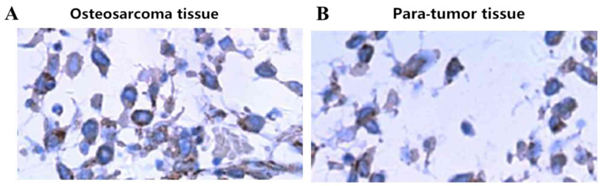
Detection of PAK5 and Ezrin expression
in osteosarcoma tissues via immunohistochemistry
The expression levels of PAK5 and Ezrin in
osteosarcoma were detected via immunohistochemistry. The para-tumor
tissue was used as the control group. Yellow staining in cytoplasm
and surrounding mesenchyme was regarded as PAK5-positive
expression, while brown staining in nucleus was regarded as
Ezrin-positive expression. The immunohistochemical results of PAK5
in osteosarcoma and para-tumor tissue are shown in Fig. 5: The expression level of PAK5 in
osteosarcoma was significantly higher than that in para-tumor
tissue, and the difference was statistically significant
(P<0.01). The immunohistochemical results of Ezrin in
osteosarcoma and para-tumor tissue are shown in Fig. 6: The expression level of Ezrin in
osteosarcoma was significantly higher than that in para-tumor
tissue, and the difference was statistically significant
(P<0.01).
Analysis of correlation between PAK5
and Ezrin expression levels in osteosarcoma patients
The CDDP resistance situation of 78 osteosarcoma
patients was recorded, the positive rates of PAK and Ezrin were
detected and correlation analysis was performed for the expressions
of PAK and Ezrin. Pearson's correlation analysis showed that the
expressions of PAK and Ezrin were positively correlated in drug
resistance of osteosarcoma (r=0.197, P=0.023). The results are
shown in Table III.
| Table III.Correlation between PAK5 and Ezrin
expression in osteosarcoma tissues. |
Table III.
Correlation between PAK5 and Ezrin
expression in osteosarcoma tissues.
|
| PAK5 |
|
|
|---|
|
|
|
|
|
|---|
| Ezrin | Positive | Negative | r | P-value |
|---|
| Positive | 38 | 13 | 0.197 | 0.023 |
| Negative | 32 | 9 |
|
|
Discussion
Osteosarcoma is the most common malignant bone tumor
in clinic at present, which often occurs in young people, and its
main clinical manifestations are progressively aggravated bone pain
and local swelling (10,11). The 5-year survival rate of
osteosarcoma patients is low, placing great economic and mental
burdens on the society and family (12). The chemotherapy drug resistance in the
treatment process of osteosarcoma is the problem puzzling
clinicians currently. There is related literature reporting that
the expression levels of some important genes and proteins not only
affect the occurrence and development of osteosarcoma, but also are
closely related to the clinical treatment effect of osteosarcoma
(13).
PAK is an evolutionarily highly conserved protein
kinase, and it plays an important role in occurrence, invasion and
metastasis of tumors (14). PAK5 has
been seldom studied, and it is a newly-discovered member in PAK
family, which is capable of activating the cell survival signaling
pathway and protecting the cell survival (15). Previous studies have shown that PAK5
may play an important role in the development and progression of
malignant tumors. Gu et al (16) detected the expression of PAK5 in tumor
tissues of osteosarcoma patients via immunohistochemistry, and
found that PAK5 was highly expressed in osteosarcoma patients,
suggesting that PAK5 may be related to the pathogenesis of
osteosarcoma. Ezrin protein is a member of the cytoskeletal
cross-linked protein ERM, which mainly mediates the connective
function of membrane and cytoskeleton with a series of functions
for relevant cells (17). The
excessive expression and activation of Ezrin can lead to abnormal
intercellular signal transmission and promote tumor cell
metastasis, thus playing a role in tumor invasion and metastasis
(18,19). Studies have shown that Ezrin protein
is closely related to pulmonary metastasis of osteosarcoma
patients, which can be used as an index of predicting the prognosis
and survival (20). In the present
study, the expression levels of PAK5 and Ezrin in tumor tissues and
para-tumor tissues of patients with osteosarcoma resistance were
analyzed by western blot analysis. The results showed that the
expression levels of PAK5 and Ezrin in osteosarcoma tissues were
significantly higher than those in para-tumor tissues, suggesting
that the drug resistance of osteosarcoma patients may be closely
related to the high expression of PAK5 and Ezrin. The
CDDP-resistance osteosarcoma cell line was constructed and the
expression of PAK5 and Ezrin was detected via semi-qPCR and western
blot analysis from the gene and protein level. The results showed
that the expression quantities of PAK5 and Ezrin in CDDP-resistance
osteosarcoma cell line were significantly higher than those in
osteosarcoma cell line, which was consistent with the results in
osteosarcoma patients. The above results revealed in vivo
and in vitro that PAK5 and Ezrin are closely related to the
drug resistance of osteosarcoma patients. The correlation study on
expression of PAK5 and Ezrin showed that the expression of PAK5 and
Ezrin are positively correlated (r=0.197, P=0.023), suggesting that
PAK5 and Ezrin may affect the drug resistance of osteosarcoma to
chemotherapy drugs through synergistic effect.
In conclusion, PAK5 and Ezrin are closely related to
the drug resistance of osteosarcoma, which provides clinical
reference for clinically clarifying drug resistance mechanism of
osteosarcoma. However, there were still some short comings in this
study: Related mechanism of PAK5 and Ezrin involved in drug
resistance of osteosarcoma was not studied deeply, the sample size
in this experiment was small and the comparative study was not
performed for healthy volunteer samples, so the results need to be
further confirmed. However, the research values of PAK5 and Ezrin
in drug resistance of osteosarcoma are beyond doubt, which can
bring a new breakthrough for clinical chemotherapy of
osteosarcoma.
References
|
1
|
Shimizu T, Kido A, Honoki K, Murata K,
Fujii H, Higuchi B, Ishihara T, Takeshita Y, Shima M, Yajima H, et
al: A successful reconstruction using a frozen autograft and a
pedicled latissimus dorsi flap after a S12345B shoulder girdle
resection in a patient with osteosarcoma. J Reconstr Microsurg.
28:155–159. 2012. View Article : Google Scholar : PubMed/NCBI
|
|
2
|
Qiu Q, Jiang J, Lin L, Cheng S, Xin D,
Jiang W, Shen J and Hu Z: Downregulation of RSK2 influences the
biological activities of human osteosarcoma cells through
inactivating AKT/mTOR signaling pathways. Int J Oncol.
48:2508–2520. 2016. View Article : Google Scholar : PubMed/NCBI
|
|
3
|
Hurley C, McCarville MB, Shulkin BL, Mao
S, Wu J, Navid F, Daw NC, Pappo AS and Bishop MW: Comparison of
(18) F-FDG-PET-CT and bone scintigraphy for evaluation of osseous
metastases in newly diagnosed and recurrent osteosarcoma. Pediatr
Blood Cancer. 63:1381–1386. 2016. View Article : Google Scholar : PubMed/NCBI
|
|
4
|
Waresijiang N, Sun J, Abuduaini R, Jiang
T, Zhou W and Yuan H: The downregulation of miR 125a 5p functions
as a tumor suppressor by directly targeting MMP 11 in osteosarcoma.
Mol Med Rep. 13:4859–4864. 2016. View Article : Google Scholar : PubMed/NCBI
|
|
5
|
Zeng W, Liu Q, Chen Z, Wu X, Zhong Y and
Wu J: Silencing of hERG1 gene inhibits proliferation and invasion,
and induces apoptosis in human osteosarcoma cells by targeting the
NF-κB Pathway. J Cancer. 7:746–757. 2016. View Article : Google Scholar : PubMed/NCBI
|
|
6
|
Sun Y, Xia P, Zhang H, Liu B and Shi Y:
P53 is required for Doxorubicin-induced apoptosis via the TGF-beta
signaling pathway in osteosarcoma-derived cells. Am J Cancer Res.
6:114–125. 2015.PubMed/NCBI
|
|
7
|
Zheng K, Yu X, Chang Z, Xu S and Xu M:
Effect of pathological fracture on limb salvage surgery with
preservation of the epiphysis in children with osteosarcoma of the
distal femur: Two case reports. Mol Clin Oncol. 4:523–526. 2016.
View Article : Google Scholar : PubMed/NCBI
|
|
8
|
Zhao J, Chen F, Zhou Q, Pan W, Wang X, Xu
J, Ni L and Yang H: Aberrant expression of microRNA-99a and its
target gene mTOR associated with malignant progression and poor
prognosis in patients with osteosarcoma. Onco Targets Ther.
9:1589–1597. 2016. View Article : Google Scholar : PubMed/NCBI
|
|
9
|
Zhai J, Qu S, Li X, Zhong J, Chen X, Qu Z
and Wu D: miR-129 suppresses tumor cell growth and invasion by
targeting PAK5 in hepatocellular carcinoma. Biochem Biophys Res
Commun. 464:161–167. 2015. View Article : Google Scholar : PubMed/NCBI
|
|
10
|
Bao ZQ, Zhang CC, Xiao YZ, Zhou JS, Tao YS
and Chai DM: Over-expression of Sox4 and β-catenin is associated
with a less favorable prognosis of osteosarcoma. J Huazhong Univ
Sci Technolog Med Sci. 36:193–199. 2016. View Article : Google Scholar : PubMed/NCBI
|
|
11
|
Maximov VV and Aqeilan RI: Genetic factors
conferring metastasis in osteosarcoma. Future Oncol. 12:1623–1644.
2016. View Article : Google Scholar : PubMed/NCBI
|
|
12
|
Zhou Q, Shen L, Liu C, Liu C, Chen H and
Liu J: The effects of estradiol and glucocorticoid on human
osteosarcoma cells: Similarities and differences. Anticancer Res.
36:1683–1691. 2016.PubMed/NCBI
|
|
13
|
Dai Y, Jiang J, Wang Y, Jin Z and Hu S:
The correlation and clinical implication of VEGF-C expression in
microvascular density and lymph node metastasis of gastric
carcinoma. Am J Transl Res. 8:5741–5747. 2016.PubMed/NCBI
|
|
14
|
Huang JF, Du WX and Chen JJ: Elevated
expression of matrix metalloproteinase-3 in human osteosarcoma and
its association with tumor metastasis. J BUON. 21:1279–1286.
2016.PubMed/NCBI
|
|
15
|
Li Y, Ke Q, Shao Y, Zhu G, Li Y, Geng N,
Jin F and Li F: GATA1 induces epithelial-mesenchymal transition in
breast cancer cells through PAK5 oncogenic signaling. Oncotarget.
6:4345–4356. 2015. View Article : Google Scholar : PubMed/NCBI
|
|
16
|
Gu X, Wang C, Wang X, Ma G, Li Y, Cui L,
Chen Y, Zhao B and Li K: Efficient inhibition of human glioma
development by RNA interference-mediated silencing of PAK5. Int J
Biol Sci. 11:230–237. 2015. View Article : Google Scholar : PubMed/NCBI
|
|
17
|
Pan D, Wang S, Ye H, Xu S and Ye G: Ezrin
expression in the primary hepatocellular carcinoma patients and
associated with clinical, pathological characteristics. J Cancer
Res Ther. 12:291–294. 2016. View Article : Google Scholar
|
|
18
|
Du P, Xu B, Zhang D, Shao Y, Zheng X, Li
X, Xiong Y, Wu C and Jiang J: Hierarchical investigating the
predictive value of p53, COX2, EGFR, nm23 in the post-operative
patients with colorectal carcinoma. Oncotarget. 8:954–966.
2017.PubMed/NCBI
|
|
19
|
Rox K, Rohde M, Chhatwal GS and Müller R:
Disorazoles block group A streptococcal invasion into epithelial
cells via interference with the host factor Ezrin. Cell Chem Biol.
24:159–170. 2017. View Article : Google Scholar : PubMed/NCBI
|
|
20
|
Hago AM, Gamallat Y, Mahmoud SA, Huang Y,
Zhang J, Mahmoud YK, Wang J, Wei Y, Wang L, Zhou S, et al: Ezrin
expression is altered in mice lymphatic metastatic hepatocellular
carcinoma and subcellular fractions upon Annexin 7 modulation
in-vitro. Biomed Pharmacother. 85:209–217. 2017. View Article : Google Scholar : PubMed/NCBI
|