Introduction
Surgical resection remains the gold standard therapy
for primary and secondary liver malignancies, but resectability
largely depends on factors such as extent of the disease,
concurrent liver steatosis or cirrhosis, amount of remaining
healthy liver tissue after resection (the future liver remnant-FLR)
as well as patient comorbidities. Therefore, thermal ablation is
widely used as an alternative or additional technique in cases were
resection deems hazardous, or in unresectable disease with multiple
lesions when only the combined ablation-and-resection (1) or two-stage hepatectomy approach seems
feasible to achieve tumor-free margins with sufficient FLR.
The two currently most common thermal ablation
modalities are radiofrequency ablation (RFA) and microwave ablation
(MWA) (2,3), both applied either percutaneously or
during open or laparoscopic surgery (‘surgical ablation’) (4). In RFA an electrical current within the
radiofrequency range is transported through either a monopolar
electrode or between two bipolar electrodes to produce heat-induced
cytotoxicity in the liver tissue (5,6). MWA
technique is different, as microwave radiation leads to high
frequency oscillation in water molecules, subsequent frictional
heating and cell death through coagulation necrosis (4,7).
Therefore, MWA requires no application of grounding pads because an
electrical circuit is not established. MWA is mostly applied
through a single coaxial electrode device (3,4,8,9).
The success of thermal ablation (complete necrosis
of tumors) generally depends on multiple factors like tumor size,
location, hepatic blood flow and equipment selection (3,7,10). In this regard, previous research
revealed several advantages of MWA compared to conventional
(monopolar) RFA, namely easier and fast use with superior heating
capacity, independency of tissue charring and only minimal
influence of the ‘heat-sink-effect’. This term describes cooling
within ablation zones next to large hepatic blood vessels which
might lead to incomplete tumor necrosis resulting in local
recurrence (8).
In general, correct image-guided insertion of
electrodes is crucial to achieve a successful ablation with
completely devitalized tumors. While in percutaneous radiological
ablation this is either achieved by computed tomography or
sonography, surgical ablation relies heavily on intraoperative
B-mode ultrasound. Although this can be a very powerful tool in
experienced hands, differentiation of viable and destructed tissue
and identification of the tumor border is quite challenging in
daily practice-mainly due to tissue scarring and gas bubble
phenomena which arise during the ablation process. Therefore
several other imaging techniques such as contrast-enhanced
sonography (11), real-time
ultrasound elastography (12) and
electrode vibration elastography (13) were already evaluated to further
enhance the security and practicability of ablations. This
experimental study for the first time examines the feasibility of
infrared thermographic monitoring in RFA and MWA.
Thermographic imaging uses radiation within the
long-infrared range of the electromagnetic spectrum (9–14 µm) that
is emitted by all objects. Hereby, colored output images with
analyzable information of surface temperatures are obtained. It is
non-invasive, easy to apply and technically well-engineered due to
its wide range use in medicine (e.g., to detect inflammation by
irregular cutaneous blood flow) and several other industries such
as construction technology (e.g., to analyze heat leaks in thermal
insulation) (14).
In this proof-of-principle study we hypothesize,
that thermography: i) is a suitable non-invasive tool for
monitoring the ablation process; and ii) is also helpful to detect
a possible heat-sink effect near large vessels. Hence, we compared
RFA to MWA using an ex vivo perfused porcine liver
model.
Materials and methods
We investigated each ablation technology in a
setting with both a heat sink and non-heat sink surrounding.
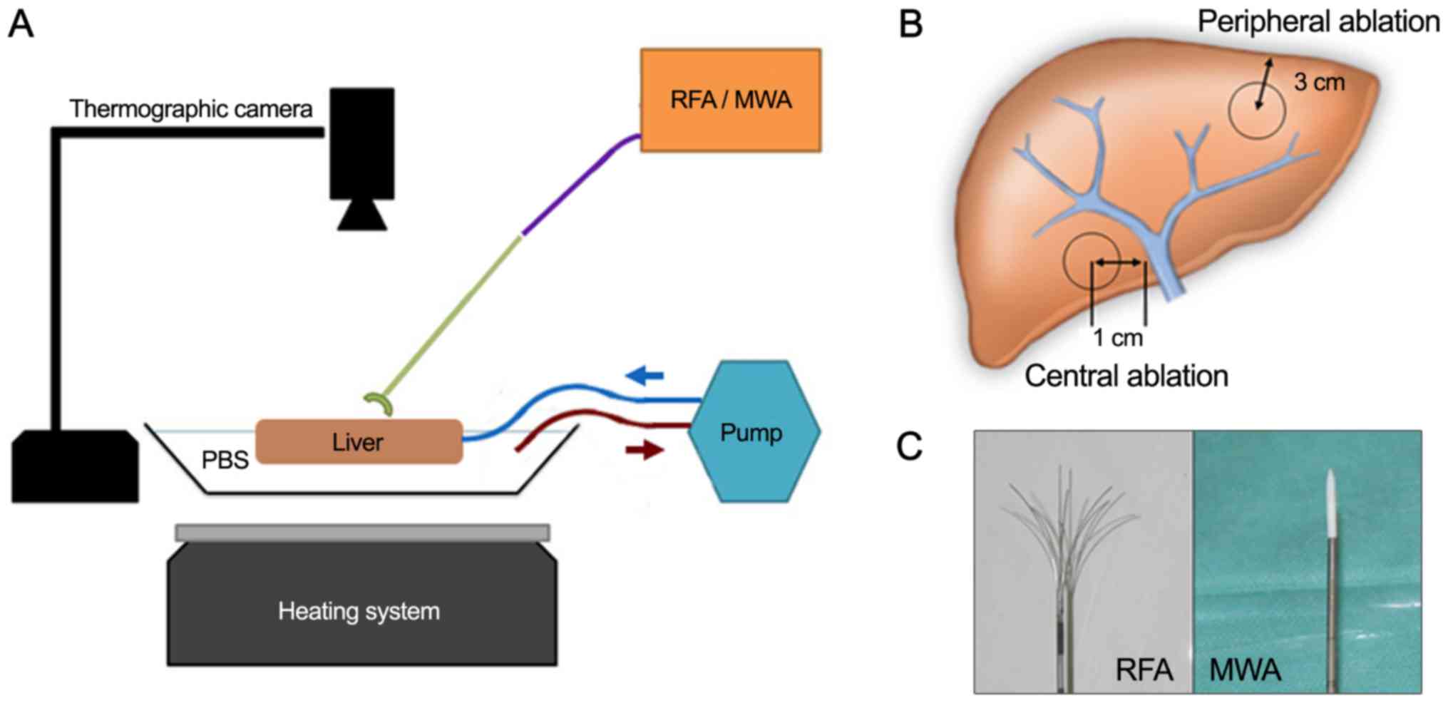
Therefore, an experimental setting using ex vivo perfused
porcine livers with hepatic flow simulation was established
(Fig. 1A and B). Since no patients or
living animals were involved no ethics approval was required
according to local regulations.
Experimental and technical setup
Four complete, freshly taken porcine livers from
adult animals with intact in- and outflow vessels were obtained
from an abattoir and instantly used after less than 1-h cooled
transport. Perfusion at body temperature was initiated by placing
the livers in a metal container half filled with 37°C PBS
(standardized sodium phosphate buffer), keeping the temperature
constant with a heating system. A temperature probe continuously
controlled temperature levels and the heating system was adjusted
accordingly. Hepatic inflow was simulated through flexible
rubber-tubes sutured to the portal vein and connected to a
perfusion pump system (Heissner P300-I; Heissner GMBH, Lauterbach,
Germany). Hereby a constant hepatic flow with 5.3 liters per min
was established to emulate the average human cardiac output. A
thermographic camera (FLIR A35sc; FLIR, Wilsonville, Oregon, USA)
was centered 50 cm above the liver in a right angle. Calibrations
for emissivity, distance, relative humidity and ambient temperature
were set according to the manufacturers recommendations.
For RFA a 250-watt radiofrequency generator (Model
1500X) was equipped with an expandable, multi-array monopolar RFA
electrode (StarBurst XL RFA Device; both RITA Medical Systems,
Fremont, CA, USA;/AngioDynamics Inc., Latham, NY, USA). MWA was
applied using a 2.45 GHz microwave generator (Sulis VpMTA Generator
with Local Control Station) with a single-monopolar
applicator-needle (Accu2i pMTA Applicator; both Microsulis Medical
Ltd., Denmead, UK/AngioDynamics Inc.) (Fig. 1C).
Experimental workflow
We sequentially performed a central and peripheral
ablation with MWA in one hepatic lobe and RFA in the other
contralateral lobe in each of the four livers. To assess the
influence of a possible heat sink effect, a central ablation with a
distance of 1 cm from the tip of the needle to the wall of the main
right or left portal vein branch was compared to a peripheral
ablation with the probe tip located 3 cm from the hepatic margin
(Fig. 1B). In both settings, the
probe tip was placed in a parenchymal depth of 3 cm below the liver
surface. Sonographic guidance was used for needle placement in both
locations and exclusion of nearby large vessels in the peripheral
setting.
Before starting and during the ablation process, the
surface temperature of non-affected central liver parenchyma and
the surrounding peripheral PBS buffer temperature were documented
as baseline values (NormV/NormP). MWA device was set to 2 min
ablation time with 100 Watt, RFA to 150 Watt for 8 min including a
maximum of 3 min pre-heating to reach operating temperature (set to
105°C). After the heat-up time of 3 min the RFA needle was extended
to the full antenna length.
All ablative procedures were constantly monitored
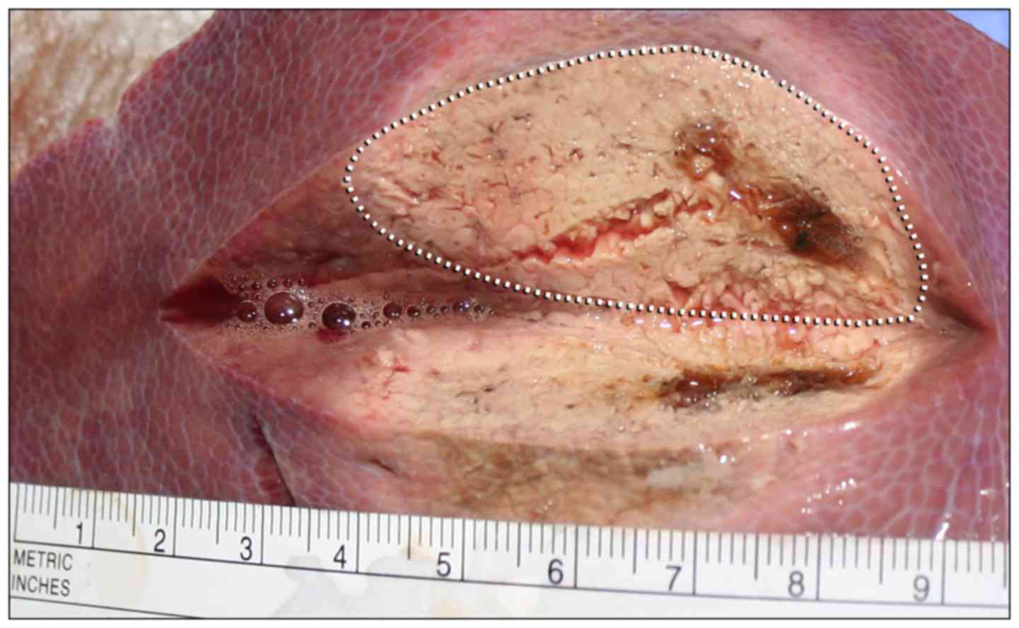
with the thermographic camera in real-time. After every ablative
run, the parenchyma was dissected vertically along the ablation
needle axis and the maximum diameter of the macroscopically clearly
marked ablation zone was measured with a linear centimeter scale
(Fig. 2). Tissue specimens of about 1
cm3 size collected from the center and the macroscopic
border were stored as formalin-fixed, paraffin-embedded (FFPE)
specimens for routine histological staining with haematoxylin and
eosin (H&E) to confirm the correct ablation process, the extent
of ablation and morphological changes, especially grade of
necrosis.
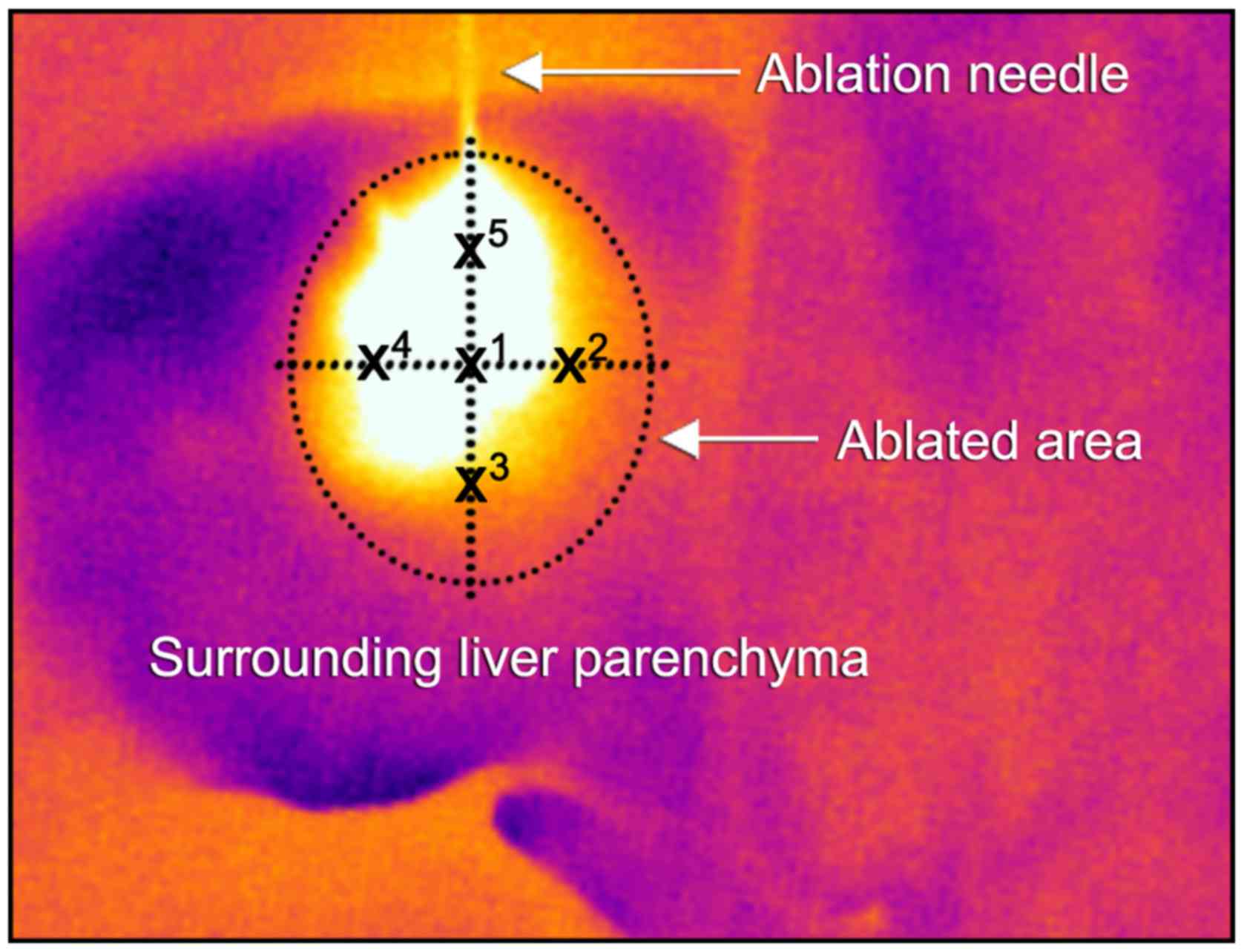
The documented thermographic images were evaluated
concerning the temperature profiles using the manufacturer's
professional infrared reporting software (FLIR Tools Plus
software). Mean temperatures (with standard deviations) were
calculated according to the scheme described in Fig. 3 with one central temperature point at
the probe tip (X1) and further four surrounding points
at 50% of the maximum visible ablation diameter (X2–5).
Temperature points were measured every 10 sec (for 120 sec) for MWA
and every 20 sec (for 480 sec) for RFA. Recorded radiometric data
were exported and further statistically analyzed with SPSS
Statistics 21 (IBM Corp., Armonk, NY, USA). Mean ablation infrared
temperature levels were compared using the Student's t-test.
Results
The experimental setup of MWA vs. RFA ablations in
general showed comparable temperatures at the surrounding
peripheral/central perivascular parenchyma of 33.43°C
(±-2.57)/32.53°C (±-1.58) in MWA and 35.6°C (±-3.16)/32.32°C
(±-1.23) in RFA, respectively. (Table
I-MWA_NormP/NormV vs. RFA_NormP/NormV). The peripheral
temperature in both techniques was higher due to increased heat
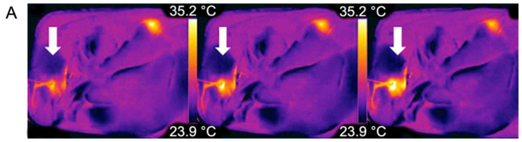
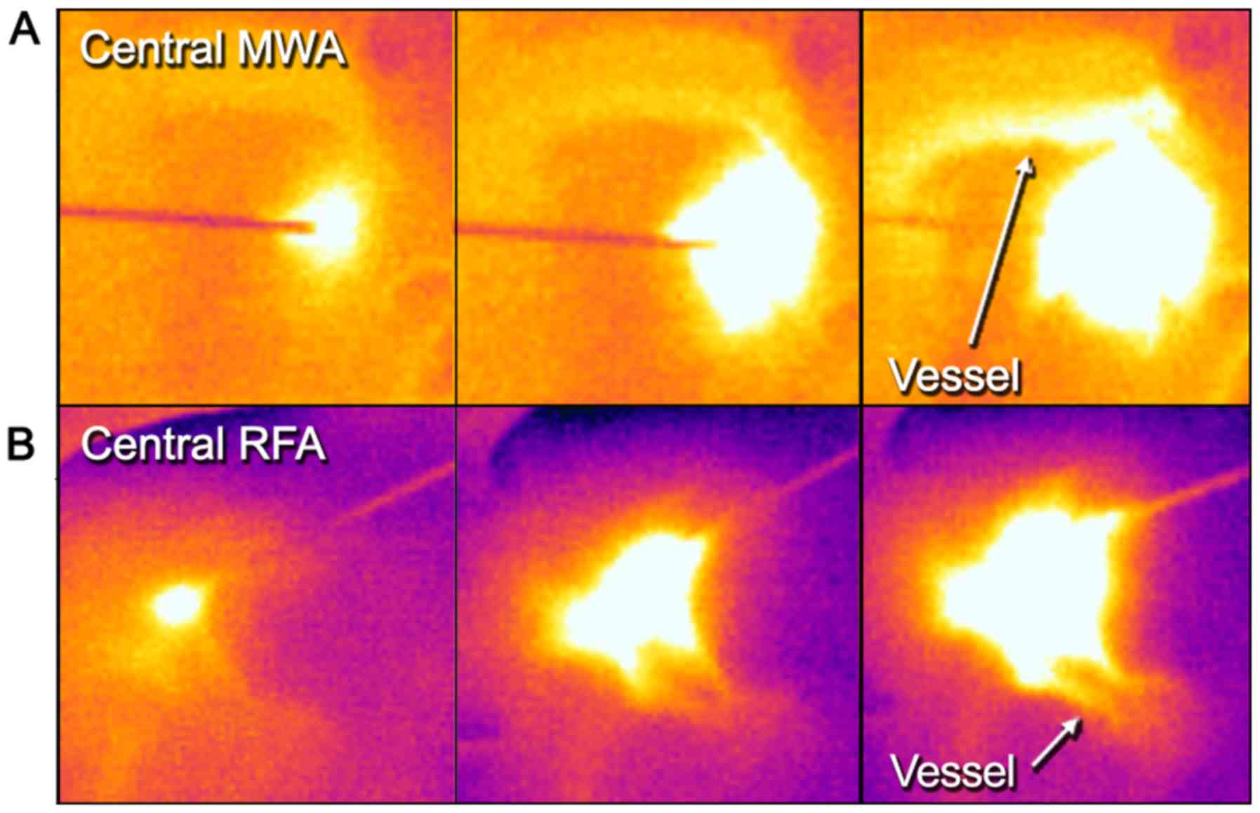
transmission in the fluid surrounding. Representative examples of
real-time thermography of an MWA and RFA ablative run are given in
Figs. 4A, B and 5A.
 | Table I.Thermographic temperature
measurements. |
Table I.
Thermographic temperature
measurements.
| Measurement | N (measuring
pointsa) | Minimum (°C) | Maximum (°C) | Mean (°C) | Standard deviation
(°C) |
|---|
| MWA_NormV | 12 | 29.37 | 34.69 | 32.53 |
1.58 |
| MWA_NormP | 12 | 29.46 | 36.73 | 33.43 |
2.57 |
| MWA_Ves | 12 | 32.72 | 60.02 | 50.52 |
8.35 |
| MWA_Peri | 12 | 33.77 | 63.45 | 50.18 | 10.35 |
| RFA_NormV | 24 | 30.17 | 33.83 | 32.32 |
1.23 |
| RFA_NormP | 24 | 29.00 | 39.75 | 35.60 |
3.16 |
| RFA_Ves | 24 | 33.87 | 57.78 | 47.11 |
8.35 |
| RFA_Peri | 24 | 35.73 | 81.15 | 68.72 | 12.70 |
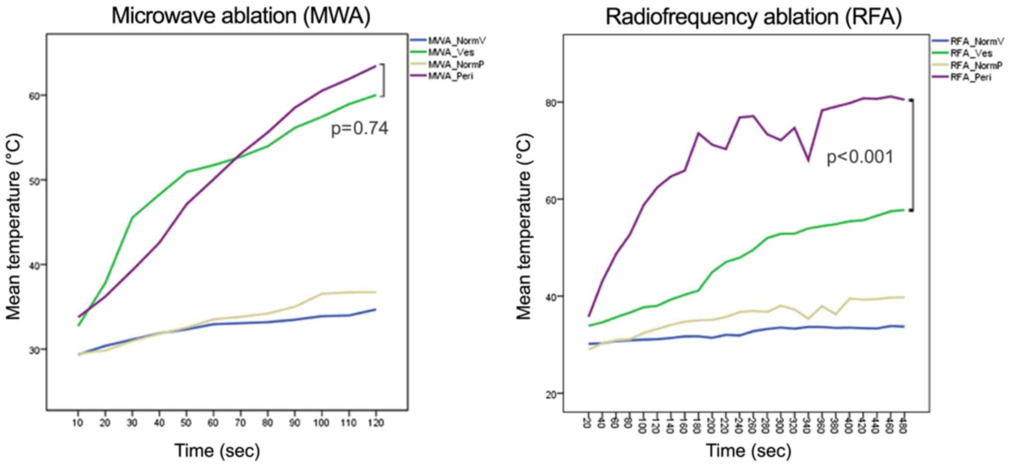
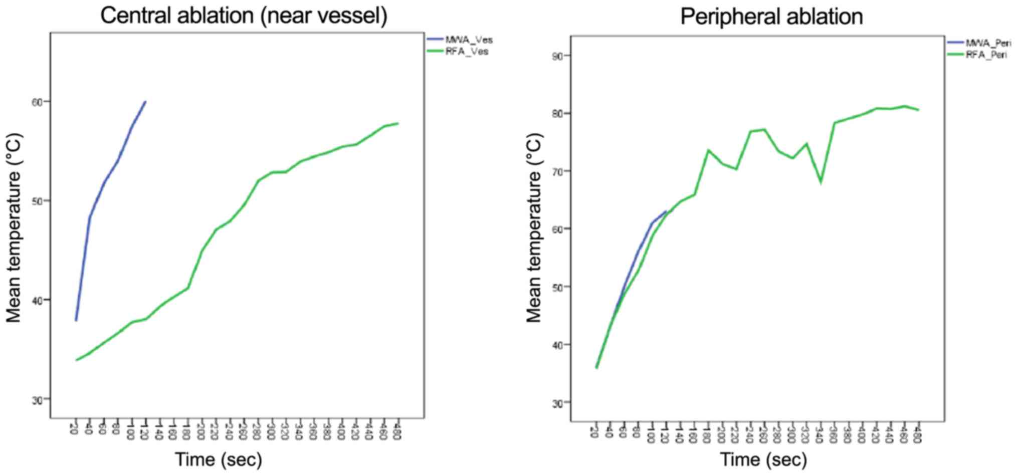
MWA data
MW ablation showed homogenous temperature profiles
and very short heat up time (Fig. 6
and Table I) in the peripheral
(non-heat-sink) environment as well as in the central (heat-sink)
location. When using the manufacturer's recommendation of 100 watts
for 120 sec to achieve a predicted spherical ablation zone of 3 cm,
the mean macroscopic ablation diameter was 3.38 cm (±0.26).
Ablation in the liver periphery resulted in a diameter of 3.19 cm
(±0.13) compared to 3.56 cm (±0.06) in proximity to a vessel. The
mean surface temperature recorded was 50.18°C (±10.35) in
peripheral vs. 50.52°C (±8.35) in central MW ablations (P=0.74).
However, the temperature increased more stable and reached a higher
end-point in the peripheral setting than in the central setting,
which might indicate a minimal heat-sink effect in MWA. In general,
the thermographic ablation zones were very clearly defined in the
thermographic documentation and showed a homogenous round or
slightly elliptic zone.
RFA data
RFA needs a considerable pre-heating time before
antenna extensions to achieve appropriate target temperatures. When
using the recommended settings of 150 Watt for 5 min (plus 3 min
pre-heat time) for an anticipated lesion of 3 cm, the mean
macroscopic ablation diameter was 3.17 cm (±0.09). Ablation in the
liver periphery resulted in a diameter of 3.10 cm (±0.36) compared
to 3.23 cm (±0.33) in proximity to a vessel. The mean surface
temperature recorded was 68.72°C (±12.70) in peripheral vs. 47.11°C
(±8.35) in central RFAs (P<0.001) (Fig. 6 and Table
I). This indicates a stronger heat sink effect than in MWA.
Furthermore, the temperature changes in peripheral and central RFA
were much more pronounced compared to MWA, suggesting increased
susceptibility of radiofrequency current to alterations in the
surrounding area, such as charring or dessication (Fig. 7).
Histology
Histology confirmed correct ablation in both
ablative techniques and microscopically verified the necrotic
border of our macroscopic measurements. H&E-staining revealed
increased cytoplasmic eosinophilia, cytoplasmic homogenization and
destruction of vessel walls and ductular epithelium indicating
liver tissue regression. These morphologic alterations could be
recognized with a much greater extent within the samples taken from
the ablation core. Although liver tissue in biopsies of healthy
tissue outside the macroscopic border mainly preserved its
histological tissue architecture some small portal tracts and
vessel walls showed the same signs of regression as samples from
the ablation core, suggesting a gradient partial cell damage beyond
the visible border.
Discussion
To the best of our knowledge, the present study is
the first to investigate the feasibility of thermography as a
monitoring tool for open surgical hepatic MWA/RFA. Both ablation
techniques induced visually well definable thermographic patterns.
Within the ablated areas, different zones of (changing)
temperatures were observed (Fig. 5):
A central, initially deformed but ultimately round to almost
elliptical hot spot zone directly around the probe tip with a clear
border. This zone is surrounded by a rather gradually colored
halo-like outer rim with lower temperatures, visually
distinguishable by two color zones (in our settings white and
yellow-orange). The overall visual thermographic appearance was
comparable between RFA and MWA but with a more pronounced shape
deformation near large vessels in RFA. This visual impression of a
possible heat sink effect was confirmed by temperature profile
analysis, where thermography depicted a significant mean surface
temperature loss in central vs. peripheral RFA, while there was no
statistical difference in MWA. Hereby, we could demonstrate that
there is indeed an observable heat sink effect using RFA next to
large, perfused vessels (Figs. 5 and
6). This further supports existing
evidence, that RFA is more dependent on vascular blood flow, than
MWA (15–18). Concerning MWA, data in the literature
are rather diverse. Some authors did not find a heat-sink-effect
(15,19), while others described it indeed
detectable but less pronounced compared to RFA (20–22).
Thorough analysis of these studies however reveals, that not only
the principal technique (MWA or RFA) used for ablation, but also
the needle design affects the magnitude of an observed
heat-sink-effect. For example, monopolar MWA and bipolar RFA show
comparably low susceptibility, while monopolar RFA seems very
strongly affected (8,10). In an experimental study Ringe et
al showed, that there is a distance- and flow-dependent
significant heat-sink-effect when using a 915 MHz MWA system with
45 W for 10 min ablations (15),
whereas others found no influence of vascular proximity or flow
rate when using a 2.45 GHz 100 W MWA system (23) identical to the equipment we used in
the present study. The present experimental data and growing
evidence for technical and procedural advantages of MWA encouraged
us to increasingly use MWA instead of RFA during surgery (4).
Although CT-/MRI-guided, software-navigated
percutaneous ablation (24) with
continuous imaging control represents the most sophisticated
ablation technique currently available in clinical radiology,
distribution of this elaborate method is still limited in most
countries. Furthermore, percutaneous ablation of a tumor located at
the liver surface or adjacent to vulnerable structures might be
technically not feasible. On the contrary, open surgical ablation
is fast and affordable and enables the surgeon to rotate the liver
within its anatomical surrounding, manually protect heat-sensitive
organs (bowel), easily conduct repeated overlapping ablations for a
clustered ablation area and also allows for instant complication
management e.g., in the case of bleeding or accidental bowel
injury.
Effective tumor ablation thereby depends on accurate
needle placement in a three-dimensional space, appropriate
tissue-destructive energy and sufficient overlapping safety margins
(4–10 mm). Local recurrence rates and hepatic progression free
survival are usually compared to the gold standard of surgical
resection, especially as researchers attribute local recurrences at
the ablation site to either insufficient imaging control or the
heat-sink-effect. This is of particular relevance when ablation is
used in a curative setting as an alternative to resection e.g., in
an ‘ablate & resect’-strategy for small and deep lesions,
aiming to preserve healthy liver parenchyma. Exemplary local
recurrence rates after open surgical MWA of colorectal cancer
metastasis are in the range of 2–4% in most studies and comparable
to those after resection, but may be higher depending on factors
such as tumor size (4). Percutaneous
and laparoscopic ablations show much more variable local recurrence
rates of usually between 5 and 14%. Occasionally, some studies
reported even higher local recurrence rates up to 52%, which is
probably a result of widely differing inclusion criteria and
obvious technical limitations of these approaches (8,25,26). In summary, preventing local recurrence
after MWA or RFA is a major issue in ablative therapies, and
several tools have been investigated for this purpose.
Most data is available on contrast-enhanced
ultrasound (CEUS), which facilitates a pictorial real-time process
of tumor vascularity. CEUS relies on bolus injection of contrast
agents consisting of microbubbles (e.g., SonoVue; Bracco SpA,
Milan, Italy). Hereby, viable tumor tissue becomes better
delineated, which is especially useful for small lesions and
steatotic livers. However, gas bubble formation and interference
with the RF generator during ablation also compromise CEUS.
(12) Furthermore, microbubbles
disrupt after some min, reducing the enhancement period and the
time frame for needle placement and successful ablation (27). As a result, appropriate application is
very much user dependent, usually requiring a well-trained
radiologist or surgeon in the operating theatre. In an experimental
rat liver model, CEUS optimally determines the maximum dimension of
the ablated zone ideally 2 h after RFA, which might further limit
its intraoperative use. (11) Other
tools such as real-time ultrasound elastography (12) and electrode vibration elastography
(13) also seem promising, but are
either technically complex, cost intensive or have so far only been
evaluated in small series.
The benefits of infrared thermography are its
non-invasive nature, the ‘user-friendly’ real-time visualization
and the reasonable cost effectiveness since it is a
well-established technology and not dependent on consumable
materials. Another possible field for the clinical use of
thermography application could be in delineating the extent of
overlapping ablations, since these are particularly challenging to
monitor with sonography due to gas bubble formation and scarring.
Thermography may also be valuable in raising alertness for heat
transmission to nearby, vulnerable structures (bile ducts, bowel
loops, diaphragm) (Fig. 5) and unmask
technical issues immediately during the ablation process.
This experimental study and the use of thermography
for ablations in general have several possible limitations.
Firstly, thermographic measurements are derived from surface
temperatures. Naturally, these do not necessarily always correlate
with the actual temperature in the whole object. However, in an
object with distinctive heat conduction such as the human liver it
may give a reasonable, reproducible approximation of nearby tissue
temperatures. Due to the anatomy of porcine livers with rather flat
hepatic parenchyma, extrapolating the investigated effect to deep
intraparenchymal ablations e.g., in segment 8 of a human liver
might be difficult. Presumably this will have a noticeable impact
on surface temperatures and consecutive infrared data and will need
further evaluation.
Secondly, we used PBS solution to simulate hepatic
blood flow, which might have different conductive properties than
whole blood, possibly affecting the results of RFA-which relies on
current flux- and might also influence heat transmission to the
liver surface and resulting thermographic images. Furthermore, a
simplified constant flow rate of 5.3 l/min may not reflect
real-life intraoperative variations in hepatic flow, which are
dependent on several factors such as heart rate, intravascular
volume, blood composition, etc (28,29).
However, similar simplified models were used in other published
studies examining heat sink effects and different ablation
techniques (8).
To overcome these limitations, the next step in
evaluating non-invasive thermographic monitoring for hepatic tumor
ablation should record the process in an in vivo clinical,
intraoperative setting. First, we suggest examining temperature
profiles in a series of superficial and deep intraparenchymal tumor
ablations to standardize surface temperatures. Comparison and/or
image-fusion of thermography with intraoperative
(contrast-enhanced) ultrasound would be preferable to assess the
practical applicability in daily routine.
In conclusion, this study for the first time
confirmed infrared thermography as a feasible tool for real-time
visualization of open MWA and RFA. Also, we have observed a
distinct heat-sink effect in RFA compared to MWA during ablation
near large vessels in real time. Further studies and in-vivo
observations are necessary to estimate its usefulness in daily
clinical routine.
Acknowledgements
This study was financially supported by the Austrian
Cancer Aid (Österreichische Krebshilfe). The results of this study
were presented at the 2014 ESSO Congress (European Society of
Surgical Oncology, Liverpool, UK) and the 38th Meeting of the
Austrian Society of Surgical Research (Salzburg, Austria).
Glossary
Abbreviations
Abbreviations:
|
CEUS
|
contrast-enhanced ultrasound
|
|
CT
|
computed tomography
|
|
FFPE
|
formalin-fixed paraffin-embedded
tissue
|
|
FLR
|
future liver remnant
|
|
MRI
|
magnetic resonance imaging
|
|
MWA
|
microwave ablation
|
|
MW
|
microwave
|
|
NormV
|
surrounding parenchyma surface
temperature (near vessel)
|
|
NormP
|
surrounding parenchyma surface
temperature (peripheral)
|
|
PBS
|
sodium phosphate buffer solution
|
|
RFA
|
radiofrequency ablation
|
References
|
1
|
Evrard S, Poston G, Kissmeyer-Nielsen P,
Diallo A, Desolneux G, Brouste V, Lalet C, Mortensen F, Stättner S,
Fenwick S, et al: Combined ablation and resection (CARe) as an
effective parenchymal sparing treatment for extensive colorectal
liver metastases. PLoS One. 9:e1144042014. View Article : Google Scholar : PubMed/NCBI
|
|
2
|
Poulou LS, Botsa E, Thanou I, Ziakas PD
and Thanos L: Percutaneous microwave ablation vs radiofrequency
ablation in the treatment of hepatocellular carcinoma. World J
Hepatol. 7:1054–1063. 2015. View Article : Google Scholar : PubMed/NCBI
|
|
3
|
Molla N, AlMenieir N, Simoneau E, Aljiffry
M, Valenti D, Metrakos P, Boucher LM and Hassanain M: The role of
interventional radiology in the management of hepatocellular
carcinoma. Curr Oncol. 21:e480–e492. 2014. View Article : Google Scholar : PubMed/NCBI
|
|
4
|
Stättner S, Primavesi F, Yip VS, Jones RP,
Öfner D, Malik HZ, Fenwick SW and Poston GJ: Evolution of surgical
microwave ablation for the treatment of colorectal cancer liver
metastasis: Review of the literature and a single centre
experience. Surg Today. 45:407–415. 2015. View Article : Google Scholar : PubMed/NCBI
|
|
5
|
Goldberg SN, Gazelle GS and Mueller PR:
Thermal ablation therapy for focal malignancy: A unified approach
to underlying principles, techniques, and diagnostic imaging
guidance. AJR Am J Roentgenol. 174:323–331. 2000. View Article : Google Scholar : PubMed/NCBI
|
|
6
|
Hänsler J, Neureiter D, Strobel D, Müller
W, Mutter D, Bernatik T, Hahn EG and Becker D: Cellular and
vascular reactions in the liver to radio-frequency thermo-ablation
with wet needle applicators. Study on juvenile domestic pigs. Eur
Surg Res. 34:357–363. 2002. View Article : Google Scholar : PubMed/NCBI
|
|
7
|
Brace CL: Microwave tissue ablation:
Biophysics, technology, and applications. Crit Rev Biomed Eng.
38:65–78. 2010. View Article : Google Scholar : PubMed/NCBI
|
|
8
|
Pillai K, Akhter J, Chua TC, Shehata M,
Alzahrani N, Al-Alem I and Morris DL: Heat sink effect on tumor
ablation characteristics as observed in monopolar radiofrequency,
bipolar radiofrequency, and microwave, using ex vivo calf liver
model. Medicine (Baltimore). 94:e5802015. View Article : Google Scholar : PubMed/NCBI
|
|
9
|
Lubner MG, Brace CL, Hinshaw JL and Lee FT
Jr: Microwave tumor ablation: Mechanism of action, clinical
results, and devices. J Vasc Interv Radiol. 21(8 Suppl): S192–S203.
2010. View Article : Google Scholar : PubMed/NCBI
|
|
10
|
Dodd GD III, Dodd NA, Lanctot AC and
Glueck DA: Effect of variation of portal venous blood flow on
radiofrequency and microwave ablations in a blood-perfused bovine
liver model. Radiology. 267:129–136. 2013. View Article : Google Scholar : PubMed/NCBI
|
|
11
|
Wu H, Wilkins LR, Ziats NP, Haaga JR and
Exner AA: Real-time monitoring of radiofrequency ablation and
postablation assessment: Accuracy of contrast-enhanced US in
experimental rat liver model. Radiology. 270:107–116. 2014.
View Article : Google Scholar : PubMed/NCBI
|
|
12
|
Wiggermann P, Brünn K, Rennert J, Loss M,
Wobser H, Schreyer AG, Stroszczynski C and Jung EM: Monitoring
during hepatic radiofrequency ablation (RFA): Comparison of
real-time ultrasound elastography (RTE) and contrast-enhanced
ultrasound (CEUS): First clinical results of 25 patients.
Ultraschall Med. 34:590–594. 2013. View Article : Google Scholar : PubMed/NCBI
|
|
13
|
Dewall RJ, Varghese T and Brace CL:
Visualizing ex vivo radiofrequency and microwave ablation zones
using electrode vibration elastography. Med Phys. 39:6692–6700.
2012. View Article : Google Scholar : PubMed/NCBI
|
|
14
|
Ring EF and Ammer K: Infrared thermal
imaging in medicine. Physiol Meas. 33:R33–R46. 2012. View Article : Google Scholar : PubMed/NCBI
|
|
15
|
Ringe KI, Lutat C, Rieder C, Schenk A,
Wacker F and Raatschen HJ: Experimental evaluation of the heat sink
effect in hepatic microwave ablation. PLoS One. 10:e01343012015.
View Article : Google Scholar : PubMed/NCBI
|
|
16
|
Chinn SB, Lee FT Jr, Kennedy GD, Chinn C,
Johnson CD, Winter TC III, Warner TF and Mahvi DM: Effect of
vascular occlusion on radiofrequency ablation of the liver: Results
in a porcine model. AJR Am J Roentgenol. 176:789–795. 2001.
View Article : Google Scholar : PubMed/NCBI
|
|
17
|
Kim YS, Rhim H, Cho OK, Koh BH and Kim Y:
Intrahepatic recurrence after percutaneous radiofrequency ablation
of hepatocellular carcinoma: Analysis of the pattern and risk
factors. Eur J Radiol. 59:432–441. 2006. View Article : Google Scholar : PubMed/NCBI
|
|
18
|
Al-Alem I, Pillai K, Akhter J, Chua TC and
Morris DL: Heat sink phenomenon of bipolar and monopolar
radiofrequency ablation observed using polypropylene tubes for
vessel simulation. Surg Innov. 21:269–276. 2014. View Article : Google Scholar : PubMed/NCBI
|
|
19
|
Brannan JD and Ladtkow CM: Modeling
bimodal vessel effects on radio and microwave frequency ablation
zones. Conf Proc IEEE Eng Med Biol Soc. 2009:pp. 5989–5992. 2009;
PubMed/NCBI
|
|
20
|
Wright AS, Sampson LA, Warner TF, Mahvi DM
and Lee FT Jr: Radiofrequency versus microwave ablation in a
hepatic porcine model. Radiology. 236:132–139. 2005. View Article : Google Scholar : PubMed/NCBI
|
|
21
|
Yu NC, Raman SS, Kim YJ, Lassman C, Chang
X and Lu DS: Microwave liver ablation: Influence of hepatic vein
size on heat-sink effect in a porcine model. J Vasc Interv Radiol.
19:1087–1092. 2008. View Article : Google Scholar : PubMed/NCBI
|
|
22
|
Bhardwaj N, Strickland AD, Ahmad F,
El-Abassy M, Morgan B, Robertson GS and Lloyd DM: Microwave
ablation for unresectable hepatic tumours: Clinical results using a
novel microwave probe and generator. Eur J Surg Oncol. 36:264–268.
2010. View Article : Google Scholar : PubMed/NCBI
|
|
23
|
Awad MM, Devgan L, Kamel IR, Torbensen M
and Choti MA: Microwave ablation in a hepatic porcine model:
Correlation of CT and histopathologic findings. HPB (Oxford).
9:357–362. 2007. View Article : Google Scholar : PubMed/NCBI
|
|
24
|
Bale R, Widmann G, Schullian P, Haidu M,
Pall G, Klaus A, Weiss H, Biebl M and Margreiter R: Percutaneous
stereotactic radiofrequency ablation of colorectal liver
metastases. Eur Radiol. 22:930–937. 2012. View Article : Google Scholar : PubMed/NCBI
|
|
25
|
Hori T, Nagata K, Hasuike S, Onaga M,
Motoda M, Moriuchi A, Iwakiri H, Uto H, Kato J, Ido A, et al: Risk
factors for the local recurrence of hepatocellular carcinoma after
a single session of percutaneous radiofrequency ablation. J
Gastroenterol. 38:977–981. 2003. View Article : Google Scholar : PubMed/NCBI
|
|
26
|
Poon RT, Ng KK, Lam CM, Ai V, Yuen J and
Fan ST: Effectiveness of radiofrequency ablation for hepatocellular
carcinomas larger than 3 cm in diameter. Arch Surg. 139:281–287.
2004. View Article : Google Scholar : PubMed/NCBI
|
|
27
|
Minami Y and Kudo M: Review of dynamic
contrast-enhanced ultrasound guidance in ablation therapy for
hepatocellular carcinoma. World J Gastroenterol. 17:4952–4959.
2011. View Article : Google Scholar : PubMed/NCBI
|
|
28
|
Schutt DJ and Haemmerich D: Effects of
variation in perfusion rates and of perfusion models in
computational models of radio frequency tumor ablation. Med Phys.
35:3462–3470. 2008. View Article : Google Scholar : PubMed/NCBI
|
|
29
|
Jakab F, Sugár I, Ráth Z, Nágy P and
Faller J: The relationship between portal venous and hepatic
arterial blood flow. I. Experimental liver transplantation. HPB
Surg. 10:21–26. 1996. View Article : Google Scholar : PubMed/NCBI
|