Introduction
Malignant melanoma tumor incidence has an annually
increasing trend worldwide. Moreover, melanoma accounts for the
largest part of skin cancer deaths (1,2). In
addition, melanoma is not sensitive to radiotherapy or
chemotherapy, and leads a serious problem to clinical treatment.
The development of new and highly effective anti-tumor compounds is
urgent and challenging. Furthermore, marine microorganisms and
secondary metabolism are promising sources for novel anti-cancer
drugs (3). Sansalvamide A, which is a
depsipeptide isolated from a marine fungi (Fusarium spp.),
exhibits anti-tumor activity on multiple cancer cell lines
(4,5)
and significant anti-proliferative effects in the National Cancer
Institute's panel of 60 cancer cell lines (6). Sansalvamide peptide has anti-tumor
activities that became the basis for developing a series of cyclic
peptide analogues with N-methylation. The synthesis of sansalvamide
A analogues received sustained attention in recent years. Bromined
sansalvamide peptide inhibits pancreatic cancer cell growth through
the G0/G1 cell-cycle arrest (7).
Methoxylized zygosporamide peptide inhibits B16 cell growth and
induces cell apoptosis (8). Cyclic
epi-pentadepis peptide induces differentiation and inhibits
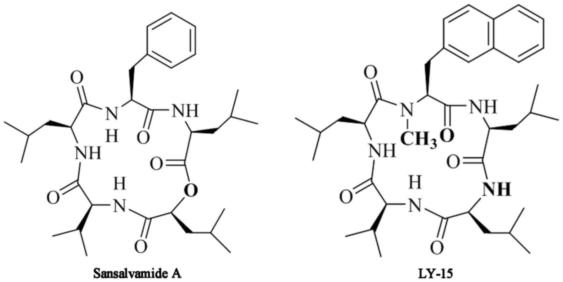
proliferation in murine melanoma B16 cells (9). Furthermore, the compound LY-15, which
was synthesized by the cyclization of the chain pentapeptide in
solution, has a molecular formula and weight of
C37H55N5O5 and 649.42,
respectively (Fig. 1). Here, we
focused on the effects of LY-15 on the growth and apoptosis of B16
cancer cell lines. Results showed that the compound has greater
potency when screened for the growth inhibition of B16 cancer
cells, suggesting that LY-15 might be a promising therapeutic
agent.
Materials and methods
Materials
The RPMI 1640 and trypsin-EDTA solution were
purchased from Gibco; Thermo Fisher Scientific, Inc., (Waltham, MA,
USA). The fetal bovine serum (FBS) was purchased from Bovogen
Biologicals Pty Ltd., (Keilor East, Victoria, Australia). The Cell
Counting Kit-8 (CCK-8) was purchased from Beijing Zoman
Biotechnology Co., Ltd., (Beijing, China) and the bicinchoninic
acid kit was purchased from Multi Sciences Co., Ltd., (Shanghai,
China). The polyvinylidene fluoride (PVDF) membranes were purchased
from Roche Applied Science (Penzberg, Germany). The antibody
against β-actin (polyclonal rabbit anti-mouse) was purchased from
Hangzhou HuaAn Biotechnology Co., Ltd., (Hangzhou, China). The
antibodies against B-cell lymphoma 2 (Bcl-2), Bcl-2-associated X
protein (Bax), caspase-3 and caspase-9 (all polyclonal rabbit
anti-mouse) were purchased from Arigo Bio (Taiwan, Xinzhu). The
secondary fluorescence anti-body (polyclonal goat anti-rabbit HRP)
was purchased from KPL, Inc., (Gaithersburg, MD, USA). The
sansalvamide analogue LY-15 was developed in Hebei Province Key
Laboratory of Molecular Chemistry for Drug (Shijiazhuang,
China).
Cancer cell line and cell culture
B16 cancer cells were selected for the present
study. The B16 cell line was obtained from the Research Center of
the Fourth Hospital of Hebei Medical University (Shijiazhuang,
China). The cells were grown in RPMI 1640 medium with 10%
heat-inactivated FBS and 100 µg/ml penicillin and streptomycin. The
cell line was grown in a humidified atmosphere of 95% O2
and 5% CO2 at 37°C and the cells were periodically
seeded into 25 cm2 flasks. The media was changed every
second or third day. For the experiments, the cells were grown to
80–90% confluence, digested with trypsin-EDTA, and plated in 25
cm2 flasks, and the media was changed every second or
third day on 6- or 96-well plates.
Concentration-dependent effect of
LY-15 on B16 cell growth inhibition
LY-15 was dissolved in DMSO and diluted with
serum-free medium to prepare solutions of 1,000, 100, 10 and 1 µM.
Single-cell suspensions of B16 cells were prepared and adjusted to
the indicated concentrations. The cells were inoculated in 96-well
plates (100 µl/well) with 5,000 cells/well. After overnight
inoculation for cell adherence, the old medium was discarded and
replaced with fresh medium with different concentrations of 100,
50, 25, 15, 10, 5 and 1 µM. Each group was placed into six wells,
and a 1% DMSO group was simultaneously prepared as the control. The
CCK-8 method was used to calculate the percentage growth of the B16
cells treated with various concentrations of LY-15 for 24 h.
Time-dependent effect of LY-15 on B16
cell growth inhibition
Upon reaching 80% confluence, the cells were
digested with trypsin-EDTA and serum-free medium was used to make a
single-cell suspension. The cells were seeded over night in 96-well
plates at a concentration of 3,000 cells/well. The wells were then
replaced with fresh complete medium and treated with 10 µM LY-15.
The percentage growth of the B16 cells treated for 24, 48, 72, 96
and 120 h was calculated.
Cell scratch test
Five uniform lines were drawn behind the 6-well
plates using a marker pen. The single-cell suspension was seeded in
the 6-well plates at a concentration of 200,000 cells/well. After 6
h, 20 µl pipette tips were used to draw through the marker lines.
The wells were washed with PBS thrice and fresh media (2 ml/well)
with different concentrations at 1, 2, 5 and 15 µM were added;
moreover, 1% DMSO was added to the last well that was
simultaneously prepared as the control. Images were captured at 0
and 24 h in the same position. We examined the effect of LY-15 on
B16 cell migration using the cell scratch test.
Flow cytometric analysis of apoptotic
cell death
At 80–90% confluence, the cells were treated with 2,
5, 10, 15 and 25 µM LY-15 for 24 h, and a control group was
prepared. The treated and untreated cells were harvested, washed
twice with PBS, and mixed with 1xbuffer 100 µl. After blending the
cells, 10 µl FITC and 5 µl propidium iodide (PI) were added. The
cells were kept in a dark place for 30 min.
Detection of caspase-3 and caspase-9
expressions using western blot analysis
The cells at 70–80% confluence were treated with 5,
15 and 25 µM of LY-15 for 24 h, and a control group was prepared.
Proteins were separated using SDS-polyacrylamide gel
electrophoresis. Equal amounts of protein (50 µg/sample) from B16
cells treated with LY-15 were loaded to 10% SDS-PAGE in an
electrophoresis buffer in a Bio-Rad slab gel apparatus. The
proteins were then transferred to a PVDF membrane under the
conditions of 80 V for 20 min, 100 V for 90 min and 200 mA for 120
min. Next, the membranes were incubated in antibody dilution
solution (rabbit anti-mouse bax, bcl-2, caspase-3 and caspase-9;
1:1,000 and β-actin; 1:3,000) overnight at 4°C. The blots were then
incubated with the secondary antibody (1:3,000) for 1 h at 37°C.
Results were obtained using the Aplegen Omega Lum C Gel Imaging
System (Gel Company, Inc., San Francisco, CA). Concetration of
protein was calculated by Image J (NIH, USA).
Statistical analysis
Statistical analysis was performed using SAS
software (SAS Institute, Inc., Cary, NC, USA) and R programming
language. Values were expressed as the mean ± standard error and
were analyzed using one-way ANOVA followed by Tukey's post hoc
test. P<0.05 was considered to indicate a statistically
significant difference. All statistical analyses were conducted
using Graphpad Prism v5 (GraphPad Software, Inc., La Jolla, CA,
USA).
Results
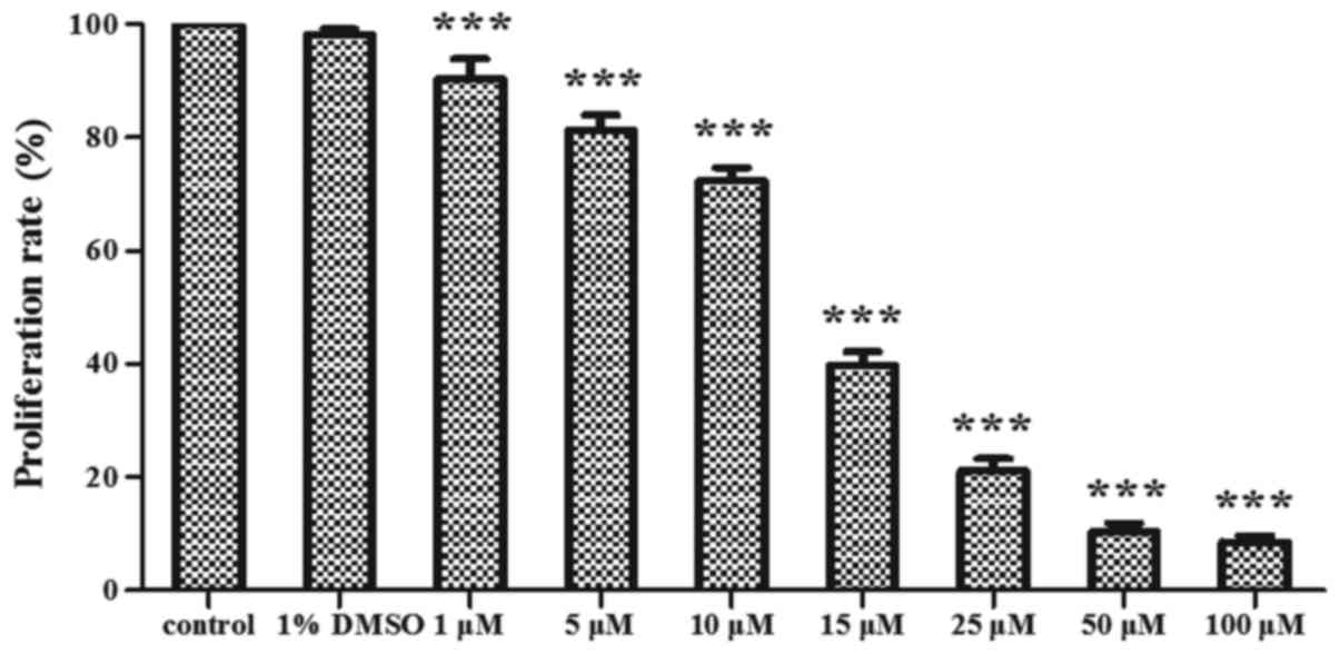
LY-15 exhibits a
concentration-dependent effect on B16 cell growth
The apoptosis and death of B16 cells were induced by
the LY-15 treatment in various concentrations of (1, 5, 10, 15, 25,
50 and 100 µM) for 24 h. Moreover, the proliferation rate of the
B16 cells showed a decreasing trend. Compared with the control
group, no significant difference was identified for the
proliferation rate of the 1% DMSO group (P>0.05), whereas those
for the treatment of the B16 cells with 100, 50, 25, 15 and 10 µM
LY-15 significantly decreased (P<0.001 for all treatment groups,
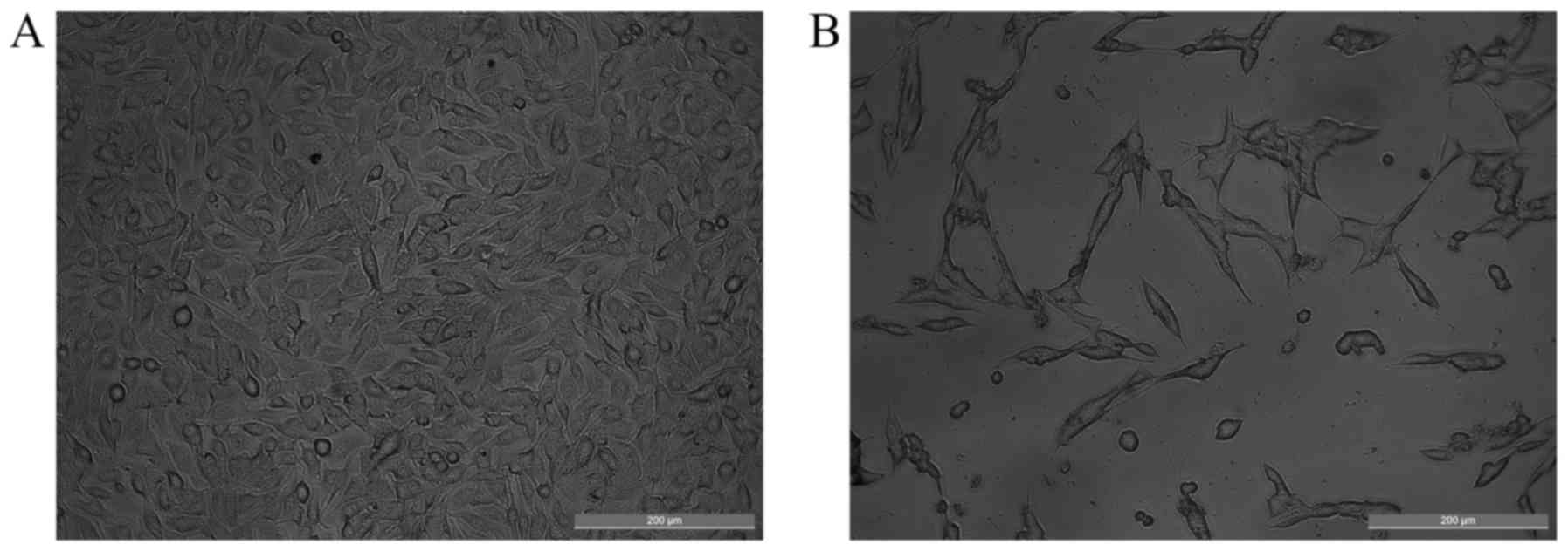
Fig. 2). The morphological changes in
B16 cells were observed via optical microscopy (Fig. 3). The B16 cells treated with 15 µM
LY-15 for 24 h exhibited morphological changes, including decreased
cell density, separation of the adjacent cells, and cell
shrinkage.
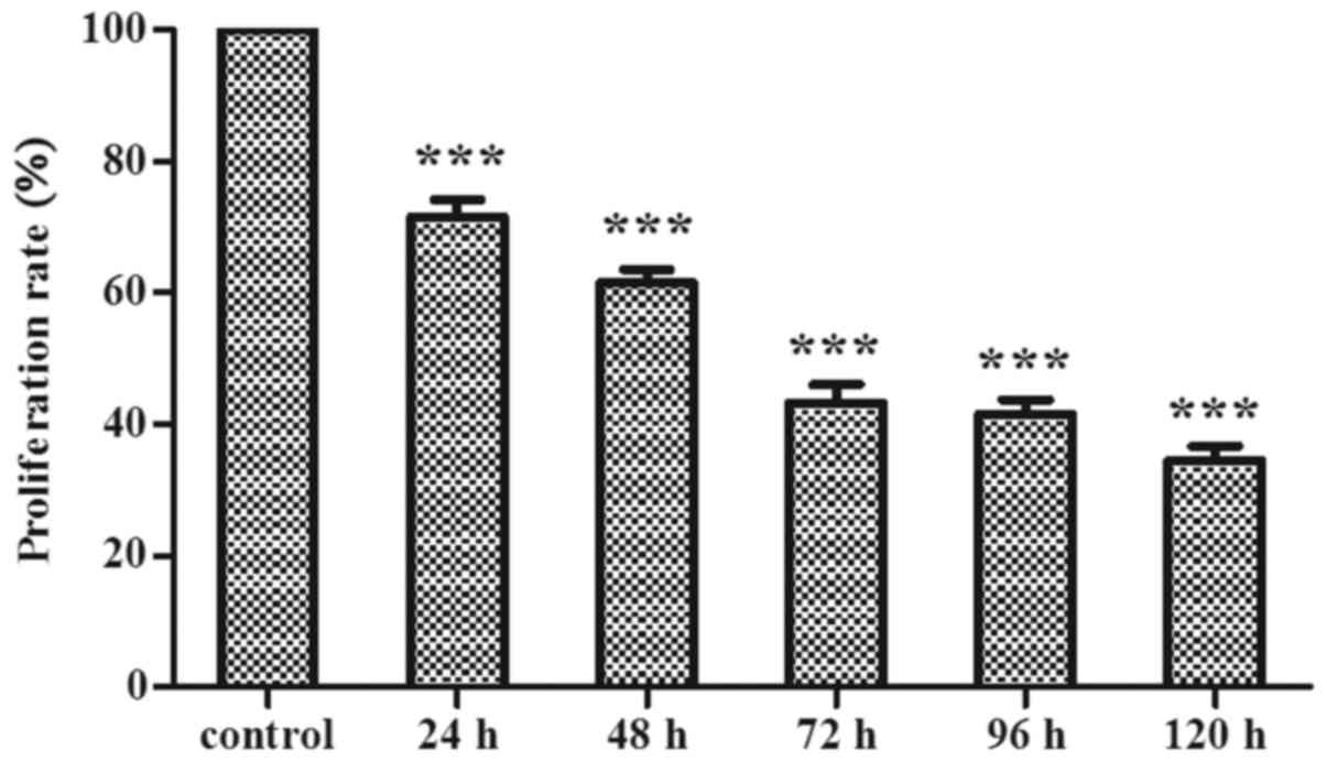
LY-15 exhibits a time-dependent effect
on B16 cell growth
The proliferation rate of the B16 cells with the
treatment of the concentration of 100, 50, 25, 15, 10, 5 and 1 µM
LY-15 was investigated. The concentration 10 µM LY-15 was
significantly reduced in a time-dependent manner compared with that
of the control group (P<0.001 for all time-dependent groups,
Fig. 4). The results showed that
LY-15 time-dependently inhibits the growth of B16 cells.
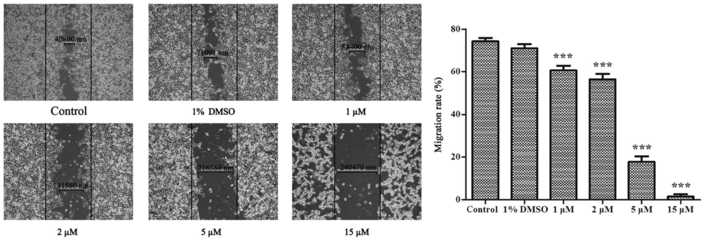
LY-15 inhibits the cell migration of
B16 cells
Compared with the control group, no obvious
difference was identified in the cell migration of the 1% DMSO
group. The cell migration of B16 was weakened by the LY-15
treatments with concentrations of 1, 2, 5 and 15 µM for 24 h. The
ability of cell migration gradually decreased as the concentration
increased. The cell migration changes were observed under optical
microscopy (Fig. 5). The cells lost
their ability to migrate at LY-15 concentration of 15 µM.
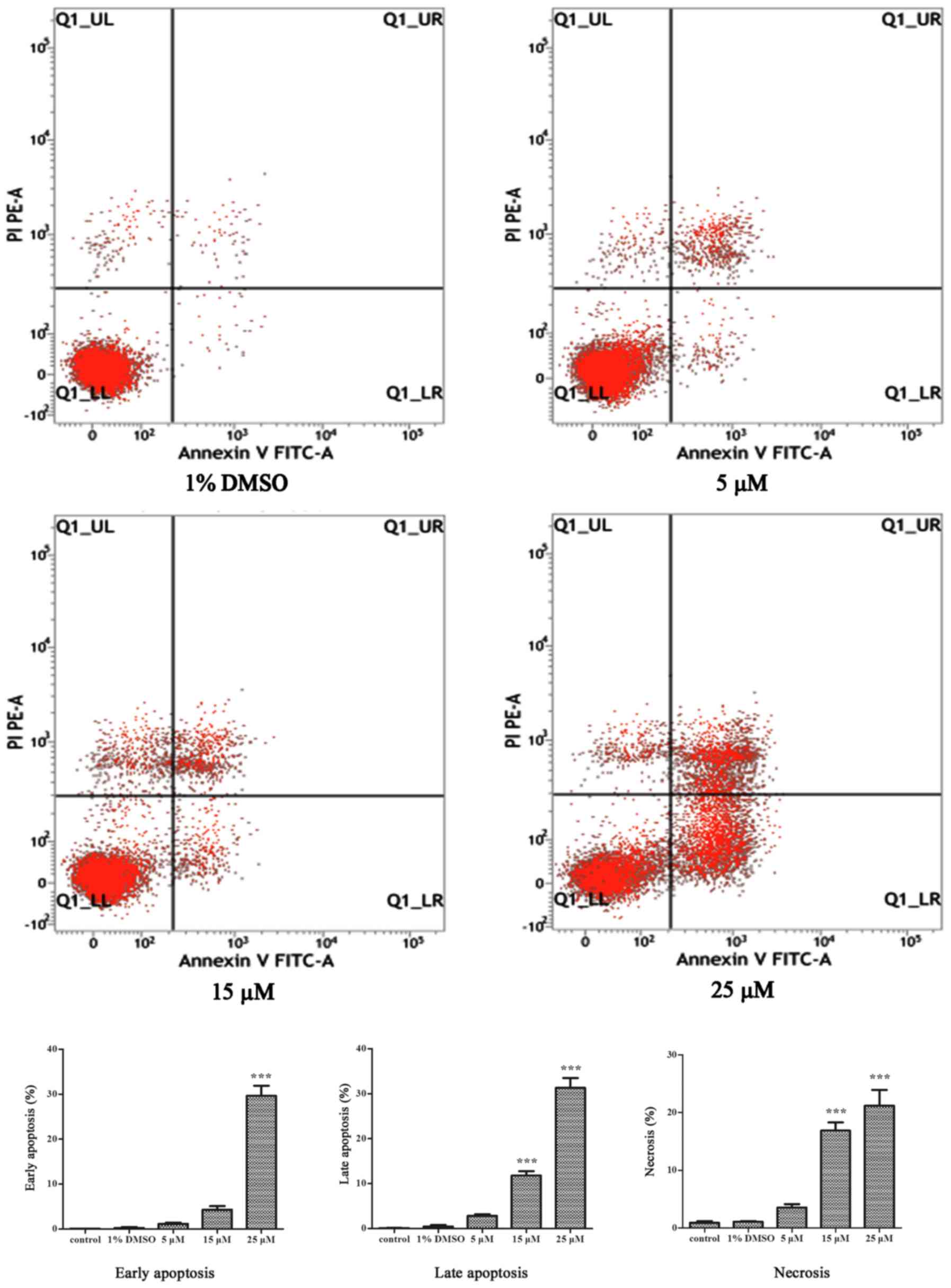
LY-15 promotes the apoptosis of B16
cells
The ability of LY-15 to induce apoptosis was
revealed by analyzing the cell samples using flow cytometry. The
cells in the early stage of apoptosis were detected by Annexin V,
whereas those in the late apoptosis were assessed by PI staining.
The 1% DMSO group did not show significant difference in apoptosis
compared with the control group. The early and late apoptosis
stages of the B16 cells gradually increased with the treatment of
LY-15 (5, 15 and 25 µM) for 24 h (Fig.
6). The percentage of apoptotic cells in the control group was
1.46; however, the proportion of apoptotic cells reached 8.57 in
the group treated with 15 µM LY-15 for 24 h.
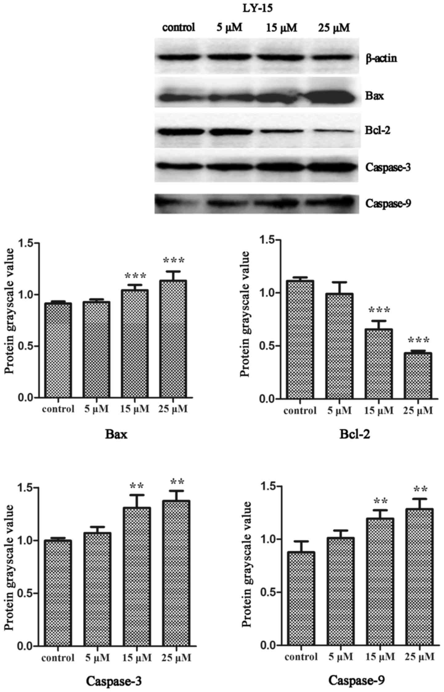
LY-15 induces apoptosis of B16 cells
through the mitochondrial pathway
At the present stage of the study, the results of
the western blot analysis showed an increasing trend in the
expression of Bax, whereas that of Bcl-2 showed the opposite.
Caspase-3 and caspase-9 expressions were analyzed using western
blot analysis and further confirmed that LY-15 induces apoptosis.
The expression levels of caspase-3 and caspase-9 in the B16 cells
increased with the treatments of 5, 15 and 25 µM LY-15 (Fig. 7). All results revealed that LY-15
induces the B16 cells apoptosis through a mitochondrial
pathway.
Discussion
Melanoma remains one of the most common cancers in
western countries and is the main contributor to skin
cancer-related deaths (10,11). Melanoma has a strong resistance and
high metastasis and mortality rates (12,13).
Studies were conducted on the synthesis and bioactivities of
various sansalvamide A derivatives (14,15). The
compound LY-15 is a novel sansalvamide A analogue that was recently
synthesized by our group. Based on the specific structures of
cyclic peptides, their bioactivities differ when the amino acid
sequences are changed; moreover, the analogues are lipophilic and
exhibit rapid membrane absorption (7). Accordingly, we studied the effects of
the compound LY-15 on melanoma cell B16 and its potential molecular
mechanisms to provide references for its clinical applications in
melanoma therapy.
Melanoma cells exhibit strong proliferation,
viability and malignancy. The curative effects of chemotherapeutic
drugs against melanoma are clinically challenged because of their
ability to resist apoptosis. Therefore, melanoma treatment studies
have focused on finding and selecting a novel effective compound.
Studies at this stage reported that the growth rate of B16 cells is
significantly inhibited by the compound LY-15. Data showed that the
effect of the compound LY-15 on the growth of B16 cells is
concentration-(100, 50, 25, 15, 10, 5 and 1 µM) and time-dependent.
Moreover, the cell proliferation rate was only 39.74% in the B16
cells cultured with 15 µM LY-15 for 24 h, indicating that the
treatment has a remarkable effect. The effect of LY-15 on
non-cancerous cells has been investigated preliminarily. The
proliferation of 293t cell still kept more than 85% even as LY-15
with the concentration 50 µM treated the cell. But, the details and
the toxicities for more non-cancerous cell need to investigate
further.
The cellular morphology of B16 treated with 15 µM
LY-15 showed that the cell density was sharply reduced over time,
and the cell exhibited a collection and spindle interstitial
substance morphology. Moreover, B16 cells were significantly and
slightly differentiated. Based on the flow cytometry data for B16
cells treated with 5 µM LY-15 for 24 h, cell apoptosis is
significant, and the values of the early and late apoptotic stages
of the B16 cells changed in a dose-dependent manner. These results
revealed that the inhibition of B16 proliferation by the compound
LY-15 is directly related to cell differentiation and apoptosis.
Scratch wound healing assay was conducted to study the effect of
LY-15 on the migration of B16 cells. The results showed that B16
cell migration was significantly inhibited by the 5 µM LY-15. In
order to describe the details of tumor cell movement, the cell
invasion and cell cycle will be key work for further investigation.
After the 15 µM LY-15 treatment, the cells turned sparse and
apoptotic.
Apoptosis is programmed cell death and is an
important part of the normal cell development and function of
organisms. This process is triggered in a cell either through an
extrinsic or intrinsic pathway (16).
Protein Bcl-2 inhibits apoptosis in various cell types (17) and Bax is a protein that promotes cell
apoptosis. Caspases play critical roles in apoptosis initiation and
execution. Caspase-9, which is from the caspase family, is an
initiator protein that drives caspase-3 to execute cell apoptosis.
In investigating the anti-apoptotic potential of LY-15, the LY-15
administration significantly elevated the levels of the apoptotic
marker proteins Bax, caspase-3 and caspase-9; whereas the
anti-apoptotic factor Bcl-2 level was reduced following the
treatment with various LY-15 concentrations (5, 15 and 25 µM).
These results support the hypothesis that LY-15 may inhibit B16
cell growth via the mitochondrial pathway, which induces
apoptosis.
In conclusion, the results of this study showed that
the compound LY-15 induces apoptosis in B16 cells and effectively
inhibits their migration. Considering the invasiveness and drug
resistance of melanoma, LY-15 provides a promising route for
improving melanoma treatments.
Acknowledgements
The authors received financial assistance from the
Basic Research Program of China (grant number 2010CB512007,
2012CB723501), the Natural Science Foundation of China (grant
number 3047204, 3087313).
References
|
1
|
Clark WH Jr, From L, Bernardino EA and
Mihm MC: The histogenesis and biologic behavior of primary human
malignant melanomas of the skin. Cancer Res. 29:705–727.
1969.PubMed/NCBI
|
|
2
|
Rigel DS, Russak J and Friedman R: The
evolution of melanoma diagnosis: 25 years beyond the ABCDs. CA
Cancer J Clin. 60:301–316. 2010. View Article : Google Scholar : PubMed/NCBI
|
|
3
|
Gupta AP, Pandotra P, Sharma R, Kushwaha M
and Gupta S: Chapter 8-Marine resource: A promising future for
anticancer drugs. Stud Nat Prod Chem. 40:229–325. 2013. View Article : Google Scholar
|
|
4
|
Styers TJ, Kekec A, Rodriguez R, Brown JD,
Cajica J, Pan PS, Parry E, Carroll CL, Medina I, Corral R, et al:
Synthesis of sansalvamide A derivatives and their cytotoxicity in
the MSS colon cancer cell line HT-29. Bioorg Med Chem.
14:5625–5631. 2006. View Article : Google Scholar : PubMed/NCBI
|
|
5
|
Otrubova K, Lushington G, Vander Velde D,
McGuire KL and McAlpine SR: Comprehensive study of sansalvamide A
derivatives and their structure-activity relationships against
drug-resistant colon cancer cell lines. J Med Chem. 51:530–544.
2008. View Article : Google Scholar : PubMed/NCBI
|
|
6
|
Belofsky GN, Jensen PR and Fenical W:
Sansalvamide: A new cytotoxic cyclic depsipeptide produced by a
marine fungus of the genus Fusarium. Tetrahedron Lett.
40:2913–2916. 1999. View Article : Google Scholar
|
|
7
|
Ujiki MB, Milam B, Ding XZ, Roginsky AB,
Salabat MR, Talamonti MS, Bell RH, Gu W, Silverman RB and Adrian
TE: A novel peptide sansalvamide analogue inhibits pancreatic
cancer cell growth through G0/G1 cell-cycle arrest. Biochem Biophys
Res Commun. 340:1224–1228. 2006. View Article : Google Scholar : PubMed/NCBI
|
|
8
|
Zhang G, Liu S, Liu Y, Wang F, Ren J, Gu
J, Zhou K and Shan B: A novel cyclic pentapeptide, H-10, inhibits
B16 cancer cell growth and induces cell apoptosis. Oncology Lett.
8:248–252. 2014. View Article : Google Scholar
|
|
9
|
Liu Y, Zhang G, Wang H, Liu S, Chen J,
Zhao L, Li J and Shan B: Novel cyclic pentapeptide H-15 induces
differentiation and inhibits proliferation in murine melanoma B16
cells. Oncology Lett. 11:1251–1255. 2016. View Article : Google Scholar
|
|
10
|
Park SY, Cho SJ, Kwon HC, Lee KR, Rhee DK
and Pyo S: Caspase-independent cell death by allicin in human
epithelial carcinoma cells: Involvement of PKA. Cancer Lett.
224:123–132. 2005. View Article : Google Scholar : PubMed/NCBI
|
|
11
|
Loescher LJ, Janda M, Soyer HP, Shea K and
Curiel-Lewandrowski C: Advances in skin cancer early detection and
diagnosis. Semin Oncol Nurs. 29:170–181. 2013. View Article : Google Scholar : PubMed/NCBI
|
|
12
|
Siegel RL, Miller KD and Jema A: Cancer
statistics. CA Cancer J Clin. 66:7–30. 2016. View Article : Google Scholar : PubMed/NCBI
|
|
13
|
Kwon SJ, Lee JH, Moon KD, Jeong IY, Ahn
DU, Lee MK and Seo KI: Induction of apoptosis by isoegomaketone
from perilla frutescens L. in B16 melanoma cells is mediated
through ROS generation and mitochondrial-dependent, -independent
pathway. Food Chem Toxicol. 65:97–104. 2014. View Article : Google Scholar : PubMed/NCBI
|
|
14
|
Pan PS, Vasko RC, Lapera SA, Johnson VA,
Sellers RP, Lin CC, Pan CM, Davis MR, Ardi VC and McAlpine SR: A
comprehensive study of Sansalvamide A derivatives: The
structure-activity relationships of 78 derivatives in two
pancreatic cancer cell lines. Bioorg Med Chem. 16:5806–5825. 2009.
View Article : Google Scholar
|
|
15
|
Pan PS, McGuire KL and McAlpine SR:
Identification of sansalvamide an analog potent against pancreatic
cancer cell lines. Bioorg Med Chem Lett. 17:5072–5077. 2007.
View Article : Google Scholar : PubMed/NCBI
|
|
16
|
Venkatesan RS and Sadiq AM: Effect of
morin-5-sulfonic acid sodium salt on the expression of apoptosis
related proteins caspase 3, Bax and Bcl 2 due to the mercury
induced oxidative stress in albino rats. Biomed Pharmacother.
85:202–208. 2017. View Article : Google Scholar : PubMed/NCBI
|
|
17
|
Zhang SD, Shan L, Li W, Li HL and Zhang
WD: Isochamaejasmin induces apoptosis in leukemia cells through
inhibiting Bcl 2 family proteins. Chin J Nat Med. 13:660–666.
2015.PubMed/NCBI
|