Introduction
Sodium citrate is widely used in the fields of
medicine (1,2) and food science as an adjuvant or
additive (3). Citrate, an important
intermediate in the tricarboxylic acid cycle, serves a notable role
in cellular metabolism (4). Under
normal physiological conditions, ATP production via oxidative
phosphorylation in the mitochondria is an efficient and primary
metabolic process, which produces far more ATP molecules from a
given amount of glucose than does glycolysis (5). By contrast, the majority of cancer cells
exhibit a high level of glycolysis for the generation of ATP to
meet their energy requirements; the metabolism of cancer cells is
often referred to as the ‘Warburg effect’ (6,7). Cancer
cells primarily metabolize glucose via glycolysis, excreting large
amounts of macromolecular precursors, including acetyl-CoA, for the
production of fatty acids, non-essential amino acids and
nucleotides (8). When glycolysis
occurs in this manner, cancer cells undergo fermentation even when
mitochondrial function is not impaired, in a process known as
‘aerobic glycolysis’ (9). Thus, any
inhibition of glycolysis may restrict oncogenic proliferation or
even halt it entirely, leading to cell death. During glycolysis,
citrate acts as an inhibitor of phosphofructokinase and may
obstruct the production of ATP or macromolecular precursors,
causing the typical cytotoxicity in cancer cells, as has been
confirmed in malignant pleural mesothelioma cells (10). Cells deficient in ATP frequently
undergo apoptosis (11,12); the induction of cell apoptosis via
citrate has been demonstrated in unicellular organisms as well as
cancer cells (13,14).
Gastric cancer is one of the most common types of
cancer in Asia (15,16). Over the last 4 decades its incidence
has decreased globally; however, higher rates of mortality have
been observed in East Asian countries than in other areas of the
world (16). Genetic and
environmental factors may serve a role in the etiology of gastric
cancer (17,18), and efforts are being made to further
understand and treat this disease.
Several micromolecular compounds have been
previously investigated for cytotoxic activity in human gastric
adenocarcinoma epithelia AGS cells, including sodium acetate
(19), sodium nitrite, and magnesium
sulfate (20,21). To varying degrees, the aforementioned
compounds exhibited cytotoxic activity, either by altering the
expression levels of pro-inflammatory cytokines in AGS cells or by
reducing cell viability. In the present study, a range of tests
were conducted to understand the anticancer activity exerted by
sodium citrate on AGS cells. The antitumor effect of citrate, an
anti-glycolytic agent inhibiting phosphofructokinase, was tested on
BGC-823 and SGC7901 cell lines (22).
Sodium citrate inhibited the growth and proliferation of MGC-803
cells by blocking the glycolytic pathway and regulating the Bcl-2
family genes to induce mitochondria-regulated apoptosis (23).
Materials and methods
The AGS cell line (Sumitomo Dainippon Pharma Co.,
Ltd., Osaka, Japan) was originally cultured from stomach
adenocarcinoma cells obtained prior to any anticancer treatment
(24). Sodium citrate was purchased
from Wako Pure Chemical Industries, Ltd. (Osaka, Japan). Quick Cell
Proliferation Assay and Annexin V-Phycoerythrin Apoptosis Detection
kits were purchased from Medical and Biological Laboratories Co.,
Ltd. (Aichi, Japan). The Lactate Dehydrogenase (LDH)-Cytotoxicity
Assay kit was purchased from Biovision, Inc., (Milpitas, CA, USA)
and the Apo Alert DNA Fragmentation Assay kit was purchased from BD
Biosciences (Franklin Lakes, NJ, USA). Caspase 3 Activity Assay kit
(cat. no. C1116), Caspase 6 Activity Assay kit (cat. no. C1136),
Caspase 8 Activity Assay kit (cat. no. C1152) and Caspase 9
Activity Assay kit (cat. no. C1158) were all purchased from
Beyotime Institute of Biotechnology (Haimen, China). Ham's F12
medium, 10% fetal bovine serum, penicillin, streptomycin,
L-glutamine and phosphate buffered saline [PBS, (pH 7.4)] were
purchased from Gibco (Thermo Fisher Scientific, Inc., Waltham, MA,
USA). The 24-well and 96-well plates were purchased from Corning
Inc. (Corning, NY, USA). Optical density was measured at 450 nm
with a microplate reader (Ceres UV 900 HD; Bio-Tek Instruments,
Inc., Winooski, VT, USA). Total RNA was extracted with Isogen
Isolation Reagent kit from Nippon Gene (Tokyo, Japan). Interleukin
(IL)-1β (Human) ELISA kit (cat. no. K4794), IL-6 (Human) ELISA kit
(cat. no., K4143), IL-8 (Human) ELISA kit (cat. no. K4169) and
TNF-α (Human) ELISA kit (cat. no. K4779) were purchased from
BioVison, Inc. (Milpitas, CA, USA). Moloney murine leukemia virus
reverse transcriptase was purchased from Invitrogen (M-MLV RT;
Thermo Fisher Scientific, Inc.). The 5X First-Strand Buffer, DTT,
dNTPs and random primers (6-mer) pd (N) 6 was purchased from Takara
Bio, Inc. (Otsu, Japan). All solvents, chemicals, and reagents were
analytical grade and were purchased from Sigma-Aldrich; Merck KGaA
(Darmstadt, Germany).
Chemical agents and cells
Experiments were performed using the human AGS cell
line, cultured in Ham's F12 nutrient mixture with L-glutamine,
supplemented with penicillin (100 U/ml), streptomycin (100 µg/ml),
and 10% fetal bovine serum (FBS). The culture plate was incubated
at 37°C in a humidified atmosphere of 5% CO2 in ambient
air. Sodium citrate was used as a stimulant.
Assessment of AGS cell viability
AGS cells were seeded into each a 24-well plate at a
density of 8×10³ cells/well and incubated for 3 h. The
Ham's F12 medium was used to dilute sodium citrate to the desired
concentration (0, 6.25, 12.5, 25, 50 and 100 mM), and the
corresponding sodium citrate-containing medium was added to each
well. The cells were then incubated at 37°C in a 5% CO2
incubator for an additional 24, 48 and 72 h. Control cells were
inoculated without sodium citrate. At the end of each incubation,
cell suspensions in PBS were treated with 0.04% Trypan blue at 37°C
for 2 min, and the dye was thereafter rinsed off with culture
medium. Under a light microscope (STZ10, at magnification, ×400)
with a CCD camera (DP70) (both from Olympus Corporation, Tokyo,
Japan), the numbers of stained (dead cells) and unstained cells
(live cells) were counted in 8 randomly chosen microscope fields
under a light microscope (magnification, ×400) using a
hemocytometer to determine cell viability.
Cell proliferation assay
Cells were seeded in 96-well plates at a density of
4×10³ cells/well and incubated at 37°C in a 5%
CO2. Simultaneously, cells were treated in triplicate
with sodium citrate, with the final sodium citrate concentrations
ranging from 0 to 50 mM (0, 1.58, 3.13, 6.25, 12.5, 25 and 50 mM).
Unstimulated controls were treated with phosphate buffered saline.
Cell proliferation was determined at 24, 48, and 72 h using the
Quick Cell Proliferation Assay kit, according to the manufacturer's
instructions.
Lactate dehydrogenase release
assay
Cell cytotoxicity was measured based on the release
of LDH from cells. Briefly, LDH levels in the supernatant of cells
pre-treated with 3.125, 6.25, 12.5, 25, or 50 mM sodium citrate for
1, 8, or 24 h were quantified using the LDH-cytotoxicity assay kit
II (BioVision, Inc.). LDH oxidizes lactate to pyruvate, which forms
a red formazan product with iodotetrazolium chloride. Dimethyl
sulfoxide was added to dissolve the formazan crystals. The amount
of formazan present in the supernatant is directly correlated with
the number of lysed cells. The optical density was then measured
492 nm using a spectrophotometer. Triton X-100 (1%)-treated cells
were used as the positive control. The cytotoxicity induced by each
dose of citrate was expressed as a percentage of LDH released by
treated cells of that released by cells treated with 1% Triton
X-100.
Caspase activity assay
Caspase-3, −6, −8 and −9 activities were determined
using a caspase colorimetric assay. Briefly, following treatment
with 0, 1.25, 2.5, 5.0, 10.0 or 20.0 mM sodium citrate for 1, 3 or
6 h, AGS cells were lysed in caspase lysis buffer (Beyotime
Institute of Biotechnology) for 15 min, followed by centrifugation
at 16,000 × g at 4°C for 15 min. Then, 50 µl extracts were
incubated with 10 µl 2 mM enzyme substrate (Ac-DEVD-pNA for
caspase-3-like proteinase, Ac-VEID-pNA for caspase-3-like
proteinase, Ac-IETD-pNA for caspase-8-like proteinase, and
Ac-LEHD-pNA for caspase-9-like proteinase, Beyotime Institute of
Biotechnology) in caspase activity assay buffer (40 µl; Beyotime
Institute of Biotechnology) in a 100-µl reaction mixture in 96-well
plates at 37°C for 4 h. The absorbance of the mixture was then
measured at a wavelength of 405 nm using a microplate reader. The
same volume lysis buffer replaced with sample extracts was used in
the control group, and other components unchanged.
Analysis of cytokine protein
levels
The levels of IL-1β, IL-6, IL-8 and tumor necrosis
factor-α (TNF-α) present in the supernatants of AGS cells exposed
to sodium citrate were detected via sandwich ELISA assay.
Supernatants were collected and stored at −20°C until the time of
assay, at which point RNA was extracted using TRIzol reagent (Life
Technologies; Thermo Fisher Scientific, Inc.) according to the
manufacturer's instructions. To the standard wells, sample wells
and control wells, standard, sample and standard diluents [(pH
7.2), 0.01 mol/l PBS], respectively, was added (50 µl in each
well). A total of 100 µl of biotin-conjugated anti-Human antibody
work solution (IL-1β, IL-6, IL-8, TNF-α) was added into the above
wells (standard, sample and control wells). The plates were sealed
with an adhesive strip and incubated for 60 min at 37°C, then
washed 5 times with washing buffer [(pH 7.2) 0.01 mol/l PBS and
0.05% Tween-20; Beyotime Institute of Biotechnology; code no.
PI305-7]. After the final wash, inverted plate, and clapped the
plate on absorbent filter papers. 100 µl HRP-conjugated secondary
antibodies (cat. no. PI305-6; Beyotime Institute of Biotechnology)
was added to each well, covered with an adhesive strip and
incubated in dark for 60 min at 37°C, then washed 4 times with
washing buffer as used above. After the final wash, the plate was
inverted and clapped on absorbent filter papers. TMB substrate (100
µl) (cat. no. PI305-8; Beyotime Institute of Biotechnology) was
added into each well, the plate was covered and incubated at 37°C
in the dark for 20 min. Then, 50 µl Stop solution (2 mol/l sulfuric
acid) was added to each well. Then, gentle mixing was performed
followed by incubated in the dark for 15 min at 37°C. The optical
density was read at 450 nm using a microplate reader within 15 min.
Origin 9.0 Software (Microcal Software Inc., Northampton, MA, USA)
was used to make a standard curve (linear regression) and calculate
the concentration of cytokine in the samples. The intensity of the
color change in the ELISA was measured at 450 nm. Results from all
experiments were included in the analysis. The minimum detectable
dose for these assays were 0.3 pg/ml for IL-1β, 5 pg/ml for IL-6, 8
pg/ml for IL-8 and 30 pg/ml for TNF-α.
Analysis of cytokine mRNA levels via
reverse transcription-polymerase chain reaction (RT-PCR)
AGS cells in culture medium were incubated for 24 h
at 37°C in the presence of sodium citrate (6.25, 12.5 and 25 mM).
Total RNA was then extracted with Isogen. Aliquots (2.5 µg) of
total RNA were incubated at 70°C for 5 min, chilled on ice, and
reverse-transcribed in a final volume of 10 µl composed of the
following components: Moloney murine leukemia virus reverse
transcriptase (cat. no. 2640A; Takara Bio, Inc.); 5X First-Strand
Buffer; 0.1 mM DTT; 2.5 mmol dNTPs; and random primers (6-mer) pd
(N)6. Reactions were performed under the following conditions: 22°C
for 10 min; 37°C for 60 min; and 80°C for 5 min. The resulting
cDNAs were stored at −20°C until use. Each cDNA (1 µl) was added to
29 µl reactions containing 3 µl 10X PCR reaction buffers (Takara
Bio, Inc.), 1 µl, 4 nmol of each primer, 0.1 µl 5 U/µl Taq DNA
polymerase, and H2O. The oligonucleotide primers
(19) are summarized in Table I. PCR was performed with an automatic
thermal cycler, Promgram Temp control system PC-701 (Biometra GmbH,
Gottingen, Germany).
 | Table I.Sequences of the 5′ and 3′ primers of
the 4 target genes. |
Table I.
Sequences of the 5′ and 3′ primers of
the 4 target genes.
| mRNA | Direction | Primer sequence | PCR fragment size,
bp |
|---|
| GAPDH | Sense |
5′-TGAAGGTCGGAGTCAACGGATTTGGT-3′ | 985 |
|
| Antisense |
5′-CATGTGGGCCATGAGGTCCACCAC-3′ |
|
| IL-1β | Sense |
5′-ATAAGCCCACTCTACAGCT-3′ | 443 |
|
| Antisense |
5′-ATTGGCCCTGAAAGGAGAGA-3′ |
|
| IL-6 | Sense |
5′-GTACCCCCAGGAGAAGATTC-3′ | 819 |
|
| Antisense |
5′-CAAACTGCATAGCCACTTTC-3′ |
|
| IL-8 | Sense |
5′-GGCACAGTGGAACAAGGACT-3′ | 585 |
|
| Antisense |
5′-GGCACAGTGGAACAAGGACT-3′ |
|
| TNF-α | Sense |
5′-TCGGGCCAATGCCCTCCTGGCCAA-3′ | 468 |
|
| Antisense |
5′-GTAGACCTGCCCAGACTCGGCAAA-3′ |
|
The amplification cycle consisted of an initial
denaturation of the template DNA at 95°C for 5 min, and then
denaturation at 94°C for 1 min, annealing at 60°C for 1 min and an
extension step at 72°C for 1 min. The final cycle included an
extension step for 7 min at 72°C to ensure full extension of the
product. Aliquots (10 µl) of each PCR product were analyzed by
electrophoresis through 1.5% agarose S (Wako Pure Chemical
Industries, Ltd.) gels containing ethidium bromide. The GAPDH gene
was used as an internal control for normalization (19). The GAPDH expression was additionally
used to establish the degree of expression of each cytokine by
dividing the cytokine mRNA expression by the level of the GAPDH
mRNA expression, which represented the average expression rate of
the cytokines.
Statistical analysis
Statistical analysis was performed using SPSS
version 16.0 (SPSS, Inc., Chicago, IL, USA). Data from three
parallel experiments are expressed as the means ± standard
deviation (SD). Student's t-test was used to perform comparisons
between two groups. Multi-group comparisons of the means were
performed by one-way analysis of variance with post hoc
Student-Newman-Keuls test. The correlation coefficient (R) was
calculated for cytokine levels and LDH release by Spearman's rank
correlation test. P<0.05 was considered to indicate a
statistically significant difference.
Results
Inhibitory effects of sodium citrate
on cell viability of AGS cells
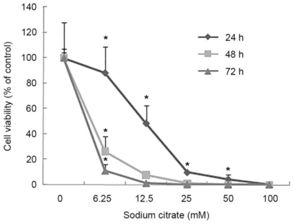
To evaluate the effect of sodium citrate on cell
viability, the viability of AGS cells was observed using a trypan
blue exclusion assay following treatment with various
concentrations of sodium citrate for 24, 48 and 72 h. Cell
viability was expressed as a percentage of the control. As
presented in Fig. 1, the viability of
AGS cells decreased in a dose- and time-dependent manner following
treatment with sodium citrate. Compared with the control
conditions, high concentrations of sodium citrate, excluding 12.5
mM, significantly decreased the viability of AGS cells. In
addition, a decrease in cell viability was associated with longer
durations of sodium citrate administration. These results were
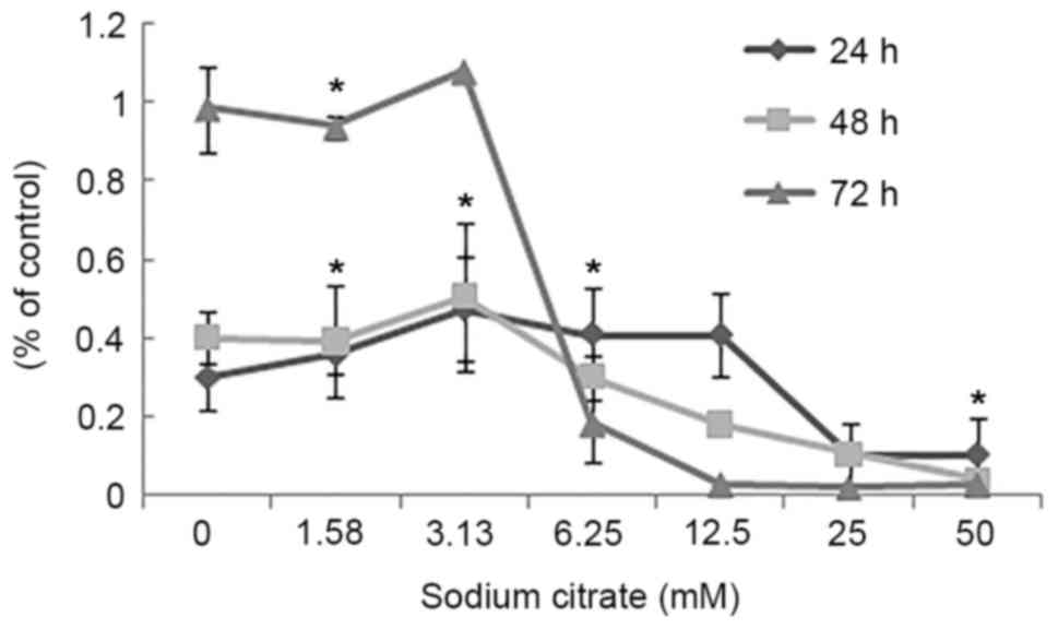
confirmed by the cell proliferation assay, which assessed the
effect of various concentrations of sodium citrate on AGS cell
proliferation. The rate of AGS cell proliferation increased in a
dose-dependent manner at the low concentration (0–3.125 mM);
however, the proliferation rate decreased when the concentration
exceeded 3.125 mM for each incubation time interval (24, 48 and 72
h; Fig. 2). Cell proliferation was
significantly inhibited at the two higher concentrations (25 and 50
mM). Compared with controls, the proliferation rate decreased
>60% (25 and 50 mM). In addition, various durations of
administration revealed a similar effect to that of variations in
dosage; low concentrations of sodium citrate (<3.125 mM)
promoted AGS cell proliferation; however, concentrations >3.125
mM inhibited AGS cell proliferation (Fig.
2). These data revealed that the toxicity of sodium citrate for
AGS cells was time- and dose-dependent from 3.125 to 50 mM for 24,
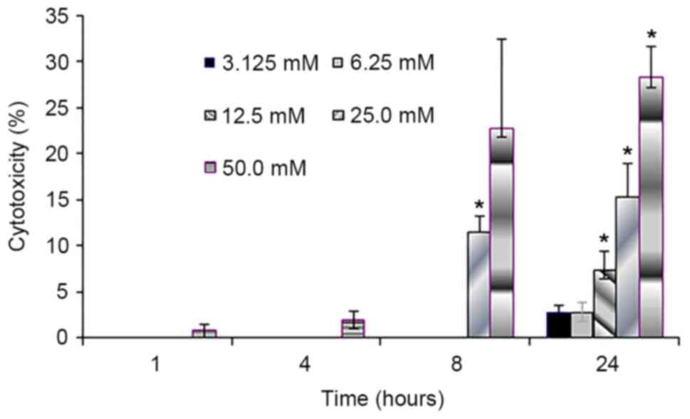
48 and 72 h. The cytotoxicity of sodium citrate was analyzed with
an LDH release assay following the incubation of AGS cells with
various concentrations of sodium citrate (Fig. 3). Administration times of 1 or 4 h had
no effect with 3.125–50.0 mM sodium citrate. However, with an
administration time of 24 h, LDH release increased in association
with the increasing concentration of sodium citrate (3.125–50.0 mM;
Fig. 3).
AGS cells cultured in the presence of 3.125 and 6.25
mM sodium citrate exhibited LDH release values of <3%. Following
a 24 h exposure to 3.125, 6.25, 12.5, 25 and 50 mM sodium citrate,
the values of LDH release were 2.72, 2.83, 7.50, 15.40 and 28.30%,
respectively. The results of the present study revealed that 1 and
4 h exposure to 3.125–50.0 mM sodium citrate had no effect on LDH
release within AGS cells; however, longer durations of sodium
citrate exposure time were associated with gradual increases in LDH
release. Treatments for 8 and 24 h revealed a positive correlation
between the increased degree of LDH release and concentration of
sodium citrate (correlation coefficient was 0.9697 for 8 h,
P<0.05; correlation coefficient 0.9906 for 24 h; P<0.05). The
result was consistent with the aforementioned cell viability and
proliferation experiment.
Effects of sodium citrate on the
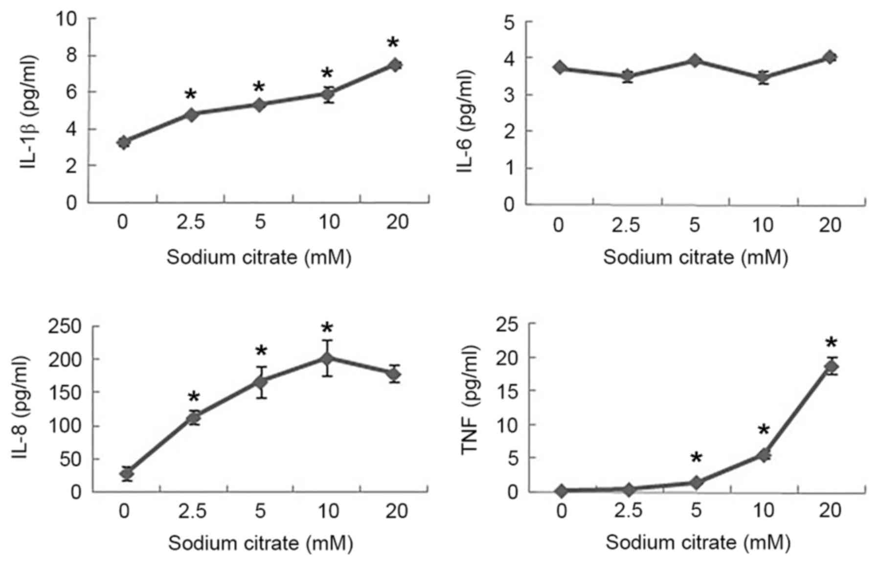
expression levels of cytokines
To investigate the effect of sodium citrate on the
expression of cytokines, supernatants harvested at 24 h were
determined by cytokine-specific ELISA, and the secretion of
pro-inflammatory cytokines (TNF-α, IL-1β, IL-6 and IL-8) from AGS
cells stimulated with sodium citrate was analyzed. In the absence
of sodium citrate, AGS cells released small amounts of IL-1β, IL-8
and TNF-α; however, in the presence of sodium citrate, the levels
of IL-1β secretion increased in a dose-dependent manner. The
highest levels of IL-1β secretion were 2.3-fold greater compared
with that in the control. The levels of IL-8 secretion increased
significantly following treatment with 2.5 mM sodium citrate, and
increased further with rising concentrations of sodium citrate. The
highest level of IL-8 secretion was 7.2-fold greater than the
control level in stimulated cells. In addition, the levels of TNF-α
increased with 10.0 mM sodium citrate; however, treatment with 20.0
mM sodium citrate further increased the levels of TNF-α to 18.8
pg/ml. Compared with the control, significant alterations in IL-6
levels were not observed (Fig.
4).
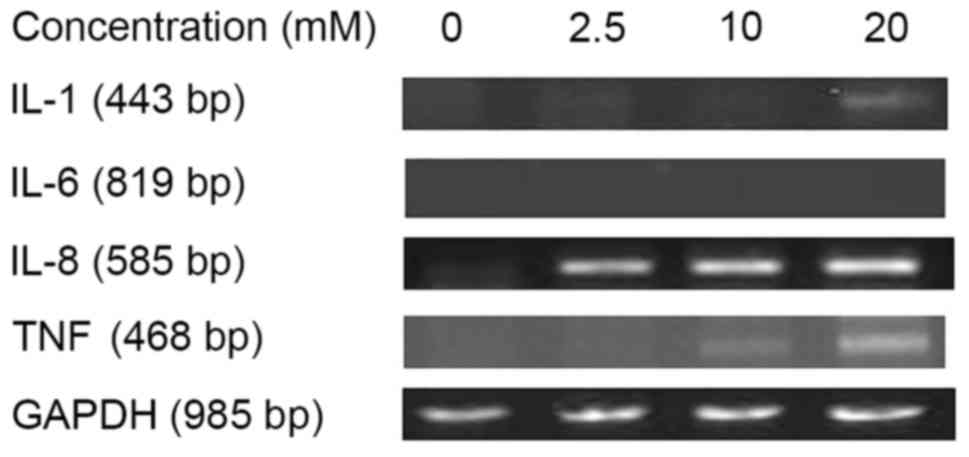
To verify the results of the present study, the
levels of cytokine mRNAs for IL-1β, IL-6, IL-8 and TNF-α were
assessed by RT-PCR. The highest levels of IL-1β mRNA was detected
with 20 mM sodium citrate treatment. The levels of IL-8 and TNF-α
mRNA expression increased with increasing doses of sodium citrate.
IL-6 mRNA expression was not detected at any dose of sodium citrate
(Fig. 5). The results of this assay
were consistent with those of the previous ELISA assay results.
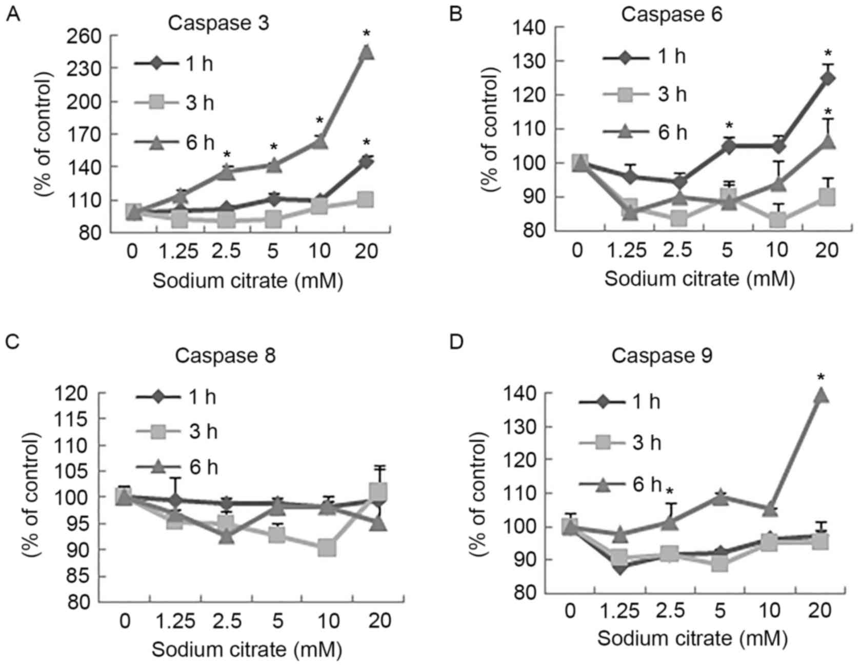
Effects of sodium citrate on caspase
activity
The activities of caspase-3 and −9 in AGS cells
increased following 6 h of treatment with different concentrations
of sodium citrate compared with the control (Fig. 4A and D). Treatment of cells with 20 mM
sodium citrate for 1 h resulted in a marked increase in the
activities of caspase-3 and −6 compared with cells treated with the
control. The highest activities of all caspases were observed in
the 20 mM treatment groups, with the exception of caspase-8, which
was not altered with varying duration or concentration, compared
with the control (Fig. 6).
Discussion
A potential strategy to inhibit cancer cell growth
may involve the targeting of glycolytic inhibitors (25). As high concentrations of citrate may
inhibit the glycolytic pathway (26,27), the
cytotoxic effects of citrate on AGS cells were investigated in the
present study. Low concentrations of citrate (<3.125 mM)
promoted AGS cell proliferation; however, higher doses were
cytotoxic. This result may be caused by citrate affecting glucose
metabolism within cancer cells (27).
Furthermore, the cytotoxic effect of citrate within cancer cells
has been reported to exert a synergistic effect with certain
anticancer drugs (10). Additionally,
as defined by the Warburg effect, LDH is an enzyme involved in
obtaining energy following aerobic glycolysis in cancer cells. The
positive correlation between the increased degree of LDH release
and concentration of sodium citrate treatment indicated that the
cytotoxic effect of sodium citrate observed in AGS cells increased
as the concentration or time of treatment increased.
Caspases are central components of the apoptotic
process, which function as initiators or executors in programmed
cell death (28). Caspase-8 and −9
have been reported to act as initiators of apoptosis, and caspase-3
and −6 as executors (29). In the
present study, the activities of caspase-3, −6, −8 and −9 were
investigated. After 6 h of treatment with various concentrations of
sodium citrate, the activities of caspase-3 and caspase-9
increased. These results are consistent with those of a previous
study (30), which demonstrated the
induction of caspase-3 and −8 cascade activation, resulting in
cancer cell apoptosis. The experiment also demonstrated that, when
AGS cells were exposed to 20 mM citrate for 1 h, caspase-6 was
likely to be involved in regulating this apoptosis, as its activity
level decreased shortly afterwards. However, the activity of
caspase-8 was not affected by any concentration of sodium citrate
(31). Therefore, treatment with
sodium citrate induced AGS cell-apoptosis via the intrinsic, and
not the extrinsic apoptotic pathway (32).
Cytokines are mediators involved in gastric
physiology, as well as pathophysiology, and may serve important
roles in the etiology of gastric cancer (33). In addition, numerous caspases serve as
critical mediators in the integration of apoptotic and inflammatory
pathways (32). However, certain
pro-inflammatory cytokines, such as IL-1β, may be activated by
caspase-8; the results of the present study revealed that IL-1β and
IL-8 levels were increased, although an increase in caspase-8
activity was not observed within AGS cells. This indicated that the
levels of IL-1β and IL-8 may be regulated by other caspases, such
as caspase-1 or −11, or by various mechanisms. Although TNF-α was
not detected in the ELISA assay, it was detected by RT-PCR. The
discrepancy between the ELISA and RT-PCR results may be due to
sodium citrate affecting only the expression of TNF-α mRNA. In
addition, the levels of IL-6 were possibly too low for detection in
the present study.
As observed in the extrinsic apoptosis pathway,
moderate levels of IL-6 and IL-8 cytokines are detected in
Fas-mediated apoptosis (33). Fas
receptor stimulation has also been demonstrated to induce phagocyte
migration in vivo (33),
indicating that it activates the apoptotic and pro-inflammatory
pathways, thus facilitating the elimination of dying cells.
Therefore, the apoptosis of AGS cells induced by sodium citrate via
the intrinsic pathway is likely to occur via the same mechanism.
This cytotoxic mechanism probably operates synergistically,
inducing apoptosis via the intrinsic pathway and altering the
cytokine expression profile.
In the present study, exposing AGS cells to higher
concentrations of sodium citrate or longer durations of treatment
may elicit a cytocidal effect, which was observed via a reduction
in cell viability and proliferation, an increase in LDH release, an
induction of the intrinsic pathway of apoptosis and alterations in
the expression levels of certain cytokines.
Acknowledgements
The present study was supported by the National
Natural Science Foundation of China (grant nos. 81473017 and
31460249).
Competing interests
The authors declare that they have no competing
interests.
References
|
1
|
Leumann E, Hoppe B, Neuhaus T and Blau N:
Efficacy of oral citrate administration in primary hyperoxaluria.
Nephrol Dial Transpl. 10 Suppl 8:S14–S16. 1995. View Article : Google Scholar
|
|
2
|
Phillips R, Hanchanale VS, Myatt A, Somani
B, Nabi G and Biyani CS: Citrate salts for preventing and treating
calcium containing kidney stones in adults. Cochrane Database Syst
Rev: CD010057. 2015. View Article : Google Scholar
|
|
3
|
Pastorino J, Hansen CL and McMahon DJ:
Effect of sodium citrate on structure-function relationships of
Cheddar cheese. J Dairy Sci. 86:3113–3121. 2003. View Article : Google Scholar : PubMed/NCBI
|
|
4
|
Westergaard N, Waagepetersen HS, Belhage B
and Schousboe A: Citrate, a ubiquitous key metabolite with
regulatory function in the CNS. Neurochem Res. 42:1583–1588. 2017.
View Article : Google Scholar : PubMed/NCBI
|
|
5
|
Wang JY, Zhu SG and Xu CF: Essential
Biochemistry. 1st. 20. Higher Education Press; Beijing: pp.
340–342. 2010
|
|
6
|
Warburg O, Wind F and Negelein E: The
metabolism of tumors in the body. J Gen Physiol. 8:519–530. 1927.
View Article : Google Scholar : PubMed/NCBI
|
|
7
|
Warburg O: On the origin of cancer cells.
Science. 123:309–314. 1956. View Article : Google Scholar : PubMed/NCBI
|
|
8
|
Moreno-Sánchez R, Rodríguez-Enríquez S,
Marín-Hernández A and Saavedra E: Energy metabolism in tumor cells.
FEBS J. 274:1393–1418. 2007. View Article : Google Scholar : PubMed/NCBI
|
|
9
|
Vander Heiden MG, Cantley LC and Thompson
CB: Understanding the Warburg effect: The metabolic requirements of
cell proliferation. Science. 324:1029–1033. 2009. View Article : Google Scholar : PubMed/NCBI
|
|
10
|
Zhang X, Varin E, Allouche S, Lu Y,
Poulain L and Icard P: Effect of citrate on malignant pleural
mesothelioma cells: A synergistic effect with cisplatin. Anticancer
Res. 29:1249–1254. 2009.PubMed/NCBI
|
|
11
|
Izyumov DS, Avetisyan AV, Pletjushkina OY,
Sakharov DV, Wirtz KW, Chernyak BV and Skulachev VP: ‘Wages of
fear’: Transient threefold decrease in intracellular ATP level
imposes apoptosis. Biochim Biophys Acta. 1658:141–147. 2004.
View Article : Google Scholar : PubMed/NCBI
|
|
12
|
Vander Heiden MG, Chandel NS, Schumacker
PT and Thompson CB: Bcl-xL prevents cell death following growth
factor withdrawal by facilitating mitochondrial ATP/ADP exchange.
Mol Cell. 3:159–167. 1999. View Article : Google Scholar : PubMed/NCBI
|
|
13
|
Yousefi S, Owens JW and Cesario TC:
Citrate shows specific, dose-dependent lympholytic activity in
neoplastic cell lines. Leuk Lymphoma. 45:1657–1665. 2004.
View Article : Google Scholar : PubMed/NCBI
|
|
14
|
Wang YS and Wang ZY: Sodium citrate
induces apoptosis in biocontrol yeast Cryptococcus laurentii. J
Appl Microbiol. 113:135–142. 2012. View Article : Google Scholar : PubMed/NCBI
|
|
15
|
Sun J, Misumi A, Shimaoka K, Aoki F and
Esaki F: Stomach cancer-related mortality. Eur J Cancer Prev.
10:61–67. 2001. View Article : Google Scholar : PubMed/NCBI
|
|
16
|
Hamashima C: Current issues and future
perspectives of gastric cancer screening. World J Gastroenterol.
20:13767–13774. 2014. View Article : Google Scholar : PubMed/NCBI
|
|
17
|
Wang XQ, Terry PD and Yan H: Review of
salt consumption and stomach cancer risk: Epidemiological and
biological evidence. World J Gastroenterol. 15:2204–2213. 2009.
View Article : Google Scholar : PubMed/NCBI
|
|
18
|
Reeves GK, Pirie K, Green J, Bull D and
Beral V; Million Women Study Collaborators, : Comparison of the
effects of genetic and environmental risk factors on in situ and
invasive ductal breast cancer. Int J Cancer. 131:930–937. 2012.
View Article : Google Scholar : PubMed/NCBI
|
|
19
|
Sun J, Bi L, Chi Y, Aoki K and Misumi J:
Effect of sodium acetate on cell proliferation and induction of
proinflammatory cytokines: A preliminary evaluation. Food Chem
Toxicol. 43:1773–1780. 2005. View Article : Google Scholar : PubMed/NCBI
|
|
20
|
Sun J, Aoki K, Wang W, Guo A and Misumi J:
Sodium nitrite-induced cytotoxicity in cultured human gastric
epithelial cells. Toxicol in Vitro. 20:1133–1138. 2006. View Article : Google Scholar : PubMed/NCBI
|
|
21
|
Zhang X, Bo A, Chi B, Xia Y, Su X and Sun
J: Magnesium sulfate induced toxicity in vitro in AGS gastric
adenocarcinoma cells and in vivo in mouse gastric mucosa. Asian Pac
J Cancer Prev. 16:71–76. 2015. View Article : Google Scholar : PubMed/NCBI
|
|
22
|
Lu Y, Zhang X, Zhang H, Lan J, Huang G,
Varin E, Lincet H, Poulain L and Icard P: Citrate induces apoptotic
cell death: A promising way to treat gastric carcinoma? Anticancer
Res. 31:797–805. 2011.PubMed/NCBI
|
|
23
|
Guo X, Zhang X, Wang T, Xian S and Lu Y:
3-Bromopyruvate and sodium citrate induce apoptosis in human
gastric cancer cell line MGC-803 by inhibiting glycolysis and
promoting mitochondria-regulated apoptosis pathway. Biochem Biophys
Res Commun. 475:37–43. 2016. View Article : Google Scholar : PubMed/NCBI
|
|
24
|
Barranco SC, Townsend CM Jr, Casartelli
JC, Macik BG, Burger NL, Boerwinkle WR and Gourley WK:
Establishment and characterization of an in vitro model system for
human adenocarcinoma of the stomach. Cancer Res. 43:1703–1709.
1983.PubMed/NCBI
|
|
25
|
Xintaropoulou C, Ward C, Wise A, Marston
H, Turnbull A and Langdon SP: A comparative analysis of inhibitors
of the glycolysis pathway in breast and ovarian cancer cell line
models. Oncotarget. 6:25677–25695. 2015. View Article : Google Scholar : PubMed/NCBI
|
|
26
|
Randle PJ, Denton RM and England PJ:
Citrate as a metabolic regulator in muscle and adipose tissue.
Biochem Soc Symp. 27:87–103. 1968.PubMed/NCBI
|
|
27
|
Icard P, Poulain L and Lincet H:
Understanding the central role of citrate in the metabolism of
cancer cells. Biochim Biophys Acta. 1825:111–116. 2012.PubMed/NCBI
|
|
28
|
Elinos-Báez CM, Maldonado V and
Meléndezzajgla J: Caspases: Apoptosis inducing molecules. Gac Med
Mex. 139:493–499. 2003.(In Spanish). PubMed/NCBI
|
|
29
|
Riedl SJ and Shi Y: Molecular mechanisms
of caspase regulation during apoptosis. Nat Rev Mol Cell Biol.
5:897–907. 2004. View
Article : Google Scholar : PubMed/NCBI
|
|
30
|
Kruspig B, Nilchian A, Orrenius S,
Zhivotovsky B and Gogvadze V: Citrate kills tumor cells through
activation of apical caspases. Cell Mol Life Sci. 69:4229–4237.
2012. View Article : Google Scholar : PubMed/NCBI
|
|
31
|
Kim GY, Park SY, Jo A, Kim M, Leem SH, Jun
WJ, Shim SI, Lee SC and Chung JW: Gecko proteins induce the
apoptosis of bladder cancer 5637 cell by inhibiting Akt and
activating intrinsic caspase cascade. BMB Rep. 48:531–536. 2015.
View Article : Google Scholar : PubMed/NCBI
|
|
32
|
Creagh EM: Caspase crosstalk: Integration
of apoptotic and innate immune signalling pathways. Trends Immunol.
35:631–640. 2014. View Article : Google Scholar : PubMed/NCBI
|
|
33
|
Persson C, Canedo P, Machado JC, El-Omar
EM and Forman D: Polymorphisms in inflammatory response genes and
their association with gastric cancer: A HuGE systematic review and
meta-analyses. Am J Epidemiol. 173:259–270. 2011. View Article : Google Scholar : PubMed/NCBI
|