Introduction
Colorectal cancer (CRC) is a worldwide health
problem being the fourth cause of death due to cancer (1). CRC tumorigenesis involves molecular
deregulation of genes related to proliferation, tumor growth,
antiapoptosis, invasiveness, metastasis and angiogenesis (2). Nuclear factor-κB (NF-κB) is a
transcriptional factor that plays an important role in biological
processes, comprises a family of five proteins grouped in homo or
heterodimers (3). NF-κB is normally
inactive, sequestered by IκBα inhibitor, but commonly has been
reported active in cancer, and plays a key role in tumorigenesis by
transcriptional regulation of multiple genes (4). Several reports have found higher
NF-κB/p65 protein expression in CRC tissue compared to normal
tissue (5–7). However, to our knowledge, the evaluation
of NF-κB/p65 and genes expression profiles, in tumor tissue
compared to adjacent normal mucosa from the same CRC patient, and
its association with clinicopathological parameters has not been
fully reported (8).
Materials and methods
Patients
Thirty patients with sporadic CRC histopathological
diagnosis who underwent to colonoscopy or surgery at Hospital Civil
de Guadalajara ‘Dr. Juan I. Menchaca’, Jalisco, Mexico, were
enrolled in this study after informed consent request, only
non-treated patients were included. Eight patients classified as
non-CRC were evaluated as comparative control group. The study was
performed according to the declaration of Helsinki and was approved
by the local Ethics Committee.
Tissues
Both, tumor tissue and its adjacent normal mucosa
were obtained from respective areas in colonic or rectal resection
specimens from the same patient according to the ‘Cancer Care
Quality Measures: Diagnosis and Treatment of Colorectal Cancer’
from the ‘Agency for Health Care Research and Quality’ (9). CRC tissue samples for RNA isolation,
were collected in RNAlater® Stabilization Solution
(AM7020; Thermo Fisher Scientific, Inc., Waltham, MA, USA), and in
10% neutral buffered formaldehyde (11–0705; Sigma-Aldrich; Merck
KGaA, Darmstadt, Germany) for immunohistochemistry analysis.
Non-CRC patient's tissue samples were collected during colonoscopy,
before pathological analysis according to the same procedure. CRC
tissues collected for RNA isolation were transported to laboratory
and processed immediately. Tissues collected in 10% of neutral
buffered formaldehyde were examined microscopically to confirm the
diagnosis and perform subsequent immunohistochemistry analysis.
Remaining tissues were stored at −80°C in case of extra
analysis.
Immunohistochemistry
Tissue samples were fixed in 10% neutral buffered
formaldehyde and embedded in paraffin wax. Each tissue sample was
sectioned at a thickness of 4–5 µm, placed on slides, and
deparaffinized by heat for 60 min at 65°C. Slides were placed in
xylene and serial alcohol solutions (100, 96, 80, and 50%). All
procedure was performed using EnVision™ FLEX kit (Dako;
Agilent Technologies, Inc. Santa Clara, CA, USA) as follows: Slides
were washed for 5 min in wash buffer, treated in epitope retrieval
solution for 20 min at 90°C, and rewashed. To block endogenous
peroxidase, slides were incubated in peroxidase-blocking reagent
for 15 min at room temperature, washed for 5 min in wash buffer,
and incubated for 1 h at room temperature with a mouse monoclonal
primary antibody raised against the N-terminus of human NF-κB/p65
(dilution 1:50) (sc-8008; Santa Cruz Biotechnology, Inc., Santa
Cruz, CA, USA). Then, the slides were washed and incubated for 30
min at room temperature with secondary antibody coupled with
peroxidase and rewashed. Visualization was made with
3,3′-diaminobenzidine and counterstaining with hematoxylin.
Finally, slides were dehydrated with serial alcohol solutions (50,
80, 96 and 100%) and xylene. Positive and negative controls were
included for each staining procedure, using a section of CRC tissue
known as strongly NF-κB/p65-positive.
NF-κB/p65 staining evaluation
Evaluation of slides was made by two pathologists
blinded to patient's characteristics. The slides were scored
according to the method recommended by Abdullah et al
(10), as follows: Intensity of
staining was classified ‘in crosses’ from 0 to 3, as 0 (−)
negative, 1 (+) weak, 2 (++) moderate, and 3 (+++) strong. The
extent of staining referred as the percentage of positive
epithelial cells in relation to the whole tumor area, was
classified from 0 to 4, as 0 (0%), 1 (≤25%), 2 (26–50%), 3 (51–75%)
and 4 (>75%). The final staining score was calculated by the
addition of staining intensity and the extent of staining. The
scores have values between 0 and 7, and scores greater than or
equal to 3 were classified as positive. For negative control,
primary antibody was replaced by distilled water, as an internal
control from each slide, staining of macrophages was
considered.
Gene expression
A group of relevant genes in CRC was selected
considering that NF-κB participates in their transcriptional
regulation and its particular role in tumoral processes as
described below: Proliferation, CCND1 (11) and PTGS2 (12); tumor growth, TNF (13), ALOX (14), and NOS2 (15); anti-apoptosis, BCL2 and
BCL2L1 (16); invasiveness and
metastasis, MMP9 (17);
angiogenesis, VEGFA (18).
Tissues were collected in
RNAlater® RNA Stabilization Solution, transported
to laboratory and processed immediately as follows: Tissue (10–20
mg) were cut into small pieces and collected in 0.5 ml of
TRIzol® Reagent (cat. no. 15596; Thermo Fisher
Scientific, Inc.). Then, each sample was homogenized in Tissue
Lyser LT (cat. no. 85600; Qiagen GmbH, Hilden, Germany) for 3
min/25 Hz. Next steps of RNA isolation were made according to
manufacturer's instructions (Life Technologies; Thermo Fisher
Scientific, Inc.). Isolated RNA samples were stored at −80°C.
Reverse transcription (RT-PCR) was performed using 1 µg of total
RNA treated with DNase1 amplification grade (cat. no. 18068; Life
Technologies; Thermo Fisher Scientific, Inc.) and the High Capacity
cDNA Reverse Transcription kit (cat. no. 4368813; Thermo Fisher
Scientific, Inc.) according to manufacturer's instructions. The
RT-PCR conditions were: 25°C/25, 37°C/120, 85°C/5 min, and infinite
hold at 4°C. RT-Quantitative-PCR (RT-qPCR) was performed using
TaqMan® Gene Expression Master Mix (cat. no. 4369016;
Thermo Fisher Scientific, Inc.), and TaqMan® Gene
Expression Assays with FAM-MGB fluorophore-quencher system. For
each gene mRNA detection, the next assays were used (PTGS2,
Hs00153133_m1; BCL2, Hs00608023_m1; BCL2L1,
Hs00236329_m1; CCND1, Hs00765553_m1; MMP9,
Hs00234579_m1; VEGFA, Hs00900055_m1; TNF,
Hs99999043_m1; NOS2, Hs01075529_m1; ALOX5,
Hs01095330_m1; GUSB, Hs00939627_m1; cat. no. 4331182, Thermo
Fisher Scientific, Inc.). RT-qPCR was performed in a 7900 HT Fast
Real-Time PCR System linked to SDS 2.4 software (Thermo Fisher
Scientific, Inc.), PCR conditions were: 50°C/2, 95°C/10 min,
95°C/15 sec and 60°C/1 min (40 cycles). Gene expression assays were
validated using β-glucoronidase (GUS), β-Actin
(ACTB), and Abelson (ABL) genes as constitutive
control (housekeeping genes). Gene-expression profiles were
calculated by relative quantification using the 2−ΔΔCq
method (19).
Statistical analysis
Data was analyzed using the SPSS 20.0 software
(SPSS, Inc., Chicago, IL, USA). The NF-κB/p65 staining scores
differences of the 3 groups were evaluated using Kruskal-Wallis
test, then differences of staining scores and positive cells
percentage pairwise comparison among groups were evaluated by
Mann-Whitney U test. Genes expression differences in tumor tissues
vs. adjacent normal mucosa were performed by Wilcoxon signed-ranked
test. To evaluate the association of NF-κB/p65 expression and
gene-expression profiles with clinicopathological parameters,
Pearson or Spearman rank correlation coefficient was used.
P<0.05 was considered to indicate a statistically significant
difference.
Results
Clinicopathological parameters in
patients
Histopathological diagnosis of CRC was confirmed by
examination of hematoxylin and eosin staining before analysis.
Twenty-three CRC patient's tissues corresponded to colon cancer and
7 to rectal cancer. Twenty-two men and 8 women were collected; none
of them were treated with radiotherapy or chemotherapy. The average
age was 60 years. Tumor staging was established by certified
pathologists according to tumor-node-metastasis (TNM)
classification. Tobacco and alcohol consumption was also
registered; most of the patients were occasional consumers. CEA, CA
19.9 and AFP tumor markers levels were higher than normal values.
Eight patients that attend ‘Colon and rectum service’, and were
diagnosed CRC negative before histopathology studies, were
evaluated as control group. CRC patients were classified according
to TNM clinicopathological parameters for association evaluation.
General features of patients are presented in Table I.
 | Table I.Clinicopathological parameters of
sporadic CRC patients (n=30). |
Table I.
Clinicopathological parameters of
sporadic CRC patients (n=30).
| Parameters | Value (%) |
|---|
|
Clinicala |
|
| Origin of
tissue |
|
|
Colonoscopy | 5 (16.7) |
|
Surgery | 25 (83.3) |
| Age (mean, 60
years) |
|
|
≤40 | 3 (10.0) |
|
41–60 | 13 (43.3) |
|
>60 | 14 (46.7) |
| Sex |
|
|
Male | 22 (73.3) |
|
Female | 8 (26.7) |
| Tobacco
consumption |
|
|
Yes | 7 (23.3) |
| No | 11 (36.7) |
|
Occasional | 12 (40.0) |
| Alcohol
consumption |
|
|
Yes | 11 (36.7) |
| No | 2 (6.7) |
|
Occasional | 17 (56.6) |
| Pathologicala |
|
| TNM stage |
|
| I | 5 (16.7) |
| II | 4 (13.3) |
|
III | 12 (40.0) |
| IV | 9 (30.0) |
| Histopathological
differentiation |
|
|
Well | 0 (0.0) |
|
Moderate | 27 (90.0) |
|
Poorly | 3 (10.0) |
| Tumor site |
|
| AC | 3 (10.0) |
| TC | 4 (13.3) |
| DC | 7 (23.3) |
| SC | 9 (30.0) |
| R | 7 (23.3) |
| Tumor depth |
|
| T1 | 3 (10.0) |
| T2 | 6 (20.0) |
| T3 | 11 (36.7) |
| T4 | 10 (33.3) |
| Lymph node
status |
|
| N0 | 9 (30.0) |
| N1 | 8 (26.7) |
| N2 | 13 (43.3) |
| Metastasis
degree |
|
| M0 | 21 (70.0) |
| M1 | 9 (30.0) |
|
Laboratorialb |
|
| Tumor markers
levels |
|
| CEA,
ng/ml 26.52 | (1.35.59.42) |
|
CA.19.9, U/ml 149.4 | (53.6.206.9) |
| AFP,
U/ml 207.1 | (27.6.276.8) |
NF-ĸB/p65 immunostaining scoring
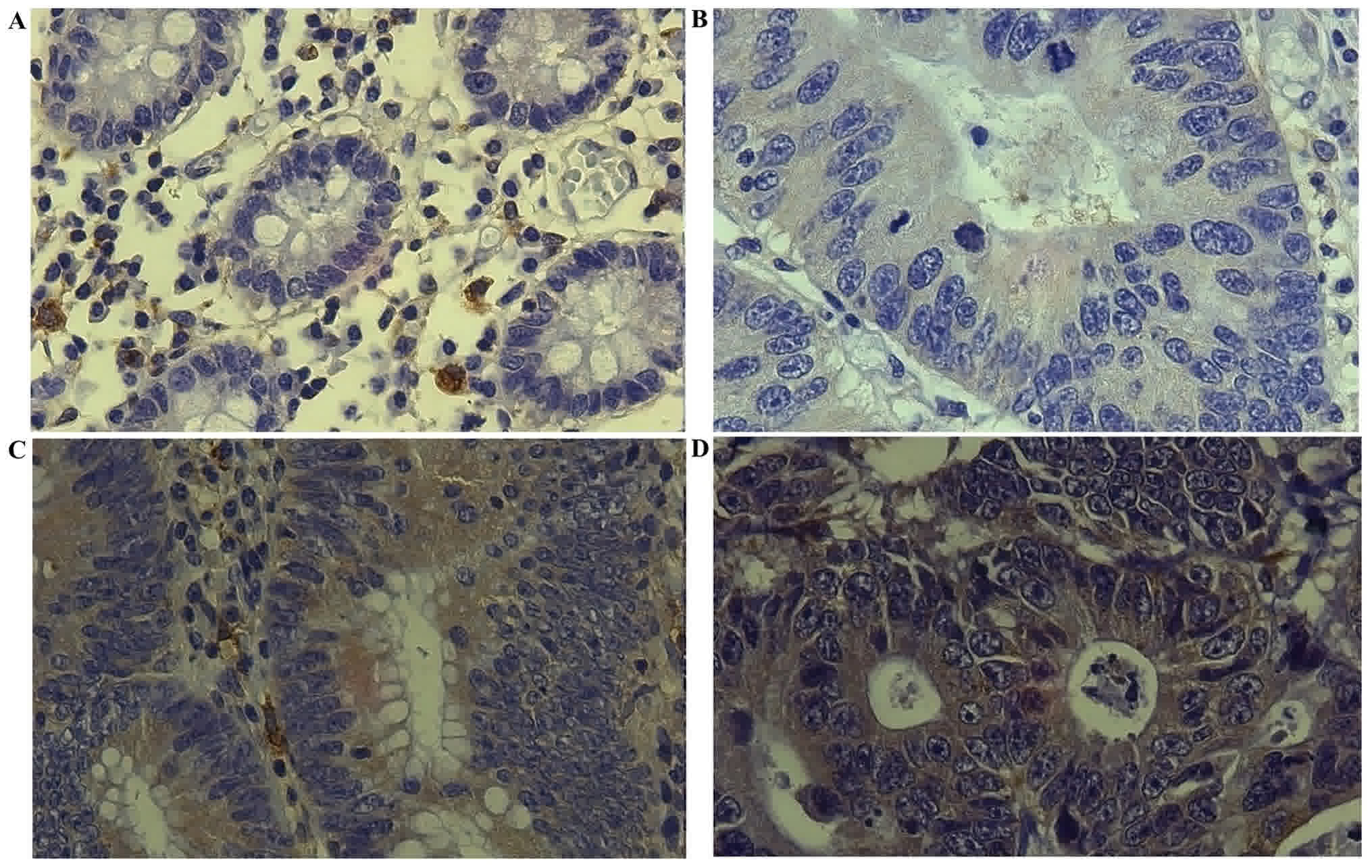
In tumor tissue group, all samples expressed
cytoplasmic NF-κB/p65, the intensities of staining were majority
‘moderate’ (15/30) and ‘strong’ (11/30), the group showed a mean
NF-κB/p65 extent of staining of 52.3% with a standard deviation of
18.2%. NF-κB/p65 intensities of staining in normal adjacent mucosa
were mostly ‘weak’ (15/30) and ‘non-staining’ (12/30), the group
showed a mean NF-κB/p65 extent of staining of 27.6% with a standard
deviation of 11.8%. In non-CRC tissues, NF-κB/p65 intensity of
staining was majority ‘non-staining’ (5/8), a mean NF-κB/p65 extent
of staining of 18.8% with a standard deviation of 7.5% was reported
for this group (Fig. 1; Table II).
| Table II.NF-κB/p65 immunostaining in CRC and
non-CRC patient's tissues. |
Table II.
NF-κB/p65 immunostaining in CRC and
non-CRC patient's tissues.
|
| Normal adjacent
mucosa/Non-CRC | Tumor tissue |
|---|
|
|
|
|
|---|
| Patients | Intensity of
staining | Extent of staining
(%) | Staining score | Intensity of
staining | Extent of staining
(%) | Staining score |
|---|
| CRC |
|
|
|
|
|
|
| 1 | 0 (−) | 0 (0) | 0 | 1 (+) | 2 (40) | 3a |
| 2 | 1 (+) | 2 (35) | 3a | 1 (+) | 2 (40) | 3a |
| 3 | 1 (+) | 2 (35) | 3a | 1 (+) | 2 (40) | 3a |
| 4 | 0 (−) | 0 (0) | 0 | 2 (++) | 3 (60) | 5a |
| 5 | 1 (+) | 1 (20) | 2 | 2 (++) | 2 (45) | 4a |
| 6 | 1 (+) | 2 (40) | 3a | 3 (+++) | 3 (75) | 6a |
| 7 | 0 (−) | 0 (0) | 0 | 1 (+) | 3 (60) | 4a |
| 8 | 3 (+++) | 2 (35) | 5a | 3 (+++) | 2 (50) | 5a |
| 9 | 2 (++) | 1 (20) | 3a | 3 (+++) | 4 (90) | 7a |
| 10 | 1 (+) | 1 (20) | 2 | 2 (++) | 3 (65) | 5a |
| 11 | 0 (−) | 0 (0) | 0 | 2 (++) | 3 (60) | 5a |
| 12 | 1 (+) | 1 (25) | 2 | 2 (++) | 2 (40) | 4a |
| 13 | 1 (+) | 1 (20) | 2 | 2 (++) | 2 (40) | 4a |
| 14 | 0 (−) | 0 (0) | 0 | 1 (+) | 2 (30) | 3a |
| 15 | 0 (−) | 0 (0) | 0 | 1 (+) | 2 (35) | 3a |
| 16 | 0 (−) | 0 (0) | 0 | 3 (+++) | 3 (75) | 6a |
| 17 | 0 (−) | 0 (0) | 0 | 2 (++) | 3 (65) | 5a |
| 18 | 1 (+) | 1 (25) | 2 | 2 (++) | 2 (45) | 4a |
| 19 | 0 (−) | 0 (0) | 0 | 1 (+) | 2 (30) | 3a |
| 20 | 1 (+) | 1 (20) | 2 | 2 (++) | 3 (60) | 5a |
| 21 | 0 (−) | 0 (0) | 0 | 2 (++) | 2 (40) | 4a |
| 22 | 1 (+) | 1 (20) | 2 | 3 (+++) | 2 (45) | 5a |
| 23 | 0 (−) | 0 (0) | 0 | 1 (+) | 1 (20) | 2 |
| 24 | 1 (+) | 1 (10) | 2 | 3 (+++) | 2 (50) | 5a |
| 25 | 2 (++) | 3 (60) | 5a | 2 (++) | 4 (85) | 6a |
| 26 | 1 (+) | 1 (20) | 2 | 3 (+++) | 3 (75) | 6a |
| 27 | 1 (+) | 2 (40) | 3a | 2 (++) | 3 (70) | 5a |
| 28 | 1 (+) | 2 (0) | 3a | 3 (+++) | 2 (50) | 5a |
| 29 | 1 (+) | 1 (25) | 2 | 2 (++) | 3 (70) | 5a |
| 30 | 0 (−) | 0 (0) | 0 | 1 (+) | 1 (20) | 2 |
| Non-CRC |
|
|
|
|
|
|
| 1 | 1 (+) | 1 (15) | 2 | – | – | – |
| 2 | 0 (−) | 0 (0) | 0 | – | – | – |
| 3 | 0 (−) | 0 (0) | 0 | – | – | – |
| 4 | 1 (+) | 1 (15) | 2 | – | – | – |
| 5 | 0 (−) | 0 (0) | 0 | – | – | – |
| 6 | 1 (+) | 2 (30) | 3a | – | – | – |
| 7 | 0 (−) | 0 (0) | 0 | – | – | – |
| 8 | 0 (+) | 0 (15) | 0 | – | – | – |
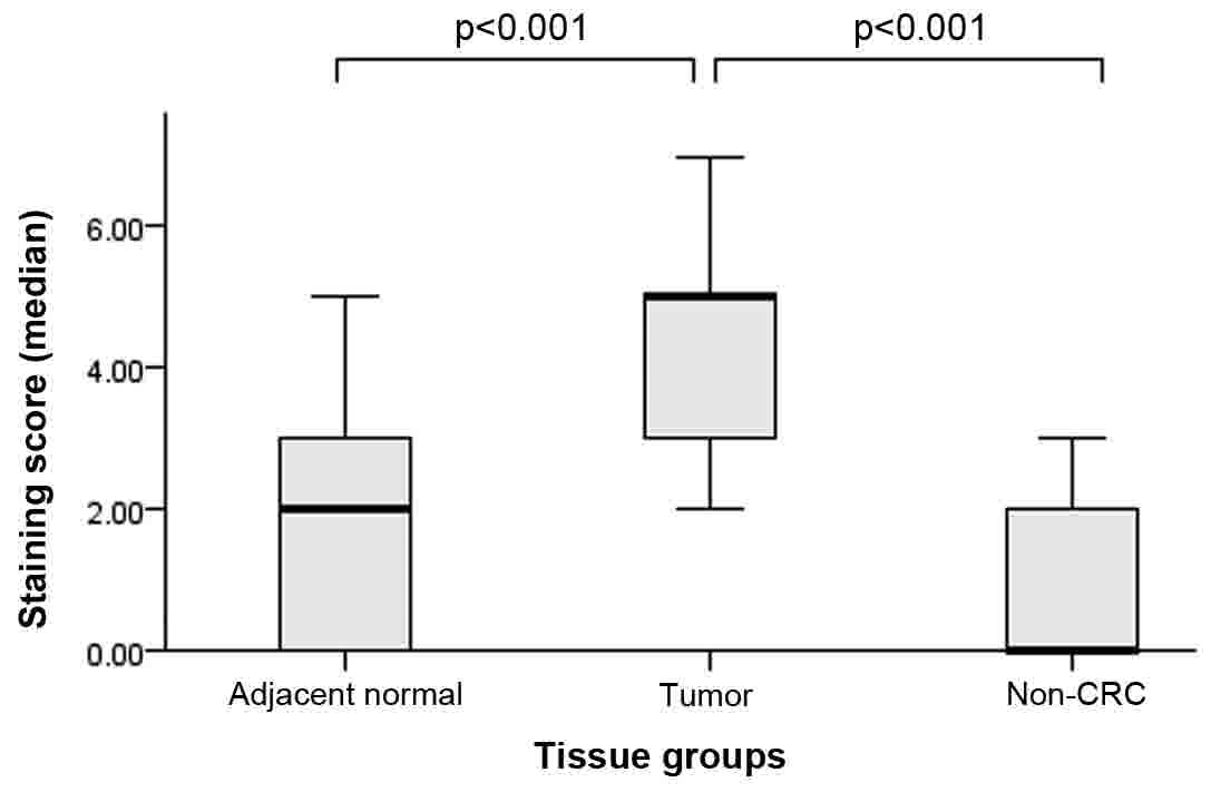
An average staining score of 4.4 in tumor tissue
group, 1.6 in normal adjacent mucosa and 0.8 in non-CRC tissues is
reported. The analysis among 3 groups showed that they were
statistically different (X2(1)=39.146, P<0.001). Pairwise comparison
of staining scores among groups showed that tumor tissue was
statistically higher than normal adjacent mucosa (z=−5.707,
P<0.001), as well it was higher when compared to non-CRC tissue
(z=−4.126, P<0.001). No difference was reported between normal
adjacent mucosa and non-CRC tissue (Fig.
2).
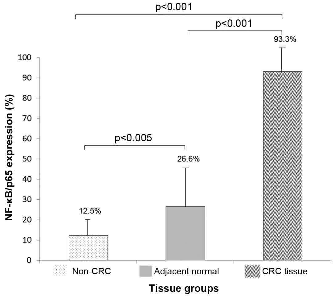
Positive samples for NF-κB/p65 expression according
to staining score (≥3), reported for each group were: 28/30 (93.3%)
in tumor tissues, 8/30 (26.6%) in adjacent normal mucosa and 1/8
(12.5%) in non-CRC tissues. For statistical analysis, positive
NF-κB/p65 samples were classified as ‘1’ and negative samples as
‘0’. NF-κB/p65 positive samples were statistically higher than
adjacent normal tissue and non-CRC tissue groups (P<0.001).
Adjacent normal tissues also showed higher NF-κB/p65 positive
samples when compared to non-CRC tissue group (P<0.05) (Fig. 3).
NF-ĸB/p65 expression and
clinicopathological parameters
NF-κB/p65 expression was analyzed according to
clinicopathological parameters; results are described below and
reported in Table III.
| Table III.NF-κB/p65 expression (%) association
with clinicopathological parameters in CRC patients (n=30). |
Table III.
NF-κB/p65 expression (%) association
with clinicopathological parameters in CRC patients (n=30).
| Parameter | NF-κB/p65
expression (%) | P-value |
|---|
| CRC tissue |
|
|
| Tumor
stage |
| <0.05 |
|
I | 27.00a |
|
|
II | 36.62b |
|
|
III | 63.51b |
|
|
IV | 70.12b |
|
|
Histopathology
differentiation |
| <0.05 |
|
Well | – |
|
|
Moderately | 46.31b |
|
|
Poorly | 79.81b |
|
| Tumor
localization |
| <0.05 |
|
Ascending
colon | 49.30b |
|
|
Transverse
colon | 45.63b |
|
|
Descending
colon | 40.74b |
|
|
Sigmoid colon | 52.72b |
|
|
Rectum | 31.33a |
|
| Tumor
depth |
| <0.05 |
|
T1 | 28.51a |
|
|
T2 | 34.33a |
|
|
T3 | 69.25b |
|
|
T4 | 74.84b |
|
| Lymph
node status |
| <0.05 |
|
N0 | 32.70a |
|
|
N1 | 49.12b |
|
|
N2 | 74.13b |
|
|
Metastasis degree |
| <0.001 |
|
M0 | 41.71b |
|
|
M1 | 73.57b |
|
| Control group |
|
|
| Non-CRC
tissue | 12.50 |
|
| Normal
adjacent tissue | 26.60a |
|
NF-κB/p65 expression analyzed by tumor stages were
reported as follows: I=27, II=36.6, III=63.5 and IV=70.1%. Stages
II, III & IV are statistically higher than control groups
(P<0.05), stage I was only statistically higher than non-CRC
tissue group (P<0.05). As well, significantly increment of
NF-κB/p65 expression in advanced stages compared to initial stages
was reported (III+IV=66.8±3.3% vs. I+II=31.75±4.8%; P<0.05),
thus NF-κB/p65 expression is positively associated to CRC
progression.
In histopathological differentiation groups,
NF-ĸB/p65 expression was: 79.8% in poorly differentiated group and
46.8% in moderately differentiated group, both were statistically
higher than control groups (P<0.05). No patients with well
differentiated tumors were collected in this study. Analysis of
poorly differentiated group vs. moderately differentiated group
showed a statistical increment of 33% (P<0.05), therefore
NF-ĸB/p65 expression is positively associated to histopathological
differentiation.
NF-κB/p65 expression analyzed by tumor localization
sites is reported as follows: 49.3% in ascending colon (AC); 45.6%
in transverse colon (TC); 40.7% in descending colon (DC); 52.7% in
sigmoid colon (SC), and 31.3% in rectum (R). AC, TC, DC, and SC
groups were statistically higher than control groups (P<0.001),
while R group was only statistically higher than non-CRC tissue
group. DC group vs. SC group NF-κB/p65 expression was statistically
different (P<0.005). No other significative difference was
reported between groups. Certainly NF-κB/p65 showed higher
expression in CRC tissues, but according to previous data, there is
no association with tumor localization.
NF-κB/p65 expression reported for tumor depth groups
were: 28.5% in T1; 34.3% in T2; 69.2% in T3, and 74.8% in T4. T3
and T4 groups were statistically higher than control groups
(P<0.05); T1 and T2 groups did not showed significative
differences when compared to normal tissue group but they were
statistically higher than non CRC group. T3 and T4 groups were also
statistically higher than T1 and T2 groups (P<0.05). NF-κB/p65
expression is positively associated to tumor depth.
NF-κB/p65 expression analysis in lymph node status
showed the next data: 32.7% in N0; 49.1% in N1, and 74.1% in N2. N1
and N2 were statistically higher than control groups (P<0.05);
N0 group did not showed significative differences when compared to
normal tissue group, but it was statistically higher than non CRC
group. N1 and N2 groups were statistically higher than N0
(P<0.05). As well, N2 was statistically higher than N1
(P<0.05). According to these results, NF-κB/p65 expression is
positively associated to lymph node status in our patients.
NF-κB/p65 expression in metastasis groups was
statistically higher than control groups: M0: 41.7 and M1: 73.5%,
(P<0.001). Likewise, M1 was statistically higher than M0
(P<0.001). NF-κB/p65 expression positively increments according
to metastasis degree.
Housekeeping genes evaluation
Ct median of selected endogenous genes in tumor
tissue compared to adjacent normal mucosa were the following:
GUSB, 30.375 vs. 29.638 (P=0.489); ACTB, 29.785 vs.
28.914 (P=0.686), and ABL, 28.726 vs. 28.278 (P=0.739).
There were not significant differences in any case. GUSB
constitutive gene, which exhibited the minimal standard deviation,
was selected as internal control for the RT-qPCR assays.
Relative quantification of gene
expression
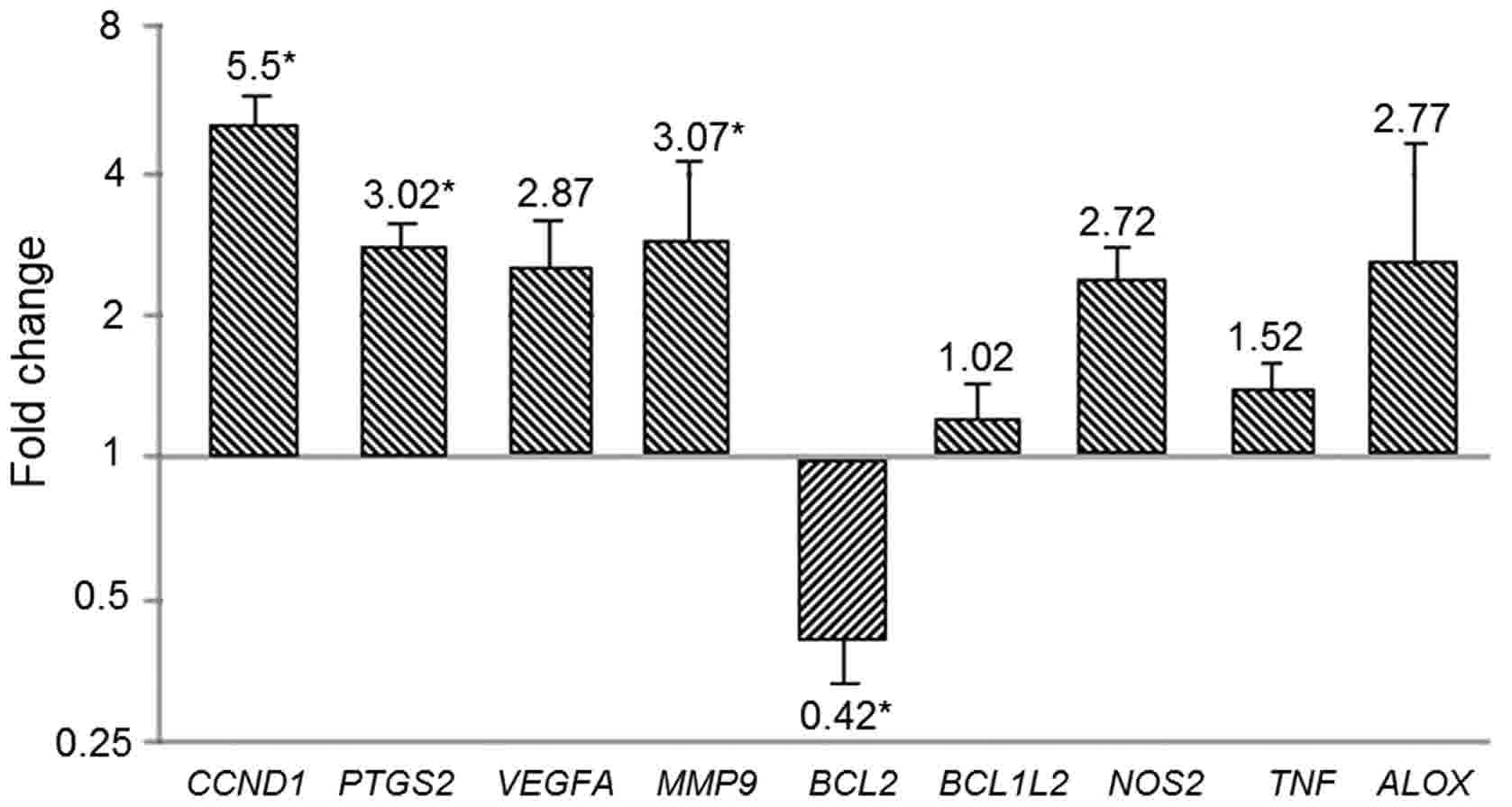
Cq data was analyzed using the 2−ΔΔCq
method to obtain the relative quantification of genes. CCND1,
PTGS2, and MMP9 were overexpressed in tumor tissue
compared to adjacent normal mucosa (5.5; 3.02; 3.07-folds,
respectively (P<0.05). While BCL2 decreased its
expression (0.42-folds, P<0.05). VEGFA, BCL2L1, NOS2,
TNF, and ALOX did not show significant differences
(Fig. 4).
Gene-expression profiles and
clinicopathological parameters
Gene's expression data was analyzed according to
clinicopathological parameters; results are described below and
reported in Table IV.
| Table IV.Gene expression association with
clinicopathological parameters in CRC patients (n=30). |
Table IV.
Gene expression association with
clinicopathological parameters in CRC patients (n=30).
|
| Relative
quantification of gene expression (mean fold difference) |
|---|
|
|
|
|---|
| Parameter | CCND1 | PTGS2 | VEGFA | MMP9 | BCL2 | BCL2L1 | NOS2 | TNF | ALOX |
|---|
| Tumor stage |
|
|
|
|
|
|
|
|
|
| I | 2.01±1.31 | 1.70±1.12 | 1.11±0.98 | 1.90±1.80 | 0.57±0.54 | 0.51±2.17 | 0.11±0.09 | 1.35±1.23 | 0.96±0.23 |
| II | 2.90±1.83 | 1.40±1.23 | 2.31±1.33 | 1.52±0.89 | 0.54±0.33 | 0.46±0.42 | 0.39±0.51 | 1.28±1.09 | 1.97±1.61 |
|
III | 7.10±2.52 | 3.22±3.12 | 3.12±1.84 | 3.10±1.22 | 0.38±0.27 | 1.01±0.81 | 4.10±1.42 | 1.57±1.21 | 3.17±1.42 |
| IV | 10.00±2.21 | 5.81±5.03 | 5.01±1.61 | 5.83±4.21 | 0.21±0.19 | 1.21±1.13 | 6.29±1.93 | 1.91±1.73 | 4.99±1.71 |
|
rs, (P-value) | 0.97
(0.02)a | 0.90 (0.94) | 0.98 (0.01)a | 0.88 (0.07) | −0.96
(0.03)a | 0.92 (0.07) | 0.95 (0.04)a | 0.89 (0.10) | 0.95
(0.01)a |
| Histological
differentiation |
|
|
|
|
|
|
|
|
|
|
Well | – | – | – | – | – | – | – | – | – |
|
Moderately | 4.71±3.73 | 2.16±2.01 | 2.66±1.91 | 1.96±1.21 | 0.55±0.72 | 0.59±0.36 | 2.46±1.12 | 1.22±1.12 | 2.13±1.22 |
|
Poorly | 6.32±1.21 | 3.92±1.10 | 3.13±0.96 | 4.21±1.53 | 0.29±0.11 | 0.99±0.51 | 3.3±2.71 | 1.82±0.96 | 3.41±1.73 |
|
rs (P-value) | 0.85 (0.05)a | 0.90 (0.11) | 0.78
(0.01)a | 0.79 (0.03) | −0.91 (0.01)a | −0.83 (0.12) | −0.54 (0.23) | −0.69 (0.27) | −0.89 (0.34) |
| Tumor
localization |
|
|
|
|
|
|
|
|
|
|
Ascending colon | 6.31±2.34 | 2.71±1.71 | 5.54±2.81 | 1.79±1.11 | 0.45±0.21 | 0.80±0.31 | 2.39±1.23 | 2.47±1.31 | 1.9±0.35 |
|
Transverse colon | 3.61±1.82 | 3.55±1.23 | 1.62±1.32 | 3.42±2.03 | 0.38±0.12 | 0.65±0.34 | 3.05±1.72 | 0.99±0.34 | 3.62±1.36 |
|
Descending colon | 6.65±4.26 | 1.63±1.12 | 3.35±2.90 | 4.66±4.23 | 0.49±0.26 | 0.91±0.61 | 1.17±0.96 | 1.32±1.02 | 2.48±1.81 |
| Sigmoid
colon | 6.98±3.81 | 5.32±4.20 | 1.83±1.12 | 4.48±3.11 | 0.49±0.24 | 0.96±0.67 | 4.41±1.23 | 1.22±0.67 | 1.05±0.43 |
|
Rectum | 3.95±1.52 | 2.05±1.71 | 2.06±1.21 | 1.05±0.76 | 0.39±0.29 | 0.63±0.31 | 3.38±1.71 | 1.67±0.97 | 4.80±1.51 |
| rs,
(P-value) | −0.40 (0.50) | −0.10 (0.87) | −0.21 (0.47) | 0.11 (0.37) | −0.10 (0.61) | −0.21 (0.87) | −0.28 (0.58) | −0.19 (0.59) | 0.23 (0.71) |
| Tumor depth |
|
|
|
|
|
|
|
|
|
| T1 | 1.98±1.42 | 1.56±1.11 | 1.45±1.21 | 2.21±1.81 | 0.54±0.22 | 0.86±0.64 | 1.21±0.78 | 1.29±0.37 | 1.21±0.56 |
| T2 | 3.92±1.80 | 2.31±1.33 | 2.88±1.80 | 1.94±1.22 | 0.49±0.18 | 0.62±0.47 | 2.24±1.35 | 1.87±0.46 | 1.69±0.79 |
| T3 | 6.38±2.01 | 2.99±1.94 | 3.34±1.23 | 3.62±1.73 | 0.34±0.28 | 0.73±0.38 | 3.62±1.92 | 1.51±0.28 | 3.87±1.23 |
| T4 | 9.72±4.33 | 5.26±1.72 | 3.85±1.71 | 4.55±2.01 | 0.31±0.171 | 0.95±0.71 | 4.45±2.41 | 1.41±0.41 | 4.31±1.54 |
|
rs, (P-value) | 0.94
(0.01)a | 0.78 (0.03)a | 0.64 (0.07) | 0.81 (0.06) | −0.89
(0.01)a | 0.54 (0.08) | 0.91 (0.01)a | 0.24 (0.35) | 0.82
(0.01)a |
| Lymph node
status |
|
|
|
|
|
|
|
|
|
| N0 | 1.91±1.22 | 1.22±0.95 | 1.23±0.67 | 1.49±1.35 | 0.74±2.73 | 0.83±0.28 | 1.29±1.32 | 1.09±0.97 | 1.96±1.13 |
| N1 | 4.62±1.85 | 3.21±1.41 | 3.01±1.44 | 3.73±1.82 | 0.32±1.73 | 0.76±0.64 | 2.37±1.93 | 1.11±0.92 | 2.59±1.51 |
| N2 | 9.97±4.21 | 4.66±2.74 | 4.43±2.91 | 4.02±2.11 | 0.20±2.12 | 0.78±0.10 | 4.98±2.61 | 2.36±1.21 | 3.76±1.44 |
|
rs, (P-value) | 0.84
(0.03)a | 0.79
(0.04)a | 0.90
(0.03)a | 0.71 (0.08) | −0.92
(0.01)a | 0.41 (0.26) | 0.78
(0.02)a | 0.62 (0.26) | 0.61 (0.14) |
| Metastasis
degree |
|
|
|
|
|
|
|
|
|
| M0 | 2.94±1.13 | 2.33±1.33 | 1.24±1.13 | 1.70±0.87 | 0.56±0.48 | 0.78±0.19 | 1.38±1.22 | 1.17±1.02 | 1.36±1.23 |
| M1 | 8.06±3.71 | 3.73±1.72 | 4.52±1.71 | 4.46±2.82 | 0.28±0.15 | 0.80±0.45 | 4.38±2.93 | 1.87±1.13 | 4.18±3.11 |
|
rs, (P-value) | 0.91
(0.04)a | 0.51 (0.12) | 0.79 (0.03)a | 0.31 (0.31) | −0.95
(0.03)a | 0.11 (0.58) | 0.81 (0.03)a | 0.21 (0.48) | 0.89 (0.06) |
Positive association of CCND1, VEGFA, NOS2,
and ALOX as well as a negative association of BCL2
with tumor stage progression was reported (P<0.05). Significant
overexpression in the advanced stages group compared to initial
stages in CCND1 (III+IV=8.55±1.45 vs. I+II=2.45±0.45;
P<0.05), VEGFA (I+II=1.2±0.3 vs. III+IV=4.55±1.3;
P<0.05), NOS2 (I+II=0.25±0.14 vs. III+IV=5.2±2.1;
P<0.05) and ALOX (I+II=1.46±0.5 vs. III+IV=4.08±0.19;
P<0.05) corroborate the positive association. No significant
difference was found in gene expression of PTGS2, MMP9,
BCL2L1, and TNF during tumor progression.
Gene expression association with histopathological
differentiation groups, was statistically positive in the case of
CCND1, MMP9 and VEGFA, while BCL2 expression
was negatively associated (P<0.05). No association of
histopathological differentiation with PTGS2, MMP9, BCL2L1,
NOS2, ALOX and TNF gene expression was reported.
No association between tumor localization site and
expression of any evaluated gene was reported.
In the case of tumor depth, positive association was
observed with CCND1, PTGS2, NOS2 and ALOX expression,
negative association with BCL2 expression was also reported
(P<0.05). VEGFA, MMP9, BCL2L1, and TNF expression
did not showed any association.
Lymph node status and gene expression was positively
associated in the case of CCND1, PTGS2, VEGFA, and
NOS2 (P<0.05). Negative association with BCL2 was
also reported (P<0.05). No association with MMP9, BCL2L1,
TNF, and ALOX was found.
Metastasis degree and gene expression was positively
associated in the case of CCND1, VEGFA and NOS2, and
negative association was reported with BCL2 (P<0.05). No
association was observed in PTGS2, MMP9, BCL2L1, TNF, and
ALOX.
Discussion
In the present study, NF-κB/p65 and genes expression
association with clinicopathological parameters of CRC patients was
investigated. We reported higher NF-κB/p65 cytoplasmic expression
in tumor tissue compared to normal adjacent mucosa; our findings
are consistent with previous studies (20). NF-κB/p65 expression in CRC showed
discrepancy in stained protein localization, nevertheless nuclear
staining has been mainly reported (20,21).
NF-κB/p65 detected in previous reports, similar as our study,
indicates released IκBα, but we were not able to confirm the
transcriptional activity. We hypothesize that NF-κB/IκBα binding
alterations could masked the nuclear localization signal in p65 as
other studies suggest (22).
NF-κB/p65 expression by CRC stages has been commonly
evaluated. Higher levels in advanced stages compared to initial
stages, evaluated by immunohistochemistry are reported in this
study. According to our results, NF-κB/p65 increment was positively
associated with tumor stage progression. We suggest that NF-κB/p65
cytoplasmic expression may play a key role in CRC progression
probably by molecular changes in its downstream pathway. In
addition, positive association between NF-κB/p65 expression with
histopathology differentiation, tumor depth, lymph node status and
metastasis degree was reported, but no association with tumor
localization was observed in any case. NF-κB/p65 association with
clinicopathological parameters has not been entirely described. A
meta-analysis in solid tumors, reported a positive association with
lymph node status and metastasis degree (23), but the conclusions are still in
contradiction. Our results suggest that NF-κB/p65 is involved in
CRC establishment and progression by promoting clinicopathological
parameters development.
Gene expression analyzed in tumor tissue vs. normal
adjacent mucosa, showed overexpression in CCND1, PTGS2 and
MMP9, decreased expression of BCL2, while no
significant differences in expression of BCL2L1, VEGFA, TNF,
ALOX and NOS2 was reported. Overexpression of
CCND1 in tumor tissue was observed in this study as in
others (24), considering that
CCND1 promotes the transition of G1- to S-phase of cell
cycle it may play a key role in tumor cells proliferation in the
evaluated tissues. PTGS2 was similarly reported
overexpressed in tumor tissue as others studies did using different
methodologies (25–27), but no alteration of PTGS2
expression has been also observed, these contradictory reports
suggest that PTGS2 activity in CRC is still unclear.
According to our results, we hypothesize that proliferation and
survival processes due to PTGS2 overexpression plays an
important role in CRC. MMP9 overexpression observed in this
study agreed with previous reports that relate its expression with
invasiveness and metastasis (28–30). In
this study we just report decreased expression of BCL2 in
CRC, higher expression in tumor tissue than in normal mucosa had
been reported (31). BCL2
promotes tumorigenesis by inhibition of apoptosis, according to our
results; we suggest that once CRC is established it began to
decrease its activity and consecutively its gene expression.
Previous studies report higher expression of BCL2L1, VEGFA, TNF,
ALOX and NOS2 in tumor tissue than in normal mucosa, in
this study we did not observed difference in expression of these
genes. BCL2L1 is associated with apoptosis and opposite of
our results its overexpression has been previously reported
(32). VEGFA overexpression
has been observed and associated with advanced tumor stages and
poor clinical outcome (33), but one
study reported no difference at protein level similar as our
results (34). TNF is involved
in tumor promotion and progression; previous reports have found
expression of TNF in 94% of tumors (35) in disagreement to our results.
ALOX activity is related with tumor growth and invasiveness
of solid tumors, opposite to our results ALOX overexpression
in CRC has been reported (36).
Similarly, NOS2 overexpression in CRC has been observed and
related to angiogenesis (37), while
according to our results, decreased expression of NOS2 in
CRC tumor was previously reported (38).
Even though we did not found significant differences
in expression of all selected genes between tumor and normal
adjacent mucosa, we analyze the association of these genes with
clinicopathological parameters due to their importance in
tumorigenesis. The results of these analyses are discussed below:
The strong positive association of CCND1 with all TNM
parameters, excluding tumor localization, confirmed its role during
CRC tumorigenesis; as well its association with tumor progression
suggests its potential as tumor marker in early stages. The
activity of MMP9 during invasiveness processes in CRC was
confirmed in this study due to its positive association with tumor
stages, but we can found any association with metastasis as other
groups did (28–30). Antiapoptosis activity of BCL2
has been associated to different cancer types, as well as CRC, in
this study BCL2 was negatively associated with all TNM
parameters, supporting the highest expression level in normal
adjacent mucosa of CRC patients we found; this data is completely
disagreeing with previous reports (31), we suggest that others antiapoptotic
factors play more important role than BCL2 does in our CRC
patients, even though we considered that is necessary to evaluate
BCL2 protein expression to reinforce our results. The role
of VEGFA in solid tumors is confirmed with the positive
association in lymph node status and metastasis degree CRC groups,
according to others reports the poor clinical outcome is related to
VEGFA expression (33), we
confirmed this data in our CRC advanced group when VEGFA is
overexpressed, so we also suggest its role as a possible tumor
marker in advanced disease. Surprisingly, TNF and
BCL1L2 genes that has been several times reported associated
with different types of solid tumors (32,35), did
not showed association with any clinicopathological parameter,
differences in their expression between tumor vs. normal adjacent
mucosa was reported neither. Further studies should analyze the
TNF and BCL1L2 proteins vs. NF-κB/p65 activity in
human CRC samples. ALOX overexpression influences in CRC
development, and it is observed according to the positive
association with tumor stages and tumor depth reported in this
study. Tumor growth mediated by inflammation, supposed to be one of
the most important processes related to ALOX overexpression
(36), this data is supported by our
results. NOS2 showed a positive association with TNM
parameters, but it was not significantly associated to
histopathological differentiation and tumor localization.
NOS2 expression was not statistical different in tumor vs.
normal adjacent mucosa, nevertheless we suggest that NOS2
activity is related to tumorigenesis, absolute quantification
analyses could help in future studies to confirm the NOS2
role in CRC establishment and development (38).
According to results discussed above, not all
selected genes regulated by NF-κB/p65 increment their expression as
the transcription factor did; probably these genes are partially
regulated by others transcription factors in CRC. These results
suggest that NF-κB activity is necessary but not sufficient in CRC
establishment. To our knowledge the present study reports for the
first time, the selected genes expression and NF-κB/p65 association
with clinicopathological parameters in sporadic CRC. Due to
NF-κB/p65 nucleus translocation is required for its transcriptional
activity; we suggest that others quantitative techniques are
necessary to statistically associate NF-κB activity with gene
expression profiles in CRC; conditions of our experiments did not
permit us to work more deeply on it because some of our patients
are already under treatment (non-inclusion criterion) or because we
are not able to take more tissue samples (the patient did not
continue attending hospital or because they passed away).
Meta-analysis association studies are indispensable to completely
understand the behavior of these molecules in CRC.
Higher NF-κB/p65 expression in CRC tissue compared
to normal adjacent mucosa from the same patient is reported, and
this increment was positively associated with clinicopathological
parameters, except for tumor localization. The monitoring and
regulation of this transcription factor may be therapeutically
useful in CRC patients.
NF-κB regulated genes showed an irregular expression
pattern when compared CRC tissue vs. normal adjacent mucosa;
CCND1, PTGS2 and MMP9 were overexpressed. VEGFA,
BCL2L1, NOS2, TNF and ALOX did not changed, while
BCL2 decreases its expression level. Results of genes
expression association with clinicopathological parameters are
summarized below; CCND1 was positively associated with all
TNM parameters. PTGS2 was associated with tumor depth and
lymph node status. VEGFA showed a positively association
with lymph node status and metastasis degree. MMP9 was
positively associated with tumor stages. NOS2 showed a
positive association with tumor stages, tumor depth, lymph node
status and metastasis degree. ALOX was positively associated
with tumor stages and tumor depth. BCL1L2 and TNF did
not showed association with any clinicopathological parameter,
while BCL2 was negatively associated with all TNM
parameters. Tumor localization site was not related with any of the
evaluated genes. According to the association of these genes with
different clinicopathological parameters, they may be considered
for the selection of proper diagnosis, treatment and follow-up for
CRC patients.
Acknowledgements
This study was partially supported by COECYTJAL
5-2010-1-1083 grant. The authors would like to thank Dr.
Bustos-Rodríguez F and Dr. Valenzuela-Pérez JA for their technical
support in this study. The authors would like to thank the Ph.D.
Program of Molecular Biology in Medicine, University of
Guadalajara.
References
|
1
|
Ferlay J, Soerjomataram I, Dikshit R, Eser
S, Mathers C, Rebelo M, Parkin DM, Forman D and Bray F: Cancer
incidence and mortality worldwide: Sources, methods and major
patterns in GLOBOCAN 2012. Int J Cancer. 136:E359–E386. 2015.
View Article : Google Scholar : PubMed/NCBI
|
|
2
|
Peddareddigari V, Wang D and Dubois RN:
The tumor microenvironment in colorectal carcinogenesis. Cancer
Microenviron. 3:149–166. 2010. View Article : Google Scholar : PubMed/NCBI
|
|
3
|
Hoesel B and Schmid JA: The complexity of
NF-κB signaling in inflammation and cancer. Mol Cancer. 12:862013.
View Article : Google Scholar : PubMed/NCBI
|
|
4
|
Sakamoto K, Maeda S, Hikiba Y, Nakagawa H,
Hayakawa Y, Shibata W, Yanai A, Ogura K and Omata M: Constitutive
NF-kappaB activation in colorectal carcinoma plays a key role in
angiogenesis, promoting tumor growth. Clin Cancer Res.
15:2248–2258. 2009. View Article : Google Scholar : PubMed/NCBI
|
|
5
|
Puvvada SD, Funkhouser WK, Greene K, Deal
A, Chu H, Baldwin AS, Tepper JE and O'Neil BH: NF-kB and Bcl-3
activation are prognostic in metastatic colorectal cancer.
Oncology. 78:181–188. 2010. View Article : Google Scholar : PubMed/NCBI
|
|
6
|
Lewander A, Gao J, Adell G, Zhang H and
Sun XF: Expression of NF-κB p65 phosphorylated at serine-536 in
rectal cancer with or without preoperative radiotherapy. Radiol
Oncol. 45:279–284. 2011. View Article : Google Scholar : PubMed/NCBI
|
|
7
|
Abdullah M, Rani AA, Sudoyo AW, Makmun D,
Handjari DR and Hernowo BS: Expression of NF-kB and COX2 in
colorectal cancer among native Indonesians: The role of
inflammation in colorectal carcinogenesis. Acta Med Indones.
45:187–192. 2013.PubMed/NCBI
|
|
8
|
Yu LL, Yu HG, Yu JP, Luo HS, Xu XM and Li
JH: Nuclear factor-kappaB p65 (RelA) transcription factor is
constitutively activated in human colorectal carcinoma tissue.
World J Gastroenterol. 10:3255–3260. 2004. View Article : Google Scholar : PubMed/NCBI
|
|
9
|
Patwardhan MB, Samsa GP, McCrory DC,
Fisher DA, Mantyh CR, Morse MA, Prosnitz RG, Cline KE and Gray RN:
Cancer care quality measures: Diagnosis and treatment of colorectal
cancer. Evid Rep Technol Assess (Full Rep). 1–116. 2006.PubMed/NCBI
|
|
10
|
Abdullah M, Sudoyo AW, Pranowo BS, Rini D,
Sutrisna B and Rani AA: Expression of NF-kappaB and COX-2 in young
versus older patients with sporadic colorectal cancer. Acta Med
Indones. 41:70–74. 2009.PubMed/NCBI
|
|
11
|
Balcerczak E, Pasz-Walczak G, Kumor P,
Panczyk M, Kordek R, Wierzbicki R and Mirowski M: Cyclin D1 protein
and CCND1 gene expression in colorectal cancer. Eur J Surg Oncol.
31:721–726. 2005. View Article : Google Scholar : PubMed/NCBI
|
|
12
|
Roelofs HM, Morsche Te RH, van Heumen BW,
Nagengast FM and Peters WH: Over-expression of COX-2 mRNA in
colorectal cancer. BMC Gastroenterol. 14:12014. View Article : Google Scholar : PubMed/NCBI
|
|
13
|
Stanilov S, Drodeva Z and Stanilova S:
Higher TNF-alpha production detected in colorectal cancer patients
monocytes. Med Biotechnol. 26:107–110. 2011.
|
|
14
|
Rao CV, Janakiram NB and Mohammed A:
Lipoxygenase and cyclooxygenase pathways and colorectal cancer
prevention. Curr Colorectal Cancer Rep. 8:316–324. 2012. View Article : Google Scholar : PubMed/NCBI
|
|
15
|
Yagihashi N, Kasajima H, Sugai S,
Matsumoto K, Ebina Y, Morita T, Murakami T and Yagihashi S:
Increased in situ expresión of nitric oxide synthase in human
colorectal cancer. Virchows Arch. 436:109–114. 2000. View Article : Google Scholar : PubMed/NCBI
|
|
16
|
Zeestraten EC, Benard A, Reimers MS,
Schouten PC, Liefers GJ, van de Velde CJ and Kuppen PJ: The
prognostic value of the apoptosis pathway in colorectal cancer: A
review of the literature on biomarkers identified by
immunohistochemistry. Biomark Cancer. 5:13–29. 2013.PubMed/NCBI
|
|
17
|
Koskensalo S, Hagström J, Linder N, Lundin
M, Sorsa T, Louhimo J and Haglund C: Lack of MMP-9 expression is a
marker for poor prognosis in Dukes' B colorectal cancer. BMC Clin
Pathol. 12:242012. View Article : Google Scholar : PubMed/NCBI
|
|
18
|
Guba M, Seeliger H, Kleespies A, Jauch KW
and Bruns C: Vascular endothelial growth factor in colorectal
cancer. Int J Colorectal Dis. 19:510–517. 2004. View Article : Google Scholar : PubMed/NCBI
|
|
19
|
Livak KJ and Schmittgen TD: Analysis of
relative gene expression data using real-time quantitative PCR and
the 2(-Delta Delta C(T)) method. Methods. 25:402–408. 2001.
View Article : Google Scholar : PubMed/NCBI
|
|
20
|
Kojima M, Morisaki T, Sasaki N, Nakano K,
Mibu R, Tanaka M and Katano M: Increased nuclear factor-kB
activation in human colorectal carcinoma and its correlation with
tumor progression. Anticancer Res. 24:675–681. 2004.PubMed/NCBI
|
|
21
|
Yu HG, Yu LL, Yang Y, Luo HS, Yu JP, Meier
JJ, Schrader H, Bastian A, Schmidt WE and Schmitz F: Increased
expression of RelA/nuclear factor-kappa B protein correlates with
colorectal tumorigenesis. Oncology. 65:37–45. 2003. View Article : Google Scholar : PubMed/NCBI
|
|
22
|
Sun SC, Ganchi PA, Béraud C, Ballar DW and
Greene WC: Autoregulation of the NF-kappa B transactivator RelA
(p65) by multiple cytoplasmic inhibitors containing ankyrin motifs.
Proc Natl Acad Sci USA. 91:1346–1350. 1994. View Article : Google Scholar : PubMed/NCBI
|
|
23
|
Wu D, Wu P, Zhao L, Huang L, Zhang Z, Zhao
S and Huang J: NF-kB expression and outcomes in solid tumors: A
systematic review and meta-analysis. Medicine (Baltimore).
94:e16872015. View Article : Google Scholar : PubMed/NCBI
|
|
24
|
Sutter T, Doi S, Carnevale KA, Arber N and
Weinstein IB: Expression of cyclins D1 and E in human colon
adenocarcinomas. J Med. 28:285–309. 1997.PubMed/NCBI
|
|
25
|
Antonacopoulou AG, Tsamandas AC, Petsas T,
Liava A, Scopa CD, Papavassiliou AG and Kalofonos HP: EGFR, HER-2
and COX-2 levels in colorectal cancer. Histopathology. 53:698–706.
2008. View Article : Google Scholar : PubMed/NCBI
|
|
26
|
Cressey R, Pimpa S, Tontrong W,
Watananupong O and Leartprasertsuke N: Expression of
cyclooxygenase-2 in colorectal adenocarcinoma is associated with
p53 accumulation and hdm2 overexpression. Cancer Lett. 233:232–239.
2006. View Article : Google Scholar : PubMed/NCBI
|
|
27
|
Eberhart CE, Coffey RJ, Radhika A,
Giardiello FM, Ferrenbach S and DuBois RN: Up-regulation of
cyclooxygenase 2 gene expression in human colorectal adenomas and
adenocarcinomas. Gastroenterology. 107:1183–1188. 1994. View Article : Google Scholar : PubMed/NCBI
|
|
28
|
Chu D, Zhao Z, Zhou Y, Li Y, Li J, Zheng
J, Zhao Q and Wang W: Matrix metalloproteinase-9 is associated with
relapse and prognosis of patients with colorectal cancer. Ann Surg
Oncol. 19:318–325. 2012. View Article : Google Scholar : PubMed/NCBI
|
|
29
|
Langers AM, Verspaget HW, Hawinkels LJ,
Kubben FJ, van Duijn W, van der Reijden JJ, Hardwick JC, Hommes DW
and Sier CF: MMP-2 and MMP-9 in normal mucosa are independently
associated with outcome of colorectal cancer patients. Br J Cancer.
106:1495–1498. 2012. View Article : Google Scholar : PubMed/NCBI
|
|
30
|
Zeng ZS, Huang Y, Cohen AM and Guillem JG:
Prediction of colorectal cancer relapse and survival via tissue RNA
levels of matrix metalloproteinase-9. J Clin Oncol. 14:3133–3140.
1996. View Article : Google Scholar : PubMed/NCBI
|
|
31
|
Sun N, Meng Q and Tian A: Expressions of
the anti-apoptotic genes Bag-1 and Bcl-2 in colon cancer and their
relationship. Am J Surg. 200:341–345. 2010. View Article : Google Scholar : PubMed/NCBI
|
|
32
|
Krajewska M, Moss SF, Krajewski S, Song K,
Holt PR and Reed JC: Elevated expression of Bcl-X and reduced Bak
in primary colorectal adenocarcinomas. Cancer Res. 56:2422–2427.
1996.PubMed/NCBI
|
|
33
|
Altomare DF, Rotelli MT, Pentimone A,
Rossiello MR, Martinelli E, Guglielmi A, De Fazio M, Marino F,
Memeo V, Colucci M and Semeraro N: Tissue factor and vascular
endothelial growth factor expression in colorectal cancer: Relation
with cancer recurrence. Colorectal Dis. 9:133–138. 2007. View Article : Google Scholar : PubMed/NCBI
|
|
34
|
Cao D, Hou M, Guan YS, Jiang M, Yang Y and
Gou HF: Expression of HIF-1alpha and VEGF in colorectal cancer:
Association with clinical outcomes and prognostic implications. BMC
Cancer. 9:4322009. View Article : Google Scholar : PubMed/NCBI
|
|
35
|
Grimm M, Lazariotou M, Kircher S,
Höfelmayr A, Germer CT, von Rahden BH, Waaga-Gasser AM and Gasser
M: Tumor necrosis factor-α is associated with positive lymph node
status in patients with recurrence of colorectal cancer indications
for anti-TNF-α agents in cancer treatment. Anal Cell Pathol (Amst).
33:151–163. 2010. View Article : Google Scholar : PubMed/NCBI
|
|
36
|
Soumaoro LT, Lida S, Uetake H, Ishiguro M,
Takagi Y, Higuchi T, Yasuno M, Enomoto M and Sugihara K: Expression
of 5-lipoxygenase in human colorectal cancer. World J
Gastroenterol. 12:6355–6360. 2006. View Article : Google Scholar : PubMed/NCBI
|
|
37
|
Moochhala S, Chhatwal VJ, Chan ST, Ngoi
SS, Chia YW and Rauff A: Nitric oxide synthase activity and
expression in human colorectal cancer. Carcinogenesis.
17:1171–1174. 1996. View Article : Google Scholar : PubMed/NCBI
|
|
38
|
Kojima M, Morisaki T, Tsukahara Y,
Uchiyama A, Matsunari Y, Mibu R and Tanaka M: Nitric oxide synthase
expression and nitric oxide production in human colon carcinoma
tissue. J Surg Oncol. 70:222–229. 1999. View Article : Google Scholar : PubMed/NCBI
|