Introduction
Lung cancer is the leading cause of cancer-related
mortality in male patients and the second leading cause in female
patients worldwide (1).
Non-small-cell lung cancer (NSCLC) accounts for ~85% of those
mortalities (2). It was predicted
that 222,500 individuals would be diagnosed with lung cancer, while
155,870 would die of the disease in America in 2017 (3). Despite rapid progress in clinical and
experimental oncology practices in recent years, the prognosis of
the majority of lung cancer patients is still not optimistic, and
the 5-year overall survival (OS) rate of such patients is ~15%
(4,5).
Moreover, the survival rate of patients with metastatic NSCLC is
~12 months, while the median progression-free survival (PFS) is
between 3 and 6 months (6).
Therefore, there is an urgent need to discover novel molecular
biomarkers related to NSCLC as therapeutic targets for the
prevention, diagnosis and treatment of NSCLC.
Human transactivation response RNA-binding proteins
(TARBPs) were initially identified as proteins that bind with HIV
type-1 (HIV-1) transactivation response RNA to activate long
terminal repeat (LTR) expression in the presence or absence of the
viral transactivator Tat (7). TARBP1
is a double-stranded RNA (dsRNA) binding protein that associates
with certain other RNA interference factors to form a minimal
RNA-induced silencing complex (RISC) (8,9), which
serves as a Dicer co-factor in the processing of ~70-nucleotide
pre-microRNAs (miRNAs) to 21–25-nucleotide mature miRNAs (10). Furthermore, TARBP1, being primarily
located in cell nuclei (11), may
play a crucial role in regulating the process of transcription. In
this study, we detected the expression of TARBP1 in NSCLC and
assessed its clinicopathological and prognostic significance.
Materials and methods
Data mining
Analysis of TARBP1 mRNA expression in NSCLC tissue
samples and normal controls (meta-analysis of TARBP1 genes) was
performed through the online cancer microarray database Oncomine
(www.oncomine.org; Compendia Biosciences, Ann
Arbor, MI, USA), which collects published cancer microarray data
and processes these data with the same criteria (12). The data regarding mRNA expression
status were organized into different types of human cancer tissue
samples defined by the original publications. Sixteen sets of
published mRNA expression data were chosen in this study. The
mining strategy of these datasets was performed according to a
method formulated by Oncomine (12).
Significantly upregulated TARBP1 in cancer tissue samples compared
with normal controls (>2-fold) were first selected. Next,
concept filters of Oncomine were employed to identify the different
expression of TARBP1 in lung carcinoma. Median rank was defined as
the estimation of unreliability values based on the failure order
number and the cumulative binomial distribution and was calculated
using the formula BetaInv (0.5, k,N-k+1), where N represents sample
size and k represents order (13).
Patients and specimens
Analyses were conducted on a total of 90
paraffin-embedded NSCLC samples from the archives of the First
Affiliated Hospital of Guangzhou Medical University. All the
patients were histopathologically diagnosed with primary lung
cancer and underwent curative surgery between July 2004 and June
2009. None of the patients had received any type of neoadjuvant
therapy. The median age of the patients was 61.5 years (range,
30–84 years), and the median tumor size was 4.6 cm (range, 1.5–9.0
cm). Among these patients, 81 of them were diagnosed
adenocarcinoma, two were diagnosed large cell carcinoma, and seven
squamous cell carcinoma. The demographic features and
clinicopathological information of the patients are summarized in
Table I. The follow-up time for the
NSCLC cohort ranged from 1 to 121 months, and the median follow-up
time was 39 months. Both tumor tissue and adjacent non-cancerous
tissue (defined as tissue at least 1-cm distance from the tumor
edge) were obtained during surgery. The expression of TARBP1
protein was detected by immunohistochemistry (IHC) in all 90
matched tissues. Another 20 paired NSCLC samples and adjacent
non-cancerous tissues were collected immediately after operation
for reverse transcription-quantitative polymerase chain reaction
(RT-qPCR) analysis (11 male and 9 female). All the patients were
histopathologically diagnosed with primary lung cancer between
January 2015 and December 2016. The histological type of all the 20
patients were NSCLC. The median age of the patients was 63 years
(range, 35–79 years), and the median tumor size was 5.2 cm (range,
4.1–8.5 cm).
 | Table I.Correlation of TARBP1 expression with
clinicopathological features. |
Table I.
Correlation of TARBP1 expression with
clinicopathological features.
|
|
| TARBP1 (%) |
|
|---|
|
|
|
|
|
|---|
|
Characteristics | Total (%) | Positive | Negative | P-value |
|---|
| Sex |
|
|
| 0.828 |
|
Male | 49 (54.4) | 38 (77.6) | 11 (22.4) |
|
|
Female | 41 (45.6) | 31 (75.6) | 10 (24.4) |
|
| Age, years |
|
|
| 0.189 |
|
≥60 | 55 (61.1) | 40 (72.7) | 15 (27.3) |
|
|
<60 | 35 (38.9) | 29 (82.9) | 6 (17.1) |
|
| Lymphatic
metastasis |
|
|
| 0.110 |
|
Positive | 48 (53.3) | 40 (83.3) | 8 (16.7) |
|
|
Negative | 42 (46.7) | 29 (69.0) | 13 (31.0) |
|
| Tumor size
(cm) |
|
|
| 0.596 |
| ≥5 | 21 (23.3) | 17 (81.0) | 4 (19.0) |
|
|
<5 | 69 (76.7) | 52 (75.4) | 17 (24.6) |
|
| Histological
grade |
|
|
| <0.001 |
| 1 | 12 (13.3) | 9 (75.0) | 3 (25.0) |
|
| 2 | 57 (63.3) | 42 (73.7) | 15 (26.3) |
|
| 3 | 21 (23.3) | 18 (85.7) | 3 (14.3) |
|
| Clinical stage |
|
|
| 0.024 |
|
I–II | 50 (55.6) | 35 (70.0) | 15 (30.0) |
|
|
III | 30 (33.3) | 26 (86.7) | 4 (13.3) |
|
| IV | 10 (11.1) | 8 (80.0) | 2 (20.0) |
|
| Pathological
type |
|
|
| <0.001 |
|
Adenocarcinoma | 81 (90.0) | 61 (75.3) | 20 (24.7) |
|
|
Other | 9 (10.0) | 1 (11.1) | 8 (88.9) |
|
This study was approved by the Ethics Committee of
the First Affiliated Hospital of Guangzhou Medical University, and
all patients provided written informed consent for the use of their
clinical samples in the present study.
RT-qPCR analysis
Total RNA was extracted from the tissue samples with
TRIzol reagent (Invitrogen; Thermo Fisher Scientific, Inc.,
Waltham, MA, USA), according to the manufacturer's protocol, and
pretreated with RNase-free DNase. cDNA synthesis was performed with
1 µg of total RNA from each sample and the use of a First-Strand
Synthesis System (Fermentas; Thermo Fisher Scientific, Inc.).
Real-time PCR was performed with a CFX96 Real-Time System and 2×
SYBR-Green Master Mix (both from Bio-Rad, Hercules, CA, USA) in a
total volume of 10 µl. The primer sequences were as follows:
TARBP1, sense, 5′-TGCAACATTTCACCCACTCAA-3′ and antisense,
5′-CCCGCAGCTAAAGGAACATC-3′; and glyceraldehyde-3-phosphate
dehydrogenase (GAPDH, internal control), sense,
5′-TGTTGCCATCAATGACCCC-3′ and antisense, 5′-CTCCACGACGTACTCAGC-3′.
All the reactions were performed in triplicate in three independent
experiments and were quantified using the 2−ΔΔCq method
(14).
IHC
IHC staining was performed according to the
manufacturer's protocol (Zymed®; Life Technologies;
Thermo Fisher Scientific, Inc.). Briefly, the paraffin-embedded
samples were heated at 60°C for 1 h and deparaffinized with xylene.
The slides were then rehydrated through a graded ethanol series,
submerged in sodium citrate buffer and heated in a microwave for
antigen retrieval. The sections were treated with 0.3%
H2O2 for 10 min to inhibit endogenous
peroxidase activity, and then incubated with normal goat serum at
room temperature for 30 min to block nonspecific staining.
Following blocking, the sections were incubated with rabbit
polyclonal anti-TARBP1 antibody (dilution, 1:100; LSBIO) at 4°C
overnight. Normal goat serum was used as a negative control. After
three washes with phosphate-buffered saline (PBS), the slides were
incubated with a biotinylated anti-rabbit secondary antibody, and
then with streptavidin-horseradish peroxidase (both from Zymed;
Thermo Fisher Scientific, Inc.) at 37°C for 30 min. After three
washes with PBS, the tissue sections were incubated in
diaminobenzidine (DAB) for color development.
The percentage and degree of positive staining were
assessed and recorded separately by two independent pathologists,
who were entirely blinded to the clinical characteristics of the
samples. Scores assigned by the two independent pathologists were
then averaged for the evaluation of TARBP1 expression. The staining
intensity of TARBP1 was classified according to the following
standards: no staining was marked as 0; weak staining (light
yellow) was marked as 1; moderate staining (yellow brown) was
marked as 2; and strong staining (brown) was marked as 3. The
proportion of positively stained tumor cells was classified
according to the following standards: no positive tumor cells was
marked as 0; 1–25% positive tumor cells was marked as 1; 26–50%
positive tumor cells was marked as 2; 51–75% positive tumor cells
was marked as 3; and >75% positive tumor cells was marked as
4.
The final score was calculated as the product of the
percentage and intensity scores. Cut-off values for TARBP1 were
chosen on account of the heterogeneity using log-rank test
concerning overall survival (OS). The optimal cut-off value was
calculated as follows: A staining index score over 8 was used to
define tumors with high TARBP1 expression and under 8 indicated low
TARBP1 expression.
Statistical analysis
All the statistical analyses were conducted using
the Statistical Software Package for the Social Sciences 20.0 (IBM
Corp., Armonk, NY, USA) and GraphPad Prism 5 (GraphPad Software,
Inc., La Jolla, CA, USA). TARBP1 mRNA levels in the NSCLC and
paired normal tissues were compared using a paired-samples t-test.
The difference in the rate of TARBP1 positive expression between
the NSCLC and adjacent non-tumor tissues was analyzed using a
Chi-square test. Survival curves were plotted by the Kaplan-Meier
method and compared with a log-rank test. The time span from the
date of each patient's randomization to either the date of the
patient's death for any reason or the date of the last follow-up
was defined as OS. The relationships between TARBP1 expression and
other clinicopathological features were evaluated using Chi-square
tests and Fisher's exact tests. The prognostic value of
clinicopathological features was analyzed by univariate and
multivariate Cox regression analyses; the enter method of the Cox
model was used for univariate analysis, while the forward method
was used for multivariate analysis. All statistical tests were
two-sided and P<0.05 was considered to indicate statistical
significance.
Results
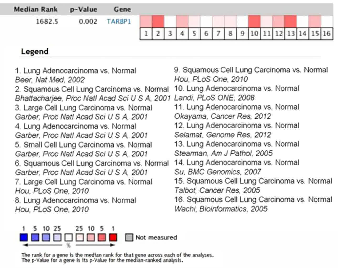
TARBP1 is overexpressed in NSCLC
patients
To determine whether TARBP1 expression was elevated
in human NSCLC samples, we first conducted a meta-analysis of the
Oncomine database, and found that TARBP1 expression was
significantly higher in lung cancer than in matched adjacent
non-tumor tissues, with a median rank of 1862.5 and a P-value of
0.002 (Fig. 1) (15–25). To
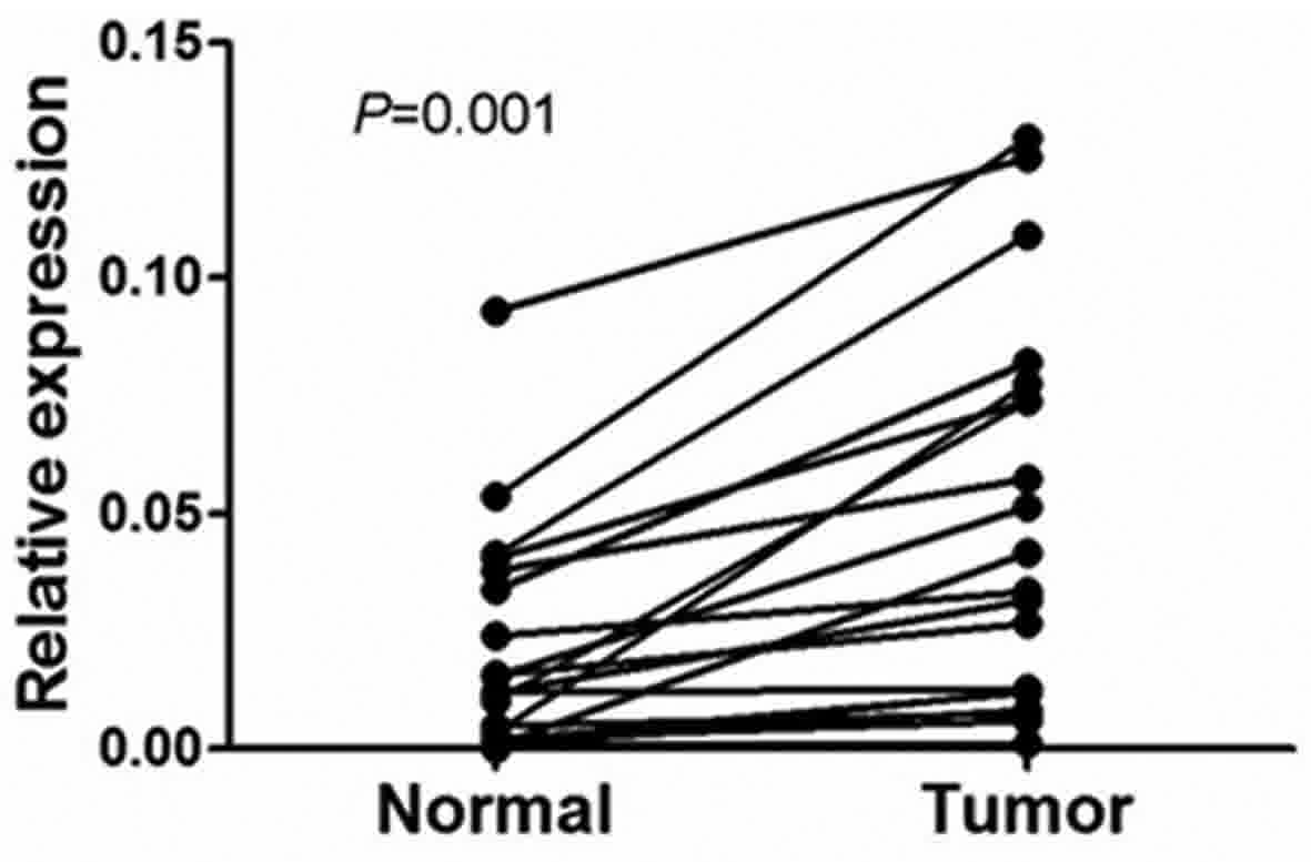
verify this result, we performed RT-qPCR analysis on 10 NSCLC
tissues and paired adjacent normal lung tissues. As illustrated in
Fig. 2, the level of TARBP1 mRNA in
the 10 NSCLC samples was significantly higher than that in the
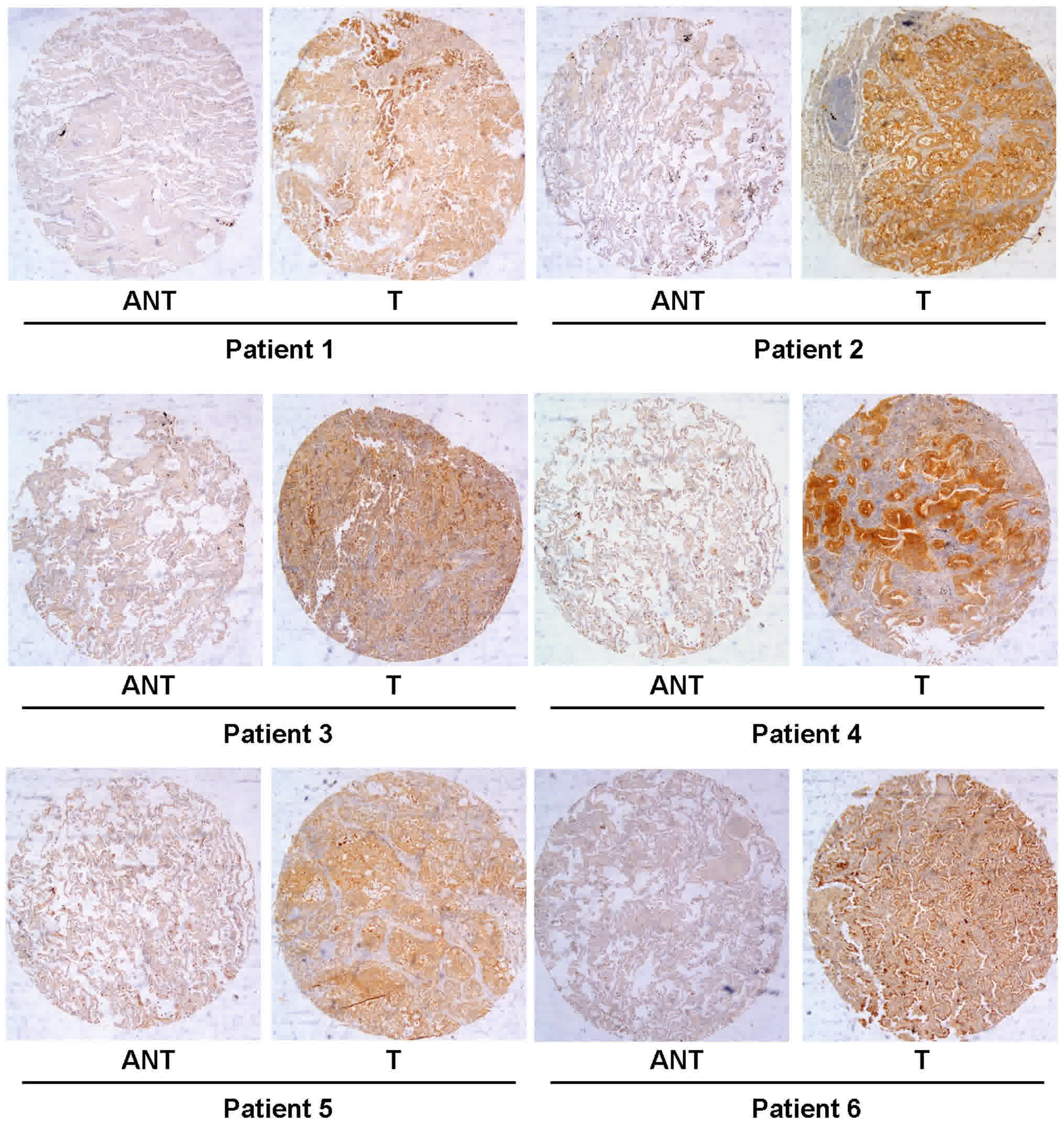
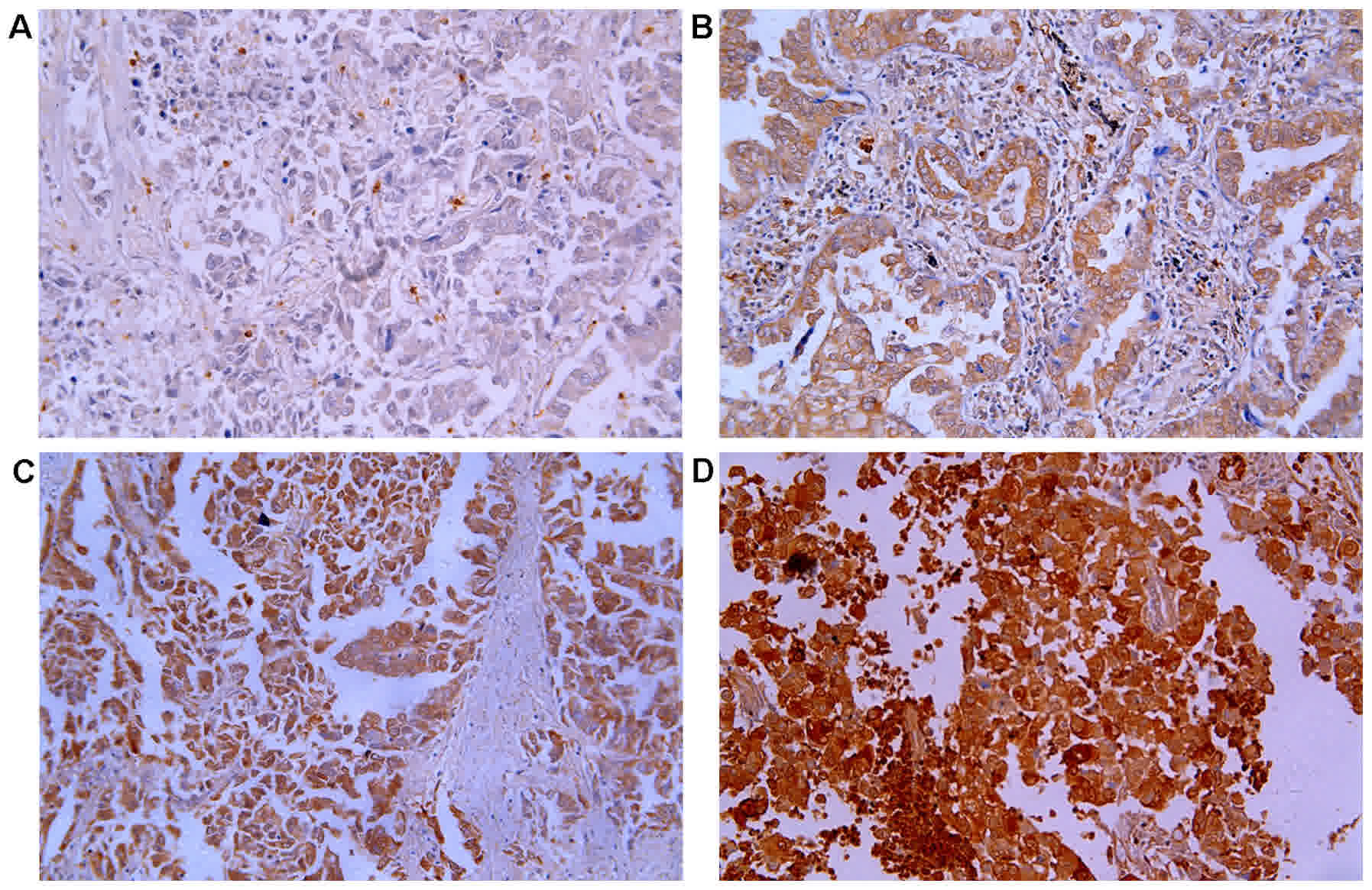
adjacent normal lung tissues (P=0.0017). Meanwhile, on IHC, the
positive expression of TARBP1 was detected in 69 out of 90 (76.67%)
of the NSCLC samples, while staining of TARBP1 protein was weak or
absent in the adjacent non-cancerous lung tissues, with a positive
expression detection rate of only 22.2% (20/90) (Figs. 3 and 4).
This difference in the rate of TARBP1 positive expression between
the NSCLC samples and non-cancerous lung tissues was statistically
significant (χ2=53.362, P<0.001).
TARBP1 expression correlates with
clinicopathological features in NSCLC
To better understand the potential functions of
TARBP1 in the development and progression of NSCLC, IHC staining
was used to investigate the expression status of TARBP1 in 90
paraffin-embedded archived NSCLC tissue samples (31 stage I tumors,
19 stage II tumors, 30 stage III and 10 stage IV tumors) (Table I). Among the 90 samples, the positive
expression of TARBP1 protein was observed in 69 cases (76.67%),
while no obvious signals were detected in the remaining 21 tumor
tissue samples (23.33%) (Table I). By
contrast, no staining or only weak staining were observed in the
adjacent normal tissue samples, and the positive expression of
TARBP1 was significantly higher in the NSCLC tissues (Figs. 3 and 4).
The predominant subcellular location of TARBP1 was the
cytoplasm.
We further investigated the relationship between
TARBP1 expression and clinicopathological indices of NSCLC
patients. As summarized in Table I,
there was no significant correlation between TARBP1 protein
expression and patient age, patient sex, lymphatic metastasis,
pathological type or tumor size. However, TARBP1 expression was
significantly associated with histological grade (P<0.001),
clinical stage (P=0.024), and pathological type (P<0.001).
Association between TARBP1 expression
and patient OS
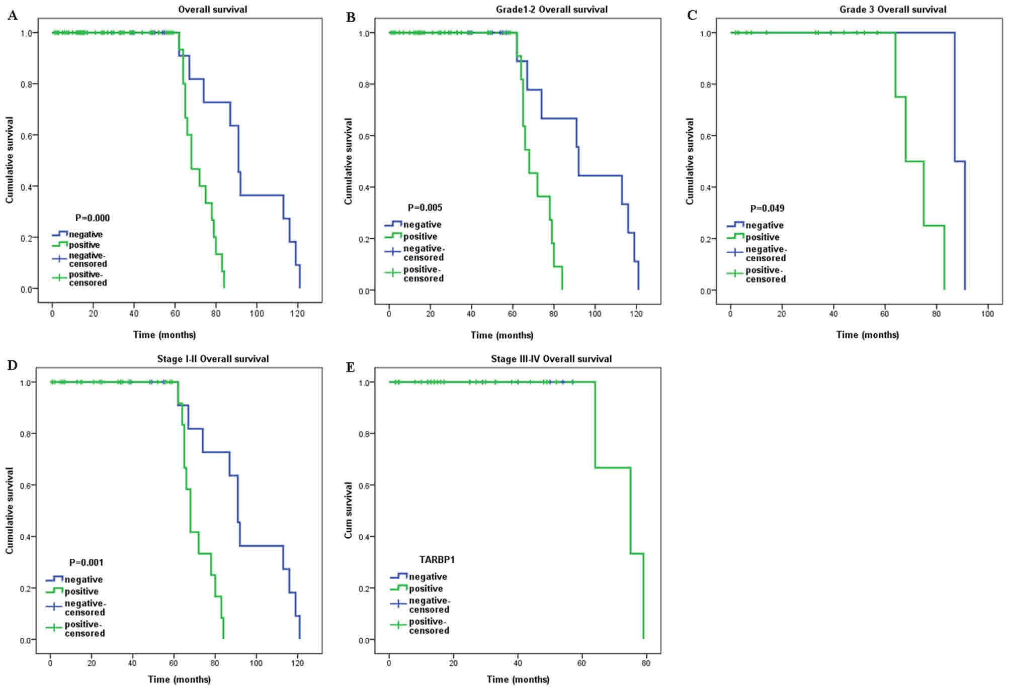
The survival analysis identified a significant
negative correlation between the expression level of TARBP1 protein
and the OS of the NSCLC patients (P<0.001) (Fig. 5A). Additionally, Cox regression
revealed that the expression of TARBP1 was an independent
prognostic factor for the OS of the NSCLC patients (Table II).
| Table II.Cox-regression analysis of various
prognostic parameters in patients for all patients. |
Table II.
Cox-regression analysis of various
prognostic parameters in patients for all patients.
|
| Univariate | Multivariate |
|---|
|
|
|
|
|---|
| Factor | HR (95% CI) | P-value | HR (95% CI) | P-value |
|---|
| Sex |
|
|
|
|
|
Male | Reference |
|
|
|
|
Female | 0.592
(0.261–1.344) | 0.210 | – | – |
| Age, years |
|
|
|
|
|
≥60 | Reference |
|
|
|
|
<60 | 0.835
(0.549–1.270) | 0.399 | – | – |
| Lymphatic
metastasis |
|
|
|
|
|
Positive | Reference |
|
|
|
|
Negative | 0.565
(0.197–1.619) | 0.288 | – | – |
| Tumor size, cm |
|
|
|
|
| ≥5 | Reference |
|
|
|
|
<5 | 4.514
(1.515–13.453) | 0.007 | – | – |
| Histological
grade |
|
|
|
|
| 1 | Reference |
|
|
|
| 2 | 0.879
(0.355–2.175) | 0.781 | – | – |
| 3 | 1.167
(0.404–3.370) | 0.775 | – | – |
| Clinical stage |
|
|
|
|
|
I–II | Reference |
|
|
|
|
III | 2.698
(0.925–7.866) | 0.069 | – | – |
| IV | 2.832
(0.729–11.001) | 0.133 | – | – |
| Pathological
type |
|
|
|
|
|
Adenocarcinoma | Reference |
|
|
|
|
Others | 1.034
(0.350–3.048) | 0.952 | – | – |
| TARBP1 |
|
|
|
|
|
Positive | Reference |
|
| Reference |
|
Negative | 0.133
(0.036–0.484) | 0.002 | 2.729
(1.471–5.061) | 0.003 |
We further investigated the prognostic value of
TARBP1 in selective patient subgroups classified by histological
grade and clinical stage, respectively. TARBP1 expression was
significantly associated with the OS of patients in the grade 1–2
subgroup (log-rank test, P=0.005) and the grade 3 subgroup
(log-rank test, P=0.049) (Fig. 5B and
C). However, when evaluated on the basis of clinical stage, the
influence of TARBP1 expression on patient outcome continued to be
significant only in the stage I–II subgroup (log-rank test,
P=0.001) (Fig. 5D), but not in the
stage III–IV subgroup (P>0.05) (Fig.
5E).
Discussion
Lung cancer is among the most prevalent malignant
tumors, with a high economic impact and rate of premature death
worldwide. In China, lung cancer is the most common type of cancer
and the leading cause of cancer-associated mortality in men
(26,27). In America, lung cancer was predicted
to be the second most common type of cancer in males and females,
as well as the leading cause of cancer-related mortality in
patients of either sex (3). Smoking
tobacco is associated with a large proportion of all lung cancer
cases (28,29). Over the last several decades,
developments in early diagnostic and treatment strategies have
considerably increased the average survival rate of cancer
patients, and some types of cancer are now considered to be
curable. However, lung cancer is typically diagnosed at a late age
(47% of patients were over 70-years-old at the time of diagnosis)
and late stage (50% of patients were diagnosed at advanced stage)
(30,31). Thus, the prognosis of lung cancer is
not optimistic. Due to the lack of apparent symptoms in early-stage
lung cancer, the majority of patients are diagnosed at an advanced
stage when the treatment options are limited (32–34). The
5-year estimated survival rates are ~6–14% in male patients and
7–18% in female patients (35). NSCLC
is the most common type of lung cancer (36,37). The
prognosis of NSCLC patients remains poor, and NSCLC accounts for
~85% of all lung cancer-associated mortalities (2). Therefore, there is a great need to
identify novel biomarkers and genetic risk factors of lung cancer,
in order to improve prognostic predictions and guide therapy.
The cellular dsRNA binding protein TARBP1 has
recently been shown to promote the replication of HIV-1 and −2
(38). Additionally, it has been
identified as a component of RISC, which serves as a Dicer cofactor
in the formation of 21–25 nucleotide mature miRNAs from
~70-nucleotide pre-miRNAs (10). In
accordance with the notion that viral infection of mammalian cells
may be restricted by miRNA regulation, the sequestration of TARBP1
by TAR RNA results in increased replication of HIV-1 in human cells
(39). Thus, TARBP1, which contains
two dsRNA-binding domains (RBD), is established as a multifarious
protein with two prominent roles (8):
First, it serves as a pre-requisite for the formation of
Dicer-containing complexes; and second, it gives assistance to
Dicer in generating miRNA (9,40).
Recently, truncating mutations of TARBP1 were found
in several human malignant cancers with microsatellite instability
(41). In Kaposi's sarcoma-associated
herpesvirus [KSHV, also known as human herpesvirus-8 (HHV-8)], the
expression of Dicer and TARBP1 is upregulated following KSHV
infection, which may promote an increase in the amount of the
miRNA-RISC loading complex available for both host and viral miRNAs
(42). In cardiomyopathy patients,
TARBP2 could regulate heart function through miRNA-mediated Sox6
repression (43). Meanwhile, Chen
et al reported that SUMOylation of TARBP2 may suppress the
progression of tumors (44).
Furthermore, knockdown of TARBP1 could reduce the accumulation of
hepatitis C virus RNA (45), and
TARBP1 has recently been proposed as a target for antiviral
therapies (38,46,47). The
function of TARBP1 may be regulated by phosphorylation via the
JNK-MAPK axis (48). TARBP1 has a
biological function in spermatogenesis and growth control during
development. It can also bind the interferon-induced
dsRNA-activated protein kinase (PKR) and with the PKR activator
(PACT), as well as with tumor suppressors (47). More recently, several diseases have
been associated with TARBP1, such as cardiac disease (43) and hepatocellular carcinoma (49), among others. In this study, we
investigated for the first time the expression of TARBP1 and its
associated clinical significance in NSCLC.
Our results clearly showed that TARBP1 was
upregulated at the mRNA level in 10 NSCLC tissue samples. In
addition, IHC staining identified a TARBP1 positive staining rate
of 76.67% (69/90) in tumor tissues, which was markedly higher than
that for adjacent normal tissues. Analogous observations have been
reported for other diseases, including HIV (47) and epithelial skin cancer (50). The differential expression of TARBP1
between tumor tissues and adjacent normal tissues indicates that
TARBP1 may be an important molecular biomarker in tumorigenesis,
which could aid to improve the precision of diagnoses. However, at
present, the exact roles of TARBP1 in malignant cancer remain
unclear, and thus further study is required to understand the
associated signaling pathways of TARBP1 in NSCLC. Our research did
not include the investigation of TARBP1 expression in tissue sample
of inflammatory lesions, because the patients with pneumonia
usually receive conservative treatment and we seldom do surgery or
biopsy for these kind of patients.
We further explored the relationship between TARBP1
expression and certain clinical indices of patients with NSCLC.
TARBP1 expression was found to be significantly correlated with
histological grade (P<0.001), clinical stage (P=0.024), and
pathological type (P<0.001). However, no associations were
identified between TARBP1 expression and patient age, patient sex,
lymphatic metastasis, pathological type or tumor size. Meanwhile,
Cox regression demonstrated that the expression of TARBP1 was an
independent prognostic factor in NSCLC patients after curative
resection. This finding indicates the potential of TARBP1
expression level as a predictor for patient prognosis and potential
therapeutic target. Interestingly, sub-group analysis implied that
TARBP1 positive expression was significantly associated with a poor
prognosis among well-differentiated and poorly-differentiated
cases, and among patients with early-stage disease. Nonetheless,
the molecular mechanism underlying the association of TARBP1 with
patient prognosis and survival remains obscure and yet to be
explored.
To conclude, to the best of our knowledge, this is
the first report on TARBP1 expression and its clinicopathological
and prognostic significance in NSCLC. Our findings indicate that
TARBP1 is upregulated and closely associated with histological
grade, clinical stage, and pathological type in NSCLC. Based on
these findings, we infer that TARBP1 is associated with aggressive
clinical features in NSCLC. Notably, multivariate analysis revealed
that TARBP1 might be an independent molecular biomarker for the
prediction of patient prognosis and survival. Combined with the
above findings, high expression of TARBP1 may cause worse prognosis
in NSCLC. Comprehensive evaluation of the expression of TARBP1 will
help the clinicians to identify patients with a worse prognosis,
paving a way of discovering effective strategies targeting TARBP1
in the treatment of NSCLC patients.
Acknowledgements
Not applicable.
Funding
The present study was supported by the Chinese
National Natural Science Foundation (no. 81672661), Guangdong
Province Natural Science Foundation (no. 2015A030310126) and
Guangzhou Medical and Health Technology Program (no.
20161A010077).
Availability of data and materials
The datasets used and/or analyzed during the current
study are available from the corresponding author on reasonable
request.
Authors' contributions
LX designed the experiments. JY and NZ carried out
the experiments. JW analyzed experimental results. YL and LT
collected the clinical samples, performed the statistical analysis
and wrote the manuscript.
Ethics approval and consent to
participate
This study was approved by the Ethics Committee of
the First Affiliated Hospital of Guangzhou Medical University, and
all patients provided written informed consent for the use of their
clinical samples in the present study.
Consent for publication
Not applicable.
Competing interests
The authors declare that they have no competing
interests.
References
|
1
|
Torre LA, Bray F, Siegel RL, Ferlay J,
Lortet-Tieulent J and Jemal A: Global cancer statistics, 2012. CA
Cancer J Clin. 65:87–108. 2015. View Article : Google Scholar : PubMed/NCBI
|
|
2
|
Goldstraw P, Crowley J, Chansky K, Giroux
DJ, Groome PA, Rami-Porta R, Postmus PE, Rusch V and Sobin L:
International Association for the Study of Lung Cancer
International Staging Committee; Participating Institutions: The
IASLC lung cancer staging project: Proposals for the revision of
the TNM stage groupings in the forthcoming (seventh) edition of the
TNM Classification of malignant tumours. J Thorac Oncol. 2:706–714.
2007. View Article : Google Scholar : PubMed/NCBI
|
|
3
|
Siegel RL, Miller KD and Jemal A: Cancer
statistics, 2017. CA Cancer J Clin. 67:7–30. 2017. View Article : Google Scholar : PubMed/NCBI
|
|
4
|
DeSantis CE, Lin CC, Mariotto AB, Siegel
RL, Stein KD, Kramer JL, Alteri R, Robbins AS and Jemal A: Cancer
treatment and survivorship statistics, 2014. CA Cancer J Clin.
64:252–271. 2014. View Article : Google Scholar : PubMed/NCBI
|
|
5
|
Travis WD: Pathology of lung cancer. Clin
Chest Med. 32:669–692. 2011. View Article : Google Scholar : PubMed/NCBI
|
|
6
|
Khan AJ and Dicker AP: On the merits and
limitations of whole-brain radiation therapy. J Clin Oncol.
31:11–13. 2013. View Article : Google Scholar : PubMed/NCBI
|
|
7
|
Kozak CA, Gatignol A, Graham K, Jeang KT
and McBride OW: Genetic mapping in human and mouse of the locus
encoding TRBP, a protein that binds the TAR region of the human
immunodeficiency virus (HIV-1). Genomics. 25:66–72. 1995.
View Article : Google Scholar : PubMed/NCBI
|
|
8
|
Daniels SM and Gatignol A: The multiple
functions of TRBP, at the hub of cell responses to viruses, stress
and cancer. Microbiol Mol Biol Rev. 76:652–666. 2012. View Article : Google Scholar : PubMed/NCBI
|
|
9
|
Takahashi T, Zenno S, Ishibashi O,
Takizawa T, Saigo K and Ui-Tei K: Interactions between the non-seed
region of siRNA and RNA-binding RLC/RISC proteins, Ago and TRBP, in
mammalian cells. Nucleic Acids Res. 42:5256–5269. 2014. View Article : Google Scholar : PubMed/NCBI
|
|
10
|
Chi YH, Semmes OJ and Jeang KT: A
proteomic study of TAR-RNA binding protein (TRBP)-associated
factors. Cell Bios. 1:92011. View Article : Google Scholar
|
|
11
|
Gagnon KT, Li L, Chu Y, Janowski BA and
Corey DR: RNAi factors are present and active in human cell nuclei.
Cell Rep. 6:211–221. 2014. View Article : Google Scholar : PubMed/NCBI
|
|
12
|
Rhodes DR, Kalyana-Sundaram S, Mahavisno
V, Varambally R, Yu J, Briggs BB, Barrette TR, Anstet MJ,
Kincead-Beal C, Kulkarni P, et al: Oncomine 3.0: Genes, pathways,
and networks in a collection of 18,000 cancer gene expression
profiles. Neoplasia. 9:166–180. 2007. View Article : Google Scholar : PubMed/NCBI
|
|
13
|
Gagnon KT, Li L, Chu Y, Janowski BA and
Corey DR: RNAi factors are present and active in human cell nuclei.
Cell Rep. 6:211–221. 2014. View Article : Google Scholar : PubMed/NCBI
|
|
14
|
Livak KJ and Schmittgen TD: Analysis of
relative gene expression data using real time quantitative PCR and
the 2(-Delta Delta C(T)) method. Methods. 25:402–408. 2001.
View Article : Google Scholar : PubMed/NCBI
|
|
15
|
Beer DG, Kardia SL, Huang CC, Giordano TJ,
Levin AM, Misek DE, Lin L, Chen G, Gharib TG, Thomas DG, et al:
Gene-expression profiles predict survival of patients with lung
adenocarcinoma. Nat Med. 8:816–824. 2002. View Article : Google Scholar : PubMed/NCBI
|
|
16
|
Bhattacharjee A, Richards WG, Staunton J,
Li C, Monti S, Vasa P, Ladd C, Beheshti J, Bueno R, Gillette M, et
al: Classification of human lung carcinomas by mRNA expression
profiling reveals distinct adenocarcinoma subclasses. Proc Natl
Acad Sci USA. 98:13790–13795. 2001. View Article : Google Scholar : PubMed/NCBI
|
|
17
|
Garber ME, Troyanskaya OG, Schluens K,
Petersen S, Thaesler Z, Pacyna-Gengelbach M, van de Rijn M, Rosen
GD, Perou CM, Whyte RI, et al: Diversity of gene expression in
adenocarcinoma of the lung. Proc Natl Acad Sci USA. 98:13784–13789.
2001. View Article : Google Scholar : PubMed/NCBI
|
|
18
|
Hou J, Aerts J, den Hamer B, van Ijcken W,
den Bakker M, Riegman P, van der Leest C, van der Spek P, Foekens
JA, Hoogsteden HC, et al: Gene expression-based classification of
non-small cell lung carcinomas and survival prediction. PLoS One.
5:e103122010. View Article : Google Scholar : PubMed/NCBI
|
|
19
|
Landi MT, Dracheva T, Rotunno M, Figueroa
JD, Liu H, Dasgupta A, Mann FE, Fukuoka J, Hames M, Bergen AW, et
al: Gene expression signature of cigarette smoking and its role in
lung adenocarcinoma development and survival. PLoS One.
3:e16512008. View Article : Google Scholar : PubMed/NCBI
|
|
20
|
Okayama H, Kohno T, Ishii Y, Shimada Y,
Shiraishi K, Iwakawa R, Furuta K, Tsuta K, Shibata T, Yamamoto S,
et al: Identification of genes upregulated in ALK-positive and
EGFR/KRAS/ALK-negative lung adenocarcinomas. Cancer Res.
72:100–111. 2012. View Article : Google Scholar : PubMed/NCBI
|
|
21
|
Selamat SA, Chung BS, Girard L, Zhang W,
Zhang Y, Campan M, Siegmund KD, Koss MN, Hagen JA, Lam WL, et al:
Genome-scale analysis of DNA methylation in lung adenocarcinoma and
integration with mRNA expression. Genome Res. 22:1197–1211. 2012.
View Article : Google Scholar : PubMed/NCBI
|
|
22
|
Stearman RS, Dwyer-Nield L, Zerbe L,
Blaine SA, Chan Z, Bunn PA Jr, Johnson GL, Hirsch FR, Merrick DT,
Franklin WA, et al: Analysis of orthologous gene expression between
human pulmonary adenocarcinoma and a carcinogen-induced murine
model. Am J Pathol. 167:1763–1775. 2005. View Article : Google Scholar : PubMed/NCBI
|
|
23
|
Su LJ, Chang CW, Wu YC, Chen KC, Lin CJ,
Liang SC, Lin CH, Whang-Peng J, Hsu SL, Chen CH and Huang CY:
Selection of DDX5 as a novel internal control for Q-RT-PCR from
microarray data using a block bootstrap re-sampling scheme. BMC
Genomics. 8:1402007. View Article : Google Scholar : PubMed/NCBI
|
|
24
|
Talbot SG, Estilo C, Maghami E, Sarkaria
IS, Pham DK, O-charoenrat P, Socci ND, Ngai I, Carlson D, Ghossein
R, et al: Gene expression profiling allows distinction between
primary and metastatic squamous cell carcinomas in the lung. Cancer
Res. 65:3063–3071. 2005. View Article : Google Scholar : PubMed/NCBI
|
|
25
|
Wachi S, Yoneda K and Wu R:
Interactome-transcriptome analysis reveals the high centrality of
genes differentially expressed in lung cancer tissues.
Bioinformatics. 21:4205–4208. 2005. View Article : Google Scholar : PubMed/NCBI
|
|
26
|
Chen W, Zheng R, Zeng H and Zhang S: The
updated incidences and mortalities of major cancers in China, 2011.
Chin J Cancer. 34:502–507. 2015. View Article : Google Scholar : PubMed/NCBI
|
|
27
|
Chen W, Zheng R, Zeng H and Zhang S: The
incidence and mortality of major cancers in China, 2012. Chin J
Cancer. 35:732016. View Article : Google Scholar : PubMed/NCBI
|
|
28
|
No authors listed: Lung cancer. Breathe
(Sheff). 12:392–399. 2016. View Article : Google Scholar : PubMed/NCBI
|
|
29
|
Torre LA, Siegel RL and Jemal A: Lung
cancer statistics. Adv Exp Med Biol. 893:1–19. 2016. View Article : Google Scholar : PubMed/NCBI
|
|
30
|
Owonikoko TK, Ragin CC, Belani CP, Oton
AB, Gooding WE, Taioli E and Ramalingam SS: Lung cancer in elderly
patients: An analysis of the surveillance, epidemiology, and end
results database. J Clin Oncol. 25:5570–5577. 2007. View Article : Google Scholar : PubMed/NCBI
|
|
31
|
Ramalingam SS, Owonikoko TK and Khuri FR:
Lung cancer: New biological insights and recent therapeutic
advances. CA Cancer J Clin. 61:91–112. 2011. View Article : Google Scholar : PubMed/NCBI
|
|
32
|
Pirozynski M: 100 years of lung cancer.
Respir Med. 100:2073–2084. 2006. View Article : Google Scholar : PubMed/NCBI
|
|
33
|
Schwartz AG, Prysak GM, Bock CH and Cote
ML: The molecular epidemiology of lung cancer. Carcinogenesis.
28:507–518. 2007. View Article : Google Scholar : PubMed/NCBI
|
|
34
|
Spiro SG and Silvestri GA: One hundred
years of lung cancer. Am J Respir Crit Care Med. 172:523–529. 2005.
View Article : Google Scholar : PubMed/NCBI
|
|
35
|
Marshall AL and Christiani DC: Genetic
susceptibility to lung cancer-light at the end of the tunnel?
Carcinogenesis. 34:487–502. 2013. View Article : Google Scholar : PubMed/NCBI
|
|
36
|
Goldstraw P, Ball D, Jett JR, Le Chevalier
T, Lim E, Nicholson AG and Shepherd FA: Non-small-cell lung cancer.
Lancet. 378:1727–1740. 2011. View Article : Google Scholar : PubMed/NCBI
|
|
37
|
Siegel R, Naishadham D and Jemal A: Cancer
statistics, 2013. CA Cancer J Clin. 63:11–30. 2013. View Article : Google Scholar : PubMed/NCBI
|
|
38
|
Christensen HS, Daher A, Soye KJ, Frankel
LB, Alexander MR, Lainé S, Bannwarth S, Ong CL, Chung SW, Campbell
SM, et al: Small interfering RNAs against the TAR RNA binding
protein, TRBP, a Dicer cofactor, inhibit human immunodeficiency
virus type 1 long terminal repeat expression and viral production.
J Virol. 81:5121–5131. 2007. View Article : Google Scholar : PubMed/NCBI
|
|
39
|
Bennasser Y, Yeung ML and Jeang KT: HIV-1
TAR RNA subverts RNA interference in transfected cells through
sequestration of TAR RNA-binding protein, TRBP. J Biol Chem.
281:27674–27678. 2006. View Article : Google Scholar : PubMed/NCBI
|
|
40
|
Redfern AD, Colley SM, Beveridge DJ, Ikeda
N, Epis MR, Li X, Foulds CE, Stuart LM, Barker A, et al:
RNA-induced silencing complex (RISC) Proteins PACT, TRBP, and Dicer
are SRA binding nuclear receptor coregulators. Proc Natl Acad Sci
USA. 110:6536–6541. 2013. View Article : Google Scholar : PubMed/NCBI
|
|
41
|
Melo SA, Ropero S, Moutinho C, Aaltonen
LA, Yamamoto H, Calin GA, Rossi S, Fernandez AF, Carneiro F,
Oliveira C, et al: A TARBP2 mutation in human cancer impairs
microRNA processing and DICER1 function. Nat Genet. 41:365–370.
2009. View
Article : Google Scholar : PubMed/NCBI
|
|
42
|
Happel C, Ramalingam D and Ziegelbauer JM:
Virus-mediated alterations in miRNA factors and degradation of
viral miRNAs by MCPIP1. PLoS Biol. 14:e20009982016. View Article : Google Scholar : PubMed/NCBI
|
|
43
|
Ding J, Chen J, Wang Y, Kataoka M, Ma L,
Zhou P, Hu X, Lin Z, Nie M, Deng ZL, et al: Trbp regulates heart
function through microRNA-mediated Sox6 repression. Nat Genet.
47:776–783. 2015. View Article : Google Scholar : PubMed/NCBI
|
|
44
|
Chen C, Zhu C, Huang J, Zhao X, Deng R,
Zhang H, Dou J, Chen Q, Xu M, Yuan H, et al: SUMOylation of TARBP2
regulates miRNA/siRNA efficiency. Nat Commun. 6:88992015.
View Article : Google Scholar : PubMed/NCBI
|
|
45
|
Zhang C, Huys A, Thibault PA and Wilson
JA: Requirements for human Dicer and TRBP in microRNA-122
regulation of HCV translation and RNA abundance. Virology.
433:479–488. 2012. View Article : Google Scholar : PubMed/NCBI
|
|
46
|
Eekels JJ, Geerts D, Jeeninga RE and
Berkhout B: Long-term inhibition of HIV-1 replication with RNA
interference against cellular co-factors. Antiviral Res. 89:43–53.
2011. View Article : Google Scholar : PubMed/NCBI
|
|
47
|
Sanghvi VR and Steel LF: The cellular TAR
RNA binding protein, TRBP, promotes HIV-1 replication primarily by
inhibiting the activation of double-stranded RNA-dependent kinase
PKR. J Virol. 85:12614–12621. 2011. View Article : Google Scholar : PubMed/NCBI
|
|
48
|
Nakamura T, Kunz RC, Zhang C, Kimura T,
Yuan CL, Baccaro B, Namiki Y, Gygi SP and Hotamisligil GS: A
critical role for PKR complexes with TRBP in Immunometabolic
regulation and eIF2α phosphorylation in obesity. Cell Rep.
11:295–307. 2015. View Article : Google Scholar : PubMed/NCBI
|
|
49
|
Ye J, Wang J, Tan L, Yang S, Xu L, Wu X,
Deng H and Tan H: Expression of protein TARBP1 in human
hepatocellular carcinoma and its prognostic significance. Int J
Clin Exp Pathol. 8:9089–9096. 2015.PubMed/NCBI
|
|
50
|
Sand M, Skrygan M, Georgas D, Arenz C,
Gambichler T, Sand D, Altmeyer P and Bechara FG: Expression levels
of the microRNA maturing microprocessor complex component DGCR8 and
the RNA-induced silencing complex (RISC) components argonaute-1,
argonaute-2, PACT, TARBP1, and TARBP2 in epithelial skin cancer.
Mol Carcinog. 51:916–922. 2012. View Article : Google Scholar : PubMed/NCBI
|