Introduction
Glioma is the most common malignant brain cancer in
adults. In patients with grade IV glioma according to the World
Health Organization (WHO) guidelines (1) their condition is similar to glioblastoma
(GBM). At present, the standard therapy is surgical excision
accompanied by chemotherapy and radiotherapy (1). Even in patients who actively cooperate
with treatment, the median overall survival (OS) time of patients
who suffer from GBM is <15 months (2), and drug resistance is partially
accountable for the poor prognostic outcome of GBM.
The epithelial-mesenchymal transition (EMT) process
serves an important function in tumor invasion (3), metastasis and drug resistance in a
number of types of cancer, including lung cancer (4) and pancreatic carcinoma (5). Conversely, the role of EMT in
gliomagenesis remains vague; however, several EMT-associated
factors, including Twist (6), zinc
finger E-box-binding homeobox (ZEB)1 (7), ZEB2, and the SNAI family, have been
confirmed to accelerate the invasion, progression and drug
resistance of glioma (8,9). Cadherin 2 (CDH2), which encodes the
N-cadherin protein, is also a marker of EMT. An increasing amount
of evidence has suggested that CDH2 has a close association with
the WHO grade of glioma (10). By
contrast, a previous study demonstrated that GBMs express lower
CDH2 levels than low-grade gliomas (11). Therefore, the association between CDH2
and glioma malignancy requires further study.
In the present study, a detailed and systematic
analysis was performed using the The Cancer Genome Atlas (TCGA),
Chinese Glioma Genome Atlas (CGGA) and Rembrandt databases, and
identified that CDH2 expression was associated with glioma grade
and may serve as a prognostic indicator for OS in patients with
glioma. In addition, in patients with GBM expressing low levels of
CDH2, temozolomide (TMZ) therapy had an improved curative effect,
among other independent prognostic factors. The results of the
present study demonstrated the prognostic and predictive value of
CDH2 for glioma patients and suggests that CDH2 levels could be
used to identify which patients are likely to benefit from TMZ
therapy in the clinical setting.
Materials and methods
Clinical samples
Clinical characteristics and CDH2 mRNA expression
data of 301 glioma specimens were obtained from the microarray data
stored in the Chinese Glioma Genome Atlas (CGGA; http://www.cgga.org.cn). The histological diagnoses
were determined according to the WHO criteria (12). Publicly available Rembrandt microarray
data were obtained online (https://wiki.nci.nih.gov/display/ICR/Rembrandt+Data+Portal)
on May 8, 2014. Any patients lost to follow-up were not included in
the survival analysis. TCGA dataset, which consists of RNA-seq
data, was downloaded from the website (https://cancergenome.nih.gov/). Nine clinical glioma
samples (fresh-frozen) were selected according to WHO grade
classification (1) and age (≤45) were
obtained from the Department of Neurosurgery of the Second
Affiliated Hospital of Harbin Medical University (Harbin, China,
Table I). All patients provided
written informed consent, and all human experiments were approved
by the Ethics Committee of the Second Affiliated Hospital of Harbin
Medical University.
 | Table I.The corresponding clinical and
pathological information of nine patients. |
Table I.
The corresponding clinical and
pathological information of nine patients.
|
| Grade | Histology | Age (years) | Sex |
|---|
| Patient 1 | II | Astrocytoma | 40 | Female |
| Patient 2 | II |
Oligodendroglioma | 30 | Female |
| Patient 3 | II | Astrocytoma | 42 | Male |
| Patient 4 | III | Anaplastic
oligodendroglioma | 38 | Male |
| Patient 5 | III | Anaplastic
oligodendroglioma | 41 | Female |
| Patient 6 | III | Anaplastic
oligodendroglioma | 44 | Female |
| Patient 7 | IV | Glioblastoma | 45 | Female |
| Patient 8 | IV | Glioblastoma | 45 | Male |
| Patient 9 | IV | Glioblastoma | 39 | Female |
Reverse transcription-polymerase chain
reaction (RT-PCR)
Total RNA was extracted from patient samples using
TRIzol® reagent (Life Technologies; Thermo Fisher
Scientific, Inc., Waltham, MA, USA). Then cDNAs were synthesized
using the PrimeScript RT Reagent kit (Promega Corporation, Madison,
WI, USA) according to the manufacturer's protocol. The following
primers (Beijing Tianyi Huiyuan Bioscience & Technology
Inc., Beijing, China) were used: CDH2 forward,
5′-ACCTTTGCCAGGAGCTGTTT-3′; CDH2 reverse,
5′-TGTGCTCCCTATGACCCAGA-3′; GAPDH forward,
5′-AGAAGGCTGGGGCTCATTTG-3′; and GAPDH reverse,
5′-AGGGGCCATCCACAGTCTTC-3′ were used for PCR. Following
amplification (denaturation 95°C for 10 secs, annealing 53°C for 10
secs and elongation 72°C for 60 secs, 40 cycles) of the PCR
product, 1% agarose gel electrophoresis (Beijing Solarbio Science
& Technology Co., Ltd., Beijing, China), DNA ladder (Beijing
Solarbio Science & Technology Co., Ltd.) and ethidium bromide
(Beijing Solarbio Science & Technology Co., Ltd.) were used to
assess the amount of CDH2. All PCR experiments were conducted in
triplicate.
Statistical analysis
Differences in OS and progression-free survival
(PFS) were evaluated using the Kaplan-Meier method and analyzed
using the log-rank test in the univariate analysis. Student's
t-test was used to examine the differences between two groups.
Multigroup comparisons of the means were carried out using a
one-way analysis of variance test with post hoc contrasts performed
using the Student-Newman-Keuls test. A χ2 test was used
to evaluate the distribution of patient characteristics between
subgroups. Cox proportional hazards regression analysis was used to
assess the prognostic value of CDH2 expression among other factors.
All statistical calculations were performed with SPSS 22.0 (IBM
Corp., Armonk, NY, USA) and GraphPad Prism version 6.01 (GraphPad
Software, Inc., La Jolla, CA, USA). P<0.05 was considered to
indicate a statistically significant difference.
Results
CDH2 is associated with WHO grade and
the prognosis of glioma patients
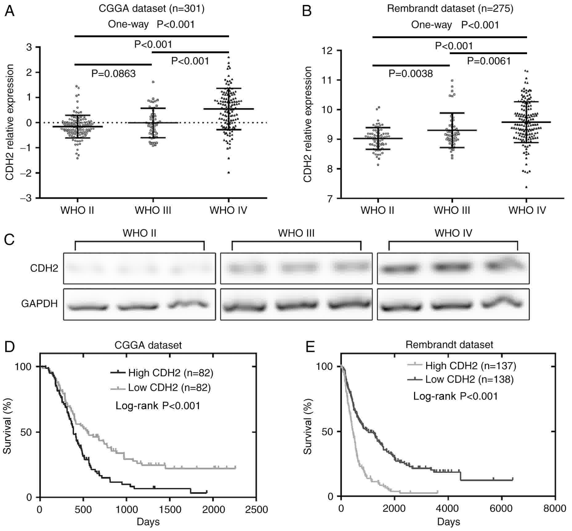
The expression of CDH2 was detected in 301 glioma
samples in the CGGA dataset (grade II, n=122; grade III, n=51;
grade IV, n=128). CDH2 expression was significantly higher in
gliomas of grade IV than in those of grades II (P<0.001) or III
(P<0.001) (Fig. 1A). However,
glioma of grade III presented no significant difference compared
with grade II (P=0.0863). Furthermore, the public dataset Rembrandt
was used to further confirm these findings. The results
demonstrated that CDH2 expression had an evident association with
the WHO grade of glioma (P<0.001; Fig.
1B). In the Rembrandt dataset, CDH2 expression in grade III
glioma was higher than that in grade II (P=0.0038). Furthermore, in
the clinical glioma tissues obtained from our hospital (n=9), the
mRNA level of CDH2 was demonstrated to be higher in grade IV than
in grade II and III glioma samples (Fig.
1C).
High expression of CDH2 confers an
unfavorable prognosis in glioma patients
The median CDH2 expression level in 164 patients
with high-grade glioma (WHO III and IV) from the CGGA data set was
used as the cut-off point to divide the patients into low CDH2
(n=82) and high CDH2 (n=82) expression groups. Kaplan-Meier
survival curves and the log-rank test were employed to identify any
associations between CDH2 expression and OS. Patients in the low
CDH2 expression group lived longer compared with those in the high
expression group (P<0.001) (Fig.
1D). The Rembrandt dataset was also analyzed for confirmation
of these findings, and the results demonstrated that the group with
a high expression of CDH2 had a significantly worse outcome
(P<0.001) (Fig. 1E). This data
demonstrated that high expression of CHD2 may be indicative of an
unfavorable survival outcome.
CDH2 is an independent prognostic
factor in patients with high-grade glioma
The clinicopathological information of 164 patients
with high-grade glioma in the CGGA dataset was investigated, and
revealed that CDH2 expression was associated with age at diagnosis
(P=0.003), isocitrate dehydrogenase 1 (IDH1) mutation status
(P=0.0136) and TCGA subtype (P=0.0326) (Table II). Univariate Cox regression
analysis was conducted to analyze the genetic and clinical
variables with respect to survival. OS was identified to be
associated with IDH1 mutation status, CDH2 expression level and
whether the patient had received chemotherapy. Subsequently,
potential prognostic factors associated with OS were evaluated
through a multivariate Cox regression model. The results
demonstrated that CDH2 expression was an independent prognostic
factor for OS [hazard ratio (HR), 1.746; 95% confidence interval
(CI), 1.211–2.518; P=0.003], following adjustment for IDH1 and
chemotherapy status (Table III).
The same statistical approach was also conducted for 275 glioma
samples in the Rembrandt dataset. The results demonstrated that
CDH2 expression remained an independent factor for predicting OS
following adjustment for sex and WHO grade (HR, 1.397; 95% CI,
1.102–1.770; P=0.006) (Tables IV and
V).
| Table II.Clinical and pathological
characteristics of 164 patients with high-grade glioma in
association with CDH2 expression. |
Table II.
Clinical and pathological
characteristics of 164 patients with high-grade glioma in
association with CDH2 expression.
|
| CDH2 expression |
|
|---|
|
|
|
|
|---|
| Variable | Low (n=82) | High (n=82) | P-value |
|---|
| Age, years |
|
| 0.0030 |
|
<45 | 52 | 33 |
|
| ≥45 | 30 | 49 |
|
| IDH1 status |
|
| 0.0136 |
|
Mutant | 15 | 50 |
|
| Not
mutant | 67 | 32 |
|
| Sex |
|
| 0.1596 |
|
Male | 50 | 49 |
|
|
Female | 32 | 33 |
|
| Chemotherapy |
|
| 0.2678 |
|
Yes | 49 | 51 |
|
| No | 27 | 31 |
|
| NA | 6 | 0 |
|
| Radiotherapy |
|
| 0.0661 |
|
Yes | 64 | 65 |
|
| No | 11 | 16 |
|
| NA | 7 | 1 |
|
| TCGA subtype |
|
| 0.0326 |
|
Neural | 9 | 20 |
|
|
Proneural | 12 | 13 |
|
|
Mesenchymal | 50 | 40 |
|
|
Classical | 11 | 9 |
|
| WHO grade |
|
| 0.1046 |
|
III | 16 | 25 |
|
| IV | 66 | 57 |
|
| Table III.Univariate and multivariate Cox
regression analyses for overall survival in 164 glioma samples of
the Chinese Glioma Genome Atlas dataset. |
Table III.
Univariate and multivariate Cox
regression analyses for overall survival in 164 glioma samples of
the Chinese Glioma Genome Atlas dataset.
|
| Univariate | Multivariate |
|---|
|
|
|
|
|---|
| Variables | HR | 95% CI | P-value | HR | 95% CI | P-value |
|---|
| Age | 1.590 | 1.116–2.265 | 0.0100 | 0.934 | 0.619–1.411 | 0.7460 |
| IDH1 status | 0.471 | 0.307–0.722 | 0.0010 | 0.566 | 0.350–0.916 | 0.0210 |
| Sex | 0.835 | 0.589–1.183 | 0.3100 | – | – | – |
| Chemotherapy | 0.640 | 0.457–0.897 | 0.0100 | 0.641 | 0.463–0.887 | 0.0070 |
| Radiotherapy | 0.774 | 0.518–1.156 | 0.2110 | – | – | – |
| TCGA subtype | 1.121 | 0.910–1.382 | 0.2830 | – | – | – |
| WHO grade | 1.872 | 1.226–2.858 | 0.0040 | 1.418 | 0.884–2.274 | 0.1470 |
| CDH2
expression | 1.910 | 1.342–2.719 | <0.0010 | 1.746 | 1.211–2.518 | 0.0030 |
| Table IV.Clinical and pathological
characteristics of 275 glioma samples in association with CDH2
expression in the Rembrandt dataset. |
Table IV.
Clinical and pathological
characteristics of 275 glioma samples in association with CDH2
expression in the Rembrandt dataset.
|
| CDH2
expression |
|
|---|
|
|
|
|
|---|
| Variables | Low (n=138) | High (n=137) | P-value |
|---|
| Sex |
|
| <0.001 |
|
Male | 47 | 34 |
|
|
Female | 80 | 67 |
|
| NA | 11 | 36 |
|
| TCGA subtype |
|
| <0.001 |
|
Neural | 30 | 5 |
|
|
Proneural | 37 | 31 |
|
|
Mesenchymal | 65 | 53 |
|
|
Classical | 6 | 48 |
|
| WHO grade |
|
| <0.001 |
| II | 53 | 14 |
|
|
III | 33 | 19 |
|
| IV | 52 | 104 |
|
| Table V.Univariate and multivariate Cox
regression analyses for overall survival in the 275 glioma
specimens of the Rembrandt dataset. |
Table V.
Univariate and multivariate Cox
regression analyses for overall survival in the 275 glioma
specimens of the Rembrandt dataset.
|
| Univariate | Multivariate |
|---|
|
|
|
|
|---|
| Variable | HR | CI | P-value | HR | CI | P-value |
|---|
| Sex | 1.673 | 1.461–1.916 | <0.001 | 1.620 | 1.415–1.856 | <0.001 |
| TCGA subtype | 1.592 | 1.374–1.844 | <0.001 | 1.027 | 0.862–1.223 | 0.768 |
| WHO grade | 1.818 | 1.525–2.167 | <0.001 | 1.297 | 1.045–1.610 | 0.018 |
| CDH2
expression | 2.034 | 1.654–2.503 | <0.001 | 1.397 | 1.102–1.770 | 0.006 |
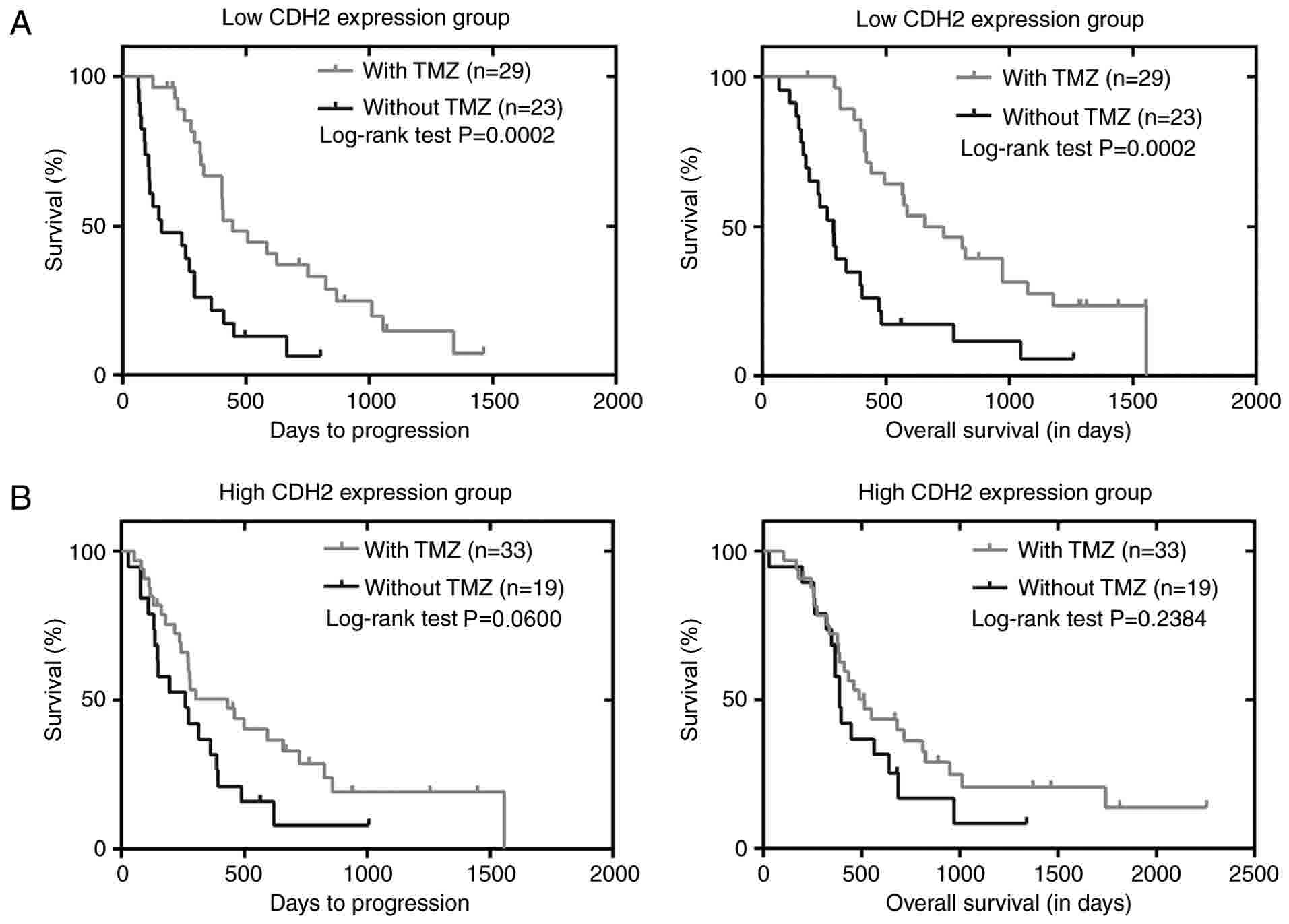
Association between CDH2 expression
and sensitivity to chemotherapy
To investigate the association between CDH2 level
and the sensitivity to chemotherapy, a primary GBM group was
enrolled from the CGGA dataset. They were divided into subgroups
depending on the median level of CDH2 and whether the patients
received TMZ chemotherapy. Kaplan-Meier survival analysis
demonstrated that, in patients with low CDH2 expression, TMZ
treatment was associated with improved OS and PFS compared with
patients not treated with TMZ (P=0.0002 and P=0.0002, respectively)
(Fig. 2A). However, no evident
survival benefit of TMZ therapy was identified for patients with
high CDH2 expression (OS, P=0.2384; PFS, P=0.0600) (Fig. 2B), indicating that low CDH2 expression
predicted a better response to TMZ. The results were also
corroborated by a Cox regression analysis (Table VI) which indicated that patients
benefited from TMZ with low expression of CDH2 after adjusting for
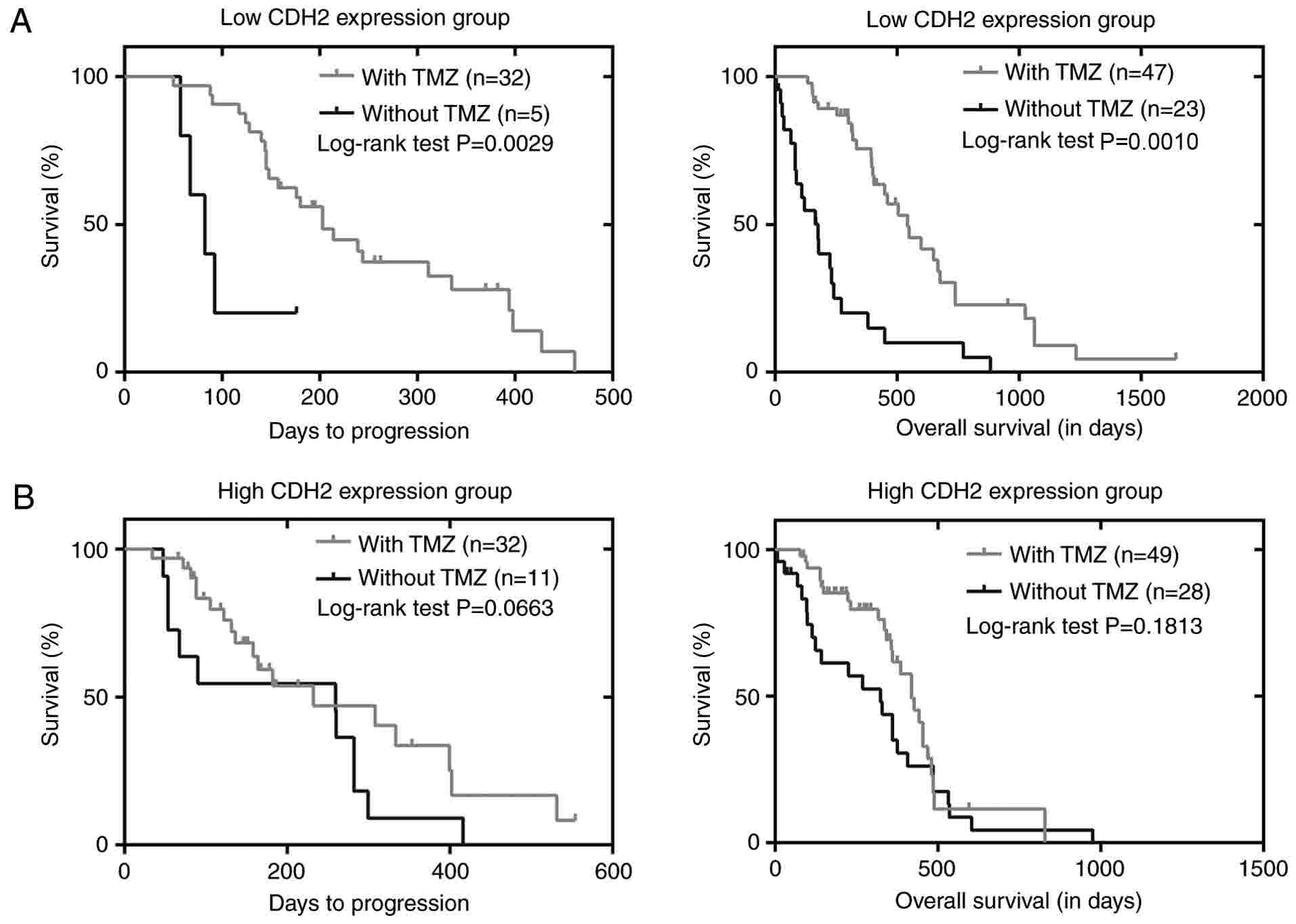
age, IDH1 status, sex and radiotherapy. Furthermore, TCGA dataset
was analyzed, as described above, which identified that patients
with low CDH2 expression and who were treated by TMZ therapy also
had better OS and PFS than patients treated without TMZ in the low
CDH2 expression group (P=0.0010 and P=0.0029, respectively)
(Fig. 3A). However, no evident
survival benefit of chemotherapy for patients with high CDH2
expression was identified (OS, P=0.1813; PFS, P=0.0663) (Fig. 3B). Cox regression analysis confirmed
these results (Table VII) which
further revealed that patients with low expression of CDH2 may
benefit from TMZ.
| Table VI.Univariate and multivariate Cox
regression analyses of overall and progression-free survival for
the low CDH2 expression group of the Chinese Glioma Genome Atlas
dataset. |
Table VI.
Univariate and multivariate Cox
regression analyses of overall and progression-free survival for
the low CDH2 expression group of the Chinese Glioma Genome Atlas
dataset.
| A, Overall
survival |
|---|
|
|---|
|
| Univariate | Multivariate |
|---|
|
|
|
|
|---|
| Variable | HR | 95% CI | P-value | HR | 95% CI | P-value |
|---|
| Age | 1.463 | 0.796–2.691 | 0.221 | 1.11 | 0.582–2.115 | 0.752 |
| IDH1 status | 0.429 | 0.196–0.943 | 0.035 | 0.312 | 0.133–0.737 | 0.008 |
| Sex | 0.943 | 0.521–1.707 | 0.846 | 1.17 | 0.629–2.178 | 0.620 |
| Chemotherapy | 0.522 | 0.284–0.958 | 0.036 | 0.322 | 0.161–0.643 | 0.001 |
| Radiotherapy | 1.273 | 0.500–3.241 | 0.613 | 3.622 | 1.051–12.490 | 0.042 |
|
| B,
Progression-free survival |
|
|
|
Univariate |
Multivariate |
|
|
|
|
|
Variable | HR | CI | P-value | HR | CI | P-value |
|
| Age | 1.246 | 0.687–2.259 | 0.211 | 1.026 | 0.546–1.927 | 0.936 |
| IDH1 status | 0.622 | 0.298–1.299 | 0.206 | 0.459 | 0.206–1.026 | 0.058 |
| Sex | 0.925 | 0.514–1.665 | 0.795 | 1.193 | 0.643–2.213 | 0.576 |
| Chemotherapy | 0.519 | 0.283–0.954 | 0.035 | 0.338 | 0.169–0.676 | 0.002 |
| Radiotherapy | 1.138 | 0.500–2.588 | 0.759 | 3.011 | 0.958–9.458 | 0.059 |
| Table VII.Univariate and multivariate Cox
regression analyses of overall and progression-free survival for
the low CDH2 group of The Cancer Genome Atlas dataset. |
Table VII.
Univariate and multivariate Cox
regression analyses of overall and progression-free survival for
the low CDH2 group of The Cancer Genome Atlas dataset.
| A, Overall
survival |
|---|
|
|---|
|
| Univariate | Multivariate |
|---|
|
|
|
|
|---|
| Variable | HR | CI | P-value | HR | CI | P-value |
|---|
| Age | 2.022 | 1.153–3.546 | 0.014 | 1.845 | 1.030–3.305 | 0.040 |
| Sex | 0.529 | 0.292–0.958 | 0.036 | 0.905 | 0.449–1.824 | 0.779 |
| Chemotherapy | 0.343 | 0.198–0.592 | <0.001 | 0.495 | 0.249–0.983 | 0.045 |
| Radiotherapy | 0.230 | 0.121–0.439 | <0.001 | 0.292 | 0.131–0.650 | 0.003 |
|
| B,
Progression-free survival |
|
|
|
Univariate |
Multivariate |
|
|
|
|
|
Variable | HR | CI | P-value | HR | CI | P-value |
|
| Age | 1.880 | 0.877–4.030 | 0.104 | 1.928 | 0.799–4.654 | 0.144 |
| Sex | 0.907 | 0.372–2.210 | 0.829 | 0.640 | 0.247–1.653 | 0.356 |
| Chemotherapy | 0.204 | 0.065–0.641 | 0.006 | 0.240 | 0.072–0.802 | 0.020 |
| Radiotherapy | 0.488 | 0.123–1.937 | 0.308 | 0.321 | 0.068–1.518 | 0.152 |
Discussion
Glioma is the most common intracranial malignant
tumor in adults. GBM is characterized by its high invasive ability,
self-renewal capability and drug resistance. Therefore, the 5-year
survival rate of patients with GBM is poor (13). Patients treated with TMZ and
radiotherapy have a favorable median survival time of 18.8 months
compared with those treated with radiotherapy alone (14.4 months)
after complete resection of GBM. Prior to the development of novel
targeted drugs for clinical glioma treatment, TMZ was considered to
be the most effective chemotherapeutic agent. However, although it
has significant effect in prolonging the lifespan of some patients
with glioma, the efficacy of TMZ for treating certain GBM patients
is limited (14), and TMZ resistance
may result in a poor prognostic outcome in patients with GBM.
Several mechanisms, including DNA repair mechanisms (15), high expression of epidermal growth
factor receptor (16), the mutation
of p53 (17) and the deficiency of
phosphatase and tensin homolog (18),
are involved in TMZ resistance. However, in a previous study,
one-third of patients exhibited hypermethylation of
methylguanine-DNA methyltransferase promoter, signifying
sensitivity toward alkylating agents (19). Effective molecular biomarkers for
glioma prognosis must be identified in order to provide a guide for
clinical treatment.
EMT-associated molecules have been reported to serve
an important role in glioma progression. Cells expressing low
levels of ZEB1 demonstrated an increased sensitivity to TMZ in GBM
(20). Our previous study
demonstrated that GBM patients with low vimentin expression had
improved survival rates when treated with TMZ (21). N-cadherin (encoded by the CDH2 gene)
is a 99.7-kDa glycoprotein and is widely distributed throughout the
central nervous system in neuronal and glial cells (22). N-cadherin appears to be upregulated
and downregulated according to the requirements of cells and
developing tissues (23). Comparable
to vimentin and matrix metallopeptidase 9, N-cadherin is
accompanied by the downregulation of epithelial cell-surface
markers, such as CDH1 (E-cadherin) (24). N-cadherin is broadly expressed in a
number of tumor types (25),
including neuroblastoma (26),
melanoma (27) and multiple myeloma
(28). Consequently, we hypothesized
that N-cadherin had a potential value to guide the clinical
application of chemotherapy.
In the present study, the level of CDH2 was
identified to be associated with glioma grade and outcome in the
CGGA and Rembrandt datasets. Patients with high-grade glioma had
high CDH2 expression compared with patients with low-grade glioma,
and patients with high CDH2 expression exhibited a worse outcome.
Statistical analysis revealed that CDH2 was an independent
prognostic factor in glioma. These results suggested that CDH2 may
serve a vital role in the molecular and pathological classification
of gliomas and may become a predictive indicator for glioma
treatment. Furthermore, patients with GBM expressing a lower level
of CDH2 may benefit to a greater extent from TMZ therapy.
In conclusion, the present study demonstrated that
CDH2 expression is significantly associated with glioma grade, and
that high CDH2 expression is an unfavorable prognostic factor for
patients with glioma and may have an important value for glioma
patients receiving TMZ. These results suggest that CDH2 may serve
as a prognostic and predictive molecular biomarker for the grading
and treatment of glioma.
Acknowledgements
Not applicable.
Funding
This study was funded by the Research Project of the
Chinese Society of Neuro-oncology, CACA (CSNO-2016-MSD12); the
Research Project of the Health and Family Planning Commission of
Heilongjiang Province (2017–201); and the Harbin Medical University
Scientific Research Innovation Fund (2017LCZX37).
Availability of data and materials
All data generated or analyzed during this study are
included in this published article.
Authors' contributions
QC performed development of methodology, acquisition
of data, analysis and interpretation of data and writing, review
and revision of the manuscript, and administrative, technical and
material support (i.e., reporting or organizing data, constructing
databases). JC and CJ performed the conception and design of the
study, along with study supervision.
Ethics approval and consent to
participate
All patients provided written informed consent, and
all human experiments were approved by the Ethics Committee of the
Second Affiliated Hospital of Harbin Medical University.
Consent for publication
Consent to publish has been obtained from all
participants.
Competing interests
The authors declare that they have no competing
interests.
Glossary
Abbreviations
Abbreviations:
|
CGGA
|
Chinese Glioma Genome Atlas
|
|
TCGA
|
The Cancer Genome Atlas
|
|
TMZ
|
temozolomide
|
References
|
1
|
Louis DN, Ohgaki H, Wiestler OD, Cavenee
WK, Burger PC, Jouvet A, Scheithauer BW and Kleihues P: The 2007
WHO classification of tumours of the central nervous system. Acta
Neuropathol. 114:97–109. 2007. View Article : Google Scholar : PubMed/NCBI
|
|
2
|
Jiang T, Mao Y, Ma W, Mao Q, You Y, Yang
X, Jiang C, Kang C, Li X, Chen L, et al: CGCG clinical practice
guidelines for the management of adult diffuse gliomas. Cancer
Lett. 375:263–273. 2016. View Article : Google Scholar : PubMed/NCBI
|
|
3
|
Kubelt C, Hattermann K, Sebens S, Mehdorn
HM and Held-Feindt J: Epithelial-to-mesenchymal transition in
paired human primary and recurrent glioblastomas. Int J Oncol.
46:2515–2525. 2015. View Article : Google Scholar : PubMed/NCBI
|
|
4
|
Fischer KR, Durrans A, Lee S, Sheng J, Li
F, Wong ST, Choi H, El Rayes T, Ryu S, Troeger J, et al:
Epithelial-to-mesenchymal transition is not required for lung
metastasis but contributes to chemoresistance. Nature. 527:472–476.
2015. View Article : Google Scholar : PubMed/NCBI
|
|
5
|
Zheng X, Carstens JL, Kim J, Scheible M,
Kaye J, Sugimoto H, Wu CC, LeBleu VS and Kalluri R:
Epithelial-to-mesenchymal transition is dispensable for metastasis
but induces chemoresistance in pancreatic cancer. Nature.
527:525–530. 2015. View Article : Google Scholar : PubMed/NCBI
|
|
6
|
Mikheeva SA, Mikheev AM, Petit A, Beyer R,
Oxford RG, Khorasani L, Maxwell JP, Glackin CA, Wakimoto H,
González-Herrero I, et al: TWIST1 promotes invasion through
mesenchymal change in human glioblastoma. Mol Cancer. 9:1942010.
View Article : Google Scholar : PubMed/NCBI
|
|
7
|
Kahlert UD, Maciaczyk D, Doostkam S, Orr
BA, Simons B, Bogiel T, Reithmeier T, Prinz M, Schubert J,
Niedermann G, et al: Activation of canonical WNT/β-catenin
signaling enhances in vitro motility of glioblastoma cells by
activation of ZEB1 and other activators of
epithelial-to-mesenchymal transition. Cancer Lett. 325:42–53. 2012.
View Article : Google Scholar : PubMed/NCBI
|
|
8
|
Cheng WY, Kandel JJ, Yamashiro DJ, Canoll
P and Anastassiou D: A multi-cancer mesenchymal transition gene
expression signature is associated with prolonged time to
recurrence in glioblastoma. PLoS One. 7:e347052012. View Article : Google Scholar : PubMed/NCBI
|
|
9
|
Peinado H, Olmeda D and Cano A: Snail, Zeb
and bHLH factors in tumour progression: An alliance against the
epithelial phenotype? Nat Rev Cancer. 7:415–428. 2007. View Article : Google Scholar : PubMed/NCBI
|
|
10
|
Wu W, Tian Y, Wan H, Ma J, Song Y, Wang Y
and Zhang L: Expression of β-catenin and E- and N-cadherin in human
brainstem gliomas and clinicopathological correlations. Int J
Neurosci. 123:318–323. 2013. View Article : Google Scholar : PubMed/NCBI
|
|
11
|
Camand E, Peglion F, Osmani N, Sanson M
and Etienne-Manneville S: N-cadherin expression level modulates
integrin-mediated polarity and strongly impacts on the speed and
directionality of glial cell migration. J Cell Sci. 125:844–857.
2012. View Article : Google Scholar : PubMed/NCBI
|
|
12
|
Louis DN, Perry A, Reifenberger G, von
Deimling A, Figarella-Branger D, Cavenee WK, Ohgaki H, Wiestler OD,
Kleihues P and Ellison DW: The 2016 World Health Organization
classification of tumors of the central nervous system: A summary.
Acta Neuropathol. 131:803–820. 2016. View Article : Google Scholar : PubMed/NCBI
|
|
13
|
Ostrom QT, Gittleman H, Liao P, Rouse C,
Chen Y, Dowling J, Wolinsky Y, Kruchko C and Barnholtz-Sloan J:
CBTRUS statistical report: Primary brain and central nervous system
tumors diagnosed in the United States in 2007–2011. Neuro Oncol. 16
Suppl 4:iv1–iv63. 2014. View Article : Google Scholar : PubMed/NCBI
|
|
14
|
Stupp R, Hegi ME, Mason WP, van den Bent
MJ, Taphoorn MJ, Janzer RC, Ludwin SK, Allgeier A, Fisher B,
Belanger K, et al: Effects of radiotherapy with concomitant and
adjuvant temozolomide versus radiotherapy alone on survival in
glioblastoma in a randomised phase III study: 5-year analysis of
the EORTC-NCIC trial. Lancet Oncol. 10:459–466. 2009. View Article : Google Scholar : PubMed/NCBI
|
|
15
|
Park CK, Kim JE, Kim JY, Song SW, Kim JW,
Choi SH, Kim TM, Lee SH, Kim IH and Park SH: The changes in MGMT
promoter methylation status in initial and recurrent glioblastomas.
Transl Oncol. 5:393–397. 2012. View Article : Google Scholar : PubMed/NCBI
|
|
16
|
Huang PH, Xu AM and White FM: Oncogenic
EGFR signaling networks in glioma. Sci Signal. 2:re62009.
View Article : Google Scholar : PubMed/NCBI
|
|
17
|
Shchors K, Persson AI, Rostker F, Tihan T,
Lyubynska N, Li N, Swigart LB, Berger MS, Hanahan D, Weiss WA and
Evan GI: Using a preclinical mouse model of high-grade astrocytoma
to optimize p53 restoration therapy. Proc Natl Acad Sci USA.
110:E1480–E1489. 2013. View Article : Google Scholar : PubMed/NCBI
|
|
18
|
Tanaka M, Koul D, Davies MA, Liebert M,
Steck PA and Grossman HB: MMAC1/PTEN inhibits cell growth and
induces chemosensitivity to doxorubicin in human bladder cancer
cells. Oncogene. 19:5406–5412. 2000. View Article : Google Scholar : PubMed/NCBI
|
|
19
|
Wick W, Weller M, van den Bent M, Sanson
M, Weiler M, von Deimling A, Plass C, Hegi M, Platten M and
Reifenberger G: MGMT testing-the challenges for biomarker-based
glioma treatment. Nat Rev Neurol. 10:372–385. 2014. View Article : Google Scholar : PubMed/NCBI
|
|
20
|
Siebzehnrubl FA, Silver DJ, Tugertimur B,
Deleyrolle LP, Siebzehnrubl D, Sarkisian MR, Devers KG, Yachnis AT,
Kupper MD, Neal D, et al: The ZEB1 pathway links glioblastoma
initiation, invasion and chemoresistance. EMBO Mol Med.
5:1196–1212. 2013. View Article : Google Scholar : PubMed/NCBI
|
|
21
|
Lin L, Wang G, Ming J, Meng X, Han B, Sun
B, Cai J and Jiang C: Analysis of expression and prognostic
significance of vimentin and the response to temozolomide in glioma
patients. Tumour Biol. 37:15333–15339. 2016. View Article : Google Scholar : PubMed/NCBI
|
|
22
|
Hatta K, Okada TS and Takeichi M: A
monoclonal antibody disrupting calcium-dependent cell-cell adhesion
of brain tissues: Possible role of its target antigen in animal
pattern formation. Proc Natl Acad Sci USA. 82:2789–2793. 1985.
View Article : Google Scholar : PubMed/NCBI
|
|
23
|
Narita Y, Nagane M, Mishima K, Huang HJ,
Furnari FB and Cavenee WK: Mutant epidermal growth factor receptor
signaling down-regulates p27 through activation of the
phosphatidylinositol 3-kinase/Akt pathway in glioblastomas. Cancer
Res. 62:6764–6769. 2002.PubMed/NCBI
|
|
24
|
Thiery JP: Epithelial-mesenchymal
transitions in tumour progression. Nat Rev Cancer. 2:442–454. 2002.
View Article : Google Scholar : PubMed/NCBI
|
|
25
|
Berx G and van Roy F: Involvement of
members of the cadherin superfamily in cancer. Cold Spring Harb
Perspect Biol. 1:a0031292009. View Article : Google Scholar : PubMed/NCBI
|
|
26
|
Lammens T, Swerts K, Derycke L, De Craemer
A, De Brouwer S, De Preter K, Van Roy N, Vandesompele J, Speleman
F, Philippé J, et al: N-cadherin in neuroblastoma disease:
Expression and clinical significance. PLoS One. 7:e312062012.
View Article : Google Scholar : PubMed/NCBI
|
|
27
|
Beasley GM, Riboh JC, Augustine CK, Zager
JS, Hochwald SN, Grobmyer SR, Peterson B, Royal R, Ross MI and
Tyler DS: Prospective multicenter phase II trial of systemic ADH-1
in combination with melphalan via isolated limb infusion in
patients with advanced extremity melanoma. J Clin Oncol.
29:1210–1215. 2011. View Article : Google Scholar : PubMed/NCBI
|
|
28
|
Volk T and Geiger B: A 135-kd membrane
protein of intercellular adherens junctions. EMBO J. 3:2249–2260.
1984.PubMed/NCBI
|