Introduction
Cervical cancer (CxCa) was the fourth most common
cancer amongst women globally in 2012 (1). The major cause of CxCa is infection with
human papillomavirus (HPV). HPV-16 and −18 are high-risk HPV
subtypes (2). The platinum-based
antitumor agent cisplatin (CDDP) is a chemotherapeutic agent for
the treatment of epithelial malignancies, including cervical
(3), lung (4), ovarian (5)
and testicular cancer (6). CDDP
modifies DNA primarily at the N7-position of guanosine, causing
inter- and intra-strand cross-links (7) and thus apoptosis (8). The clinical use of CDDP is often limited
owing to its severe adverse effects (9) and the generation of chemoresistance
(10).
Plants are major sources of phytochemicals.
Polyphenolic compounds are involved in induction of apoptosis,
growth arrest, inhibition of DNA synthesis and modulation of signal
transduction pathways in cancer cells (11–13). Prior
studies have demonstrated that phytochemicals have anti-oxidative
(14), anti-inflammatory (15) and anticancer (16) activities. The use of phytochemicals
for the treatment of cancer may enhance the efficacy of
chemotherapy, lowering toxicity to normal cells. Therefore,
phytochemicals in combination with CDDP may reduce the side effects
caused by CDDP treatment alone. A previous study reported that tea
polyphenols enhance the effect of CDDP in cervical cancer cells via
the induction of apoptosis (17).
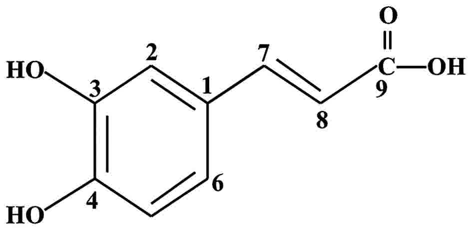
Caffeic acid (CFC) is a simple phenolic compound (Fig. 1) identified primarily in coffee and
specific herbs, particularly thyme, sage and spearmint (18). It was reported that CFC interacted
synergistically with 5-fluorouracil (5-FU), leading to a reduction
of apoptosis in HeLa cell line with minimum amount of hemolytic
activity (19). Considering the
aforementioned, CDDP and CFC were selected and their CxCa
anticancer activity was assessed in combination.
Materials and methods
Reagents
CDDP was obtained from Masu Co., Ltd. (Bangkok,
Thailand). CFC was obtained from Merck KGaA (Darmstadt, Germany).
DMEM-high glucose medium, fetal bovine serum (FBS) and trypsin-EDTA
were obtained from Gibco; Thermo Fisher Scientific, Inc. (Waltham,
MA, USA).
Cell lines and cell culture
Four cell lines, HeLa (HPV-18-positive), SiHa and
CaSki (HPV-16-positive), and C33A (HPV-negative) cervical cancer
cells and normal monkey epithelial kidney Vero cells were
maintained at 37°C with 5% CO2 in DMEM-high glucose
medium supplemented with 10% FBS. Once the cells were ~80%
confluent, they were trypsinized with 1 ml of 1X trypsin-EDTA,
incubated at 37°C for 5 min and centrifuged at 250 × g for 5 min at
room temperature. The supernatant was then removed, and 200 µl of
seeding cells were resuspended in 4 ml of DMEM-high glucose medium.
Under these conditions, cells reached confluence in 3 days. Cells
were then treated with the test compounds.
In vitro cytotoxicity assay
The in vitro cytotoxic effect of the test
compounds was determined using a sulforhodamine B (SRB) assay
(20). Briefly, cell lines
(6×10³ cells/well) were seeded in a 96-well plate for 24
h (day 0). Next, these cells were treated with various
concentrations of CDDP (2, 4, 6, 8, 10, 12, 14, 16, 18 and 24 µM)
and CFC (20, 50, 100, 200, 400 and 800 µM) for 24 h. Cells treated
with 1% dimethyl sulfoxide (DMSO) were used as a negative control.
Following this, cells were incubated at 37 °C with 5%
CO2 for 24 h, medium was replaced with 100 µl cold 10%
(w/v) trichloroacetic acid in each well, and plates were incubated
at 4°C for 1 h. Next, the plates were washed four times with tap
water and excess water was removed by paper towels and was
completed dried using a blow dryer or air-dried at room
temperature. Next, 100 µl of 0.057% (w/v) SRB solution was added to
each well and left at room temperature for 1 h. Following this, the
plates were quickly rinsed four times with 1% (v/v) acetic acid,
200 µl of 10 mM Tris base solution (pH 10.5) was added to each well
and the plates were shaken on a gyratory shaker for 1 h. Finally,
the optical density (OD) of solution in the plates was measured
using a microplate reader at 510 nm. Each concentration of drug
treatment was repeated for three independent experiments. Cell
viability was calculated by using the following formula: Cell
viability (%)=[(mean ODsample-mean
ODday0)/(mean OD negative control-mean
ODday0)] ×100.
For the half-maximal inhibitory concentration
determination (IC50), a dose-response curve between the
compound concentration and percent cell viability was plotted. The
cytotoxicity of the test compounds was compared between the CxCa
and Vero cell lines.
Estimation of combination index
(CI)
To estimate the CI of CDDP-CFC, the concentration of
CDDP and CFC used in this experiment was a series of 1.5-fold
dilutions of IC50 values. In the present study, HeLa
cells were treated with CDDP-CFC at various concentrations (3.25
and 88.88, 4.88 and 133.31, 7.32 and 200, 11 and 300, and 16.5 and
450 µM CDDP and CFC, respectively), CaSki cells were treated with
CDDP-CFC at the following concentrations: (3.25 and 59.27, 4.88 and
88.88, 7.32 and 133.31, 11 and 200, and 16.5 and 300 µM CDDP and
CFC, respectively), and Vero cells were treated with CDDP-CFC at
various concentrations (3.25 and 88.88, 4.88 and 133.31, 7.32 and
200, 11 and 300, 16.5 and 450, and 24.7 and 675 µM CDDP and CFC,
respectively). After 24 h, cell growth was examined using the SRB
assay. The effect of CDDP-CFC, quantified by determining CI, was
performed using the Chou-Talalay algorithm (21) using CalcuSyn software (version 1.1;
Biosoft, Cambridge, UK). A CI value of 1 indicates an additive
effect, CI<1 represents synergism and CI>1 represents
antagonism. The dose reduction index (DRI), which is defined as the
degree of dose reduction possible in a combination for a given
degree of effect, compared with the dose of each drug alone, was
also calculated using this software.
Caspases activity assay
Apoptosis pathway analysis was performed by
observing caspase activity using Caspase-Glo-3/7, −8 and −9 assay
kits (Promega Corporation, Madison, WI, USA). Cell lines
(6×10³ cells) in 100 µl of media were seeded into
96-well plates. CDDP alone (11 µM), CFC (300 µM) and CDDP (11 µM)
or CFC alone (200 µM) was added to HeLa and CaSki cells, which were
incubated at 37°C for 24 h. A total of 100 µl Caspase-Glo-3/7, −8
and −9 reagents were then added, the plates were shaken for 30 sec,
followed by incubation at room temperature for 1 h. For the
negative control, no CDDP or CFC was added. The blank control
contained Caspase-Glo-3/7, −8 and −9 reagents without cells and
CDDP-CFC. Following this, luminescent signal was detected by using
a SpectraMax L Luminescence microplate reader (Molecular Devices,
LLC, Sunnyvale, CA, USA). The data was analyzed using Soft
Max® Pro software (version 6.2.2; Molecular Devices,
LLC, CA, USA).
Statistical analysis
Data are expressed as the mean ± standard deviation
(SD). The differences between testing groups were determined using
Tukeys post hoc test following one-way analysis of variance.
P<0.05 was considered to indicate a statistically significant
difference. All analyses were performed using SPSS (version 17;
SPSS Inc., Chicago, IL, USA). The correlation coefficient, CI and
DRI were calculated using CalcuSyn software (version 1.1; Biosoft,
Cambridge, UK).
Results
Cytotoxicity of CDDP and CFC on
cervical cancer cell lines
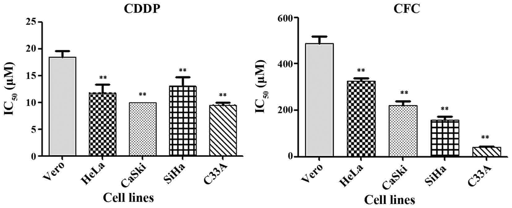
CDDP and CFC significantly inhibited the growth of
CxCa cell lines (Fig. 2).
IC50 of CDDP in HeLa, CaSki, SiHa, C33A and Vero cells
was 12±1.57, 10±0.00, 13±13.32, 10±0.50 and 18±1.22 µM,
respectively. All CxCa cell lines, other than HeLa, had a
significant lower IC50 for CDDP than did Vero cells. The
results in Fig. 2 show that CFC
significantly inhibited the growth of HeLa, CaSki, SiHa, C33A and
Vero cells, with IC50 values of 327±11.55, 220±18.03,
157±15.28, 40±3.21 and 487±30.55 µM, respectively. In the present
study, a 1% concentration of DMSO was tested in each of the cell
lines. The results demonstrated that the percentage of cell
viability in Vero, HeLa, CaSki, SiHa and C33A cell lines at 1% DMSO
were 101.09, 95.18, 96.00, 96.47 and 98.46, respectively.
Therefore, this concentration was safe for experimentation. Prior
studies have also reported that the maximum tolerated DMSO
percentage in cell culture is 1% (v/v) (22,23).
Effects of cisplatin in combination
with CFC on cervical cancer cells
The effects of CDDP and CFC in combination on
inhibition of HeLa, CaSki and Vero cell viability was determined
using isobologram analysis, as described previously (16). The proxies for the combined effects
were i) the dose-reduction index (DRI), ii) the combination index
(CI) and iii) the dose-effect levels of cell growth inhibition at
the IC50, IC75 and IC90 (Table I). CDDP and CFC exerted a synergistic
effect on HeLa and CaSki cells, but an antagonistic effect on Vero
cells (Fig. 3). The highest
synergistic effect dose of CDDP and CFC was found in HeLa cells, at
the IC50, IC75 and IC90 gave a CI
of 0.88, 0.77 and 0.69, respectively (Table I). The lowest synergistic effect dose
of CDDP-CFC was found in CaSki cells (CI value at
IC50=0.92) (Table I).
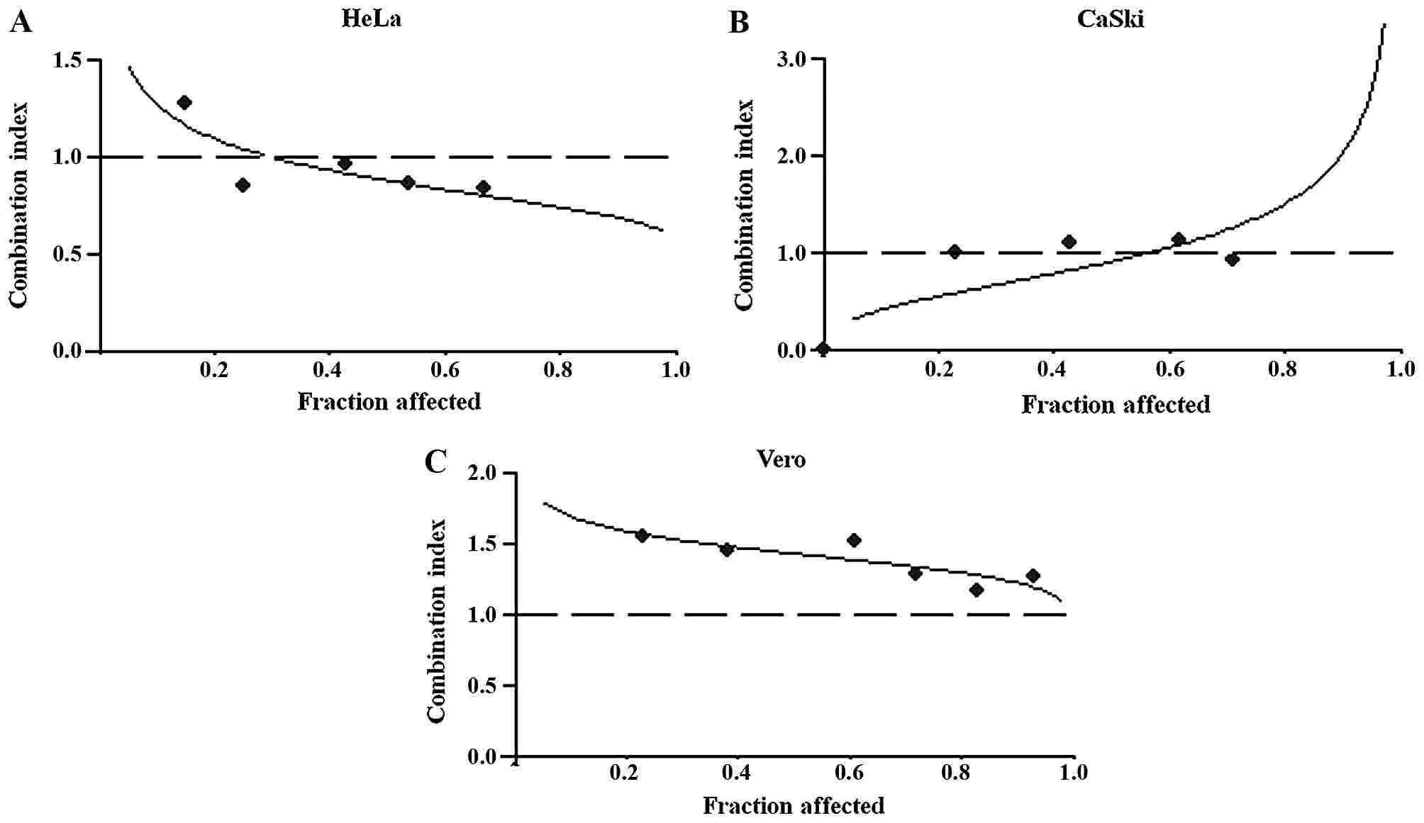 | Figure 3.Cytotoxicity of CDDP and CFC in
combination on (A) HeLa, (B) CaSki and (C) Vero. The cells were
treated with the appropriate concentrations of CDDP (2, 4, 6, 8,
10,12, 14, 16, 18 and 24 µM) and CFC (20, 50, 100, 200, 400 and 800
µM) for 24 h. Plots of the combination index vs. fraction of cells
affected were obtained using the median-effect analysis program.
Dashed lines indicate a CI of 1. CDDP, cisplatin; CFC, caffeic
acid. |
 | Table I.Dose-response association of CDDP and
CFC alone or in combination on HeLa and CaSki cells. |
Table I.
Dose-response association of CDDP and
CFC alone or in combination on HeLa and CaSki cells.
|
|
|
| CI value | DRI value |
|---|
|
|
|
|
|
|
|---|
| Cell line | Compound | Parameter, r |
IC50 |
IC75 |
IC90 |
IC50 |
IC75 |
IC90 |
|---|
| HeLa | CDDP | 0.93 |
|
|
| 2.18 | 2.28 | 2.21 |
|
| CFC | 0.99 |
|
|
| 2.35 | 3.04 | 4.14 |
|
| CDDP-CFC | 0.99 | 0.88 | 0.77 | 0.69 |
|
|
|
|
|
(1:27.3)a |
|
|
|
|
|
|
|
| CaSki | CDDP | 0.99 |
|
|
| 1.98 |
|
|
|
| CFC | 0.99 |
|
|
| 2.41 |
|
|
|
| CDDP-CFC | 0.83 | 0.92 | 1.36 | 2.02 |
|
|
|
|
|
(1:18.21)a |
|
|
|
|
|
|
|
As a result of the observed synergistic effect of
CDDP and CFC, there was a considerable reduction in the DRI. At a
dose level corresponding to synergistic drug combinations, the DRI
indicated that the IC50 of CDDP could be decreased
2.18-fold (HeLa) and 1.98-fold (CaSki) (Table I).
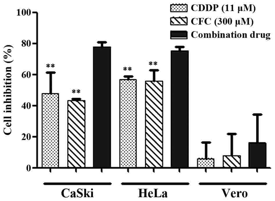
CDDP, CFC and a combination of CDDP and CFC were
tested for their cytotoxicity in CaSki (HPV-16-positive) and HeLa
(HPV-18-positive) cell lines, compared with the Vero cell line.
CaSki and HeLa cells treated with CDDP and CFC (11 and 300 µM,
respectively) had statistically significant higher percentage of
cell inhibition than those treated with CDDP or CFC alone (Fig. 4). No significance in percentage of
cell inhibition was found when Vero cells were treated with
CDDP-CFC, compared to CDDP and CFC alone.
CDDP-CFC treatment on cervical cancer
cells induced apoptosis
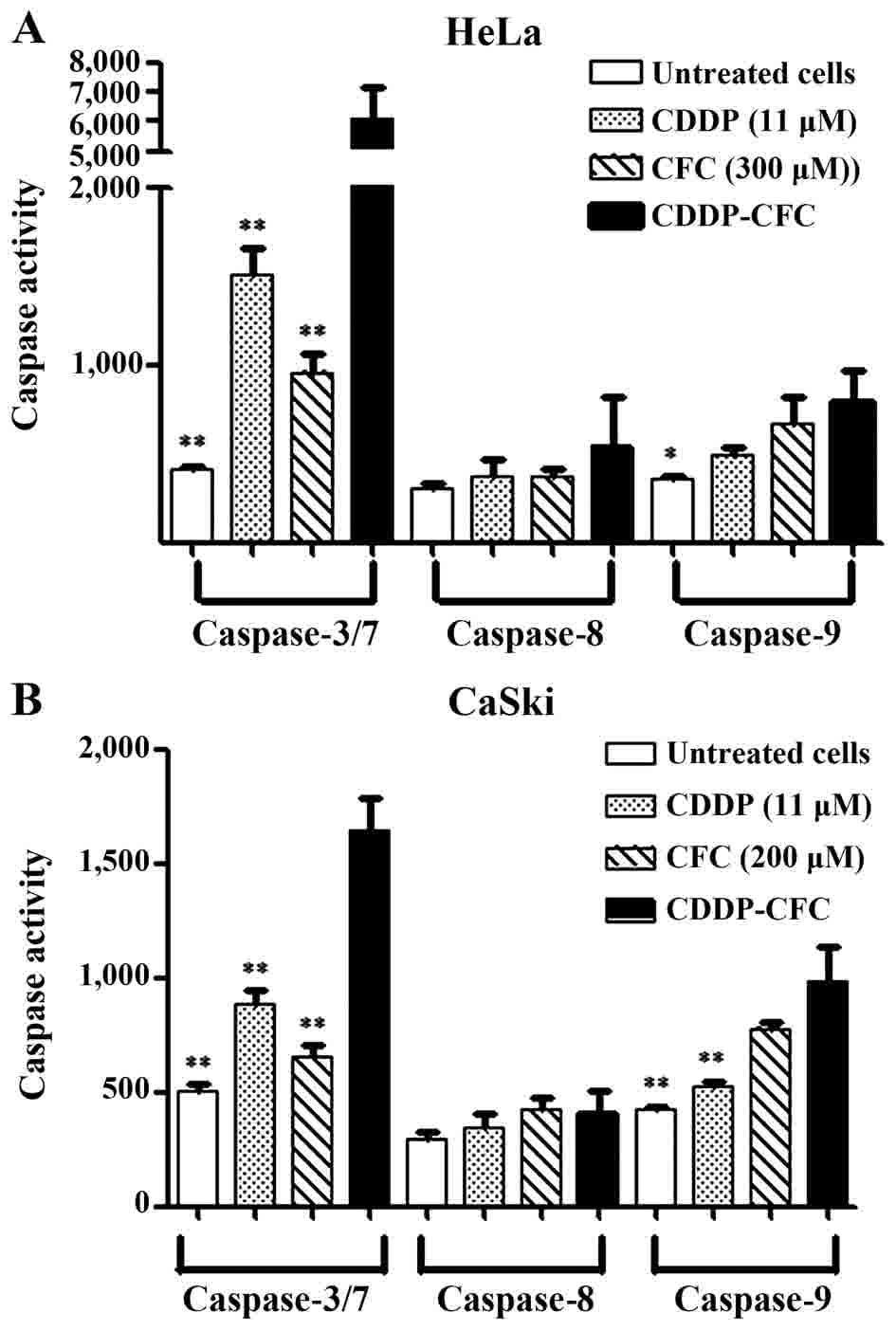
CDDP and CFC in combination increased caspase-3, −7
and −9 activity (apoptosis via the intrinsic pathway) to greater
degree than using either CDDP or CFC alone (Fig. 5). In Fig.
5A, CDDP and CFC, at 11 and 300 µM, respectively, significantly
activated the expression of the caspase-3 and −7 in HeLa cells by
4.02- and 6.34-fold compared with CDDP and CFC alone, respectively.
Similarly, caspase-9 expression was significantly increased by CDDP
and CFC combination treatment, compared with CDDP or CFC alone.
Similar results were obtained for CaSki cells
(Fig. 5B). The increase in caspase-3
and −7 expression upon treatment with CDDP-CFC, was 1.87-fold
higher than CDDP treatment alone and was 2.51-fold higher than CFC
treatment alone. Caspase-9 was increased by 2.29-fold, when treated
with a combination of CDDP and CFC, which was significantly higher
than that of cells treated with 11 µM CDDP (1.88-fold) and treated
with CFC (1.26-fold). In addition, CDDP, CFC and CDDP-CFC affected
caspase-8 activity less than caspase-3, −7 and −9.
In the present study, when a combination of CDDP and
CFC was used to treat HeLa and CaSki cells, the expression of
caspase-3 and −7 was increased compared with the expression of
caspase-9 by 7.70-fold (black bar; Fig.
5A) and by 1.67-fold (black bar; Fig.
5B), respectively. For HeLa cells treated with CFC, the
expression of caspase-3 and −7 was 1.43-fold higher than the
expression of caspase-9 (diagonal bar; Fig. 5A), but in CaSki cells, the expression
of caspase-9 was 0.84-fold higher than the expression caspase-3 or
−7 (diagonal bar; Fig. 5B).
Discussion
Chemoresistance is one of the major problems
encountered in CxCa therapy. CDDP is an anticancer drug used for
the treatment of CxCa; however, there are numerous side effects and
drug resistance is frequently developed (24,25). These
issues require the investigation of a novel anticancer agent,
potentially derived from natural sources. In the present study, the
CxCa CaSki, SiHa, HeLa and C33A cell lines were selected for the
experiments owing to their different properties. CaSki and SiHa are
HPV-16-positive, whereas HeLa is HPV-18-positive (26). C33A is HPV-negative with mutations to
tumor protein p53 (hereafter p53) (27). It was reported that SiHa cells were
more resistant to drug treatment than CaSki cells (28) and that HeLa is more sensitive to CDDP
than CaSki (29). C33A is the most
responsive of the cell lines examined in the present study to a
combination of chemotherapy and radiation (28). Vero cells, isolated from African green
monkey kidney epithelial cells, was used as normal control instead
of normal human cervical cells due to ethical issues, which is a
limitation in the present study. However, previous studies
(30,31) have also used Vero cells as control.
DMSO is often used in biological studies for solubilizing drugs or
studied compounds.
The combination of phytochemicals with anticancer
drugs may result in a synergistic, antagonistic or additive effect
in the treatment of cancer. The advantage of synergism is that it
can increase the efficacy of therapy; it can also decrease dosage
of the compound used which may lead to a reduction in drug toxicity
(32). CFC (Fig. 1) is a phytochemical with anticancer
properties in CxCa cells (16).
Previous studies revealed that CFC altered the development of
tumors by inhibiting cell growth and modifying the levels of
estrogen and insulin-like growth factor I receptors in human breast
cancer (33), exhibiting a potent
anticancer effect in the human fibrosarcoma HT-1080 cell line
(34), alteration of the
mitochondrial membrane potential and induction of mitochondrial
collapse (35). The results indicated
that CFC exhibited lower cytotoxicity than CDDP. In C33A cells, the
IC50 value of CFC was 4-fold higher than that of CDDP
(40±3.21 vs. 10±0.50 µM). The IC50 of CFC and CDDP
combined, were obtained for SiHa, CaSki, HeLa and Vero cells as
follows: 12, 22, 27 and 27-fold compared to CDDP, respectively.
Therefore, CaSki and HeLa cells were selected for assessing the
combination of CDDP and CFC, compared with Vero cells.
CDDP and CFC exhibited a higher synergistic effect
in HeLa cells than in Caski cell lines (CI value at
IC75=0.77 vs. 1.36) (Table
I and Fig. 3). A similar study
reported that 5-FU in combination with CFC (5-FU and CFC), at the
IC75 in HeLa cells, exhibited a strong synergistic
effect (14). The result of DRI
(Table I) demonstrated that CDDP and
CFC in combination can reduce the effective dose of CDDP for HeLa
cells by 2.28-fold and 2.21-fold at the IC75 and
IC90, respectively, and for CaSki cells by 1.98-fold at
IC50. The findings of the present study indicated that
CDDP and CFC in combination increased the cytotoxicity of each
agent against the studied cell lines (Fig. 4). The concentrations of CDDP and CFC
used in the present study were based on the IC50 values
of HeLa cells. CDDP (11 µM), CFC (300 µM) and the same
concentration of CDDP combined with CFC could inhibit CaSki and
HeLa cell growth; however, no effect was found on Vero cells.
CDDP is often used in combination with natural
compounds for enhancing treatment of cancer (6). According to the success of 5-FU and CFC
in combination as anticancer agents in HeLa cells (19), the present study assumes that the
carboxylic (-COOH) group of CFC binds with the ammonia
(-NH3) group in CDDP, forming CDDP-CFC. A previous study
revealed that CDDP cross-linked with DNA and altered DNA
conformation, leading to DNA damage (36).
The mechanism of action of CDDP and CFC combined in
the CxCa cell lines in the present study was investigated via the
apoptotic pathway. Under normal condition, p53 initiates apoptosis
in response to cellular stress. Previous reports state that CDDP
increased expression of p53 protein in HeLa (37) and CaSki cell lines (38). The CxCa cells might be more sensitive
to CDDP as a result of dormant p53 tumor suppressor pathways
(39). These previous studies support
the findings of the present study, indicating that the return of
p53 expression may contribute to the chemosensitivity of CxCa
cells. CDDP and CFC in combination may induce apoptosis by
activating DNA damage. The present study is in agreement with that
of Ye et al (40), which found
that expression of p53 could lead to the induction of apoptosis by
activating DNA damage in osteosarcoma cell lines. One limitation of
the present study is that the expression of proteins involved in
apoptotic pathways was not determined. Measuring the expression of
p53, anti-apoptosis [B-cell lymphoma-2 (Bcl-2) and Bcl-xl] and
pro-apoptosis proteins (Bcl-associated X and Bcl-2 homologous
antagonist/killer Bak) and should be further investigated to aid
elucidation the mechanism of CDDP-CFC-induced apoptosis. The
results of the present study (Fig. 5)
indicated that CDDP and CFC in combination induced apoptosis via
the intrinsic pathway. CDDP and CFC in combination significantly
increased the expression of caspase-3, −7 and −9 in HeLa and CaSki
cells compared with treatment with either alone. CDDP may induce
apoptosis better compared with CFC as the expression of caspase-3
and 7 in HeLa and CaSki cells, which was induced by CDDP, was
significantly increased compared with that induced by CFC (Fig. 4). Previous studies reported that CDDP
induced apoptosis through the caspase cascade pathway (41) and CFC induced apoptosis via the
intrinsic pathway (16). In addition,
CDDP, CFC and CDDP and CFC in combination affected caspase-8
activity less than that of the other caspase (caspase-3, −7 and −9)
(Fig. 5). As aforementioned, the
expression of caspase-8 was lower than that of caspase-3, −7 and
−9, as the expression of caspase-3, −7 and −9, but not that of
caspase-8, was detected at 12 h following drug treatment.
In conclusion, the results of the present study
demonstrated that CDDP and CFC in combination synergistically
inhibited the growth of cells and induced apoptosis in HeLa and
CaSki cells. The mechanism that is most likely to be behind the
efficacy of this treatment is the modulation of apoptosis-regulated
expression (i.e. the activation of caspase-3, −7 and −9). No
cytotoxicity induced by CDDP and CFC in combination was observed
for Vero cells, indicating the feasibility of using CDDP in
combination with CFC as an adjunct to chemotherapy for the
management of CxCa. Further in vivo and clinical studies
should be conducted to determine the effectiveness of CDDP and CFC
in combination for the treatment of CxCa.
Acknowledgements
The present study was supported by the Thailand
Research Fund (grant no. RSA5880036) and Khon Kaen University
(grant no. 581205). The authors would like to thank the Centre for
Research and Development of Medical Diagnostic Laboratories,
Faculty of Associated Medical Sciences and Graduate School, Khon
Kaen University for providing research facilities.
Glossary
Abbreviations
Abbreviations:
|
CxCa
|
cervical cancer
|
|
CDDP
|
cisplatin
|
|
CFC
|
caffeic acid
|
|
SRB
|
sulforhodamine B
|
References
|
1
|
Ferlay J, Soerjomataram I, Dikshit R, Eser
S, Mathers C, Rebelo M, Parkin DM, Forman D and Bray F: Cancer
incidence and mortality worldwide: Sources, methods and major
patterns in GLOBOCAN 2012. Int J Cancer. 136:E359–E386. 2015.
View Article : Google Scholar : PubMed/NCBI
|
|
2
|
Arbyn M, Castellsagué X, Sanjosé SD, Bruni
L, Saraiya M, Bray F and Ferlay J: Worldwide burden of cervical
cancer in 2008. Ann Oncol. 22:2675–2686. 2011. View Article : Google Scholar : PubMed/NCBI
|
|
3
|
Matsuki M, Takahashi A, Katou S,
Takayanagi A, Takagi Y and Kamata K: Pathological complete response
to gemcitabine and cisplatin chemotherapy for advanced upper tract
urothelial carcinoma: A case report. Nihon Hinyokika Gakkai Zasshi.
104:33–37. 2013.(In Japanese). PubMed/NCBI
|
|
4
|
Pignon JP, Tribodet H, Scagliotti GV,
Douillard JY, Shepherd FA, Stephens RJ, Dunant A, Torri V, Rosell
R, Seymour L, et al: Lung adjuvant cisplatin evaluation: A pooled
analysis by the LACE collaborative group. J Clin Oncol.
20:3552–3559. 2008. View Article : Google Scholar
|
|
5
|
Agarwal R and Kaye SB: Ovarian cancer:
Strategies for overcoming resistance to chemotherapy. Nat Rev
Cancer. 3:502–516. 2003. View
Article : Google Scholar : PubMed/NCBI
|
|
6
|
Dasari S and Tchounwou PB: Cisplatin in
cancer therapy: Molecular mechanisms of action. Eur J Pharmacol.
740:364–378. 2014. View Article : Google Scholar : PubMed/NCBI
|
|
7
|
Wang D and Lippard SJ: Cellular processing
of platinum anticancer drugs. Nat Rev Drug Discov. 4:307–320. 2005.
View Article : Google Scholar : PubMed/NCBI
|
|
8
|
Hernandez-Flores G, Ortiz-Lazareno PC,
Lerma-Diaz JM, Dominguez-Rodriguez JR, Jave-Suarez LF, Adel
Aguilar-Lemarroy C, de Celis-Carrillo R, del Toro-Arreola S,
Castellanos-Esparza YC and Bravo-Cuellar A: Pentoxifylline
sensitizes human cervical tumor cells to cisplatin-induced
apoptosis by suppressing NF-kappa B and decreased cell senescence.
BMC Cancer. 11:4832011. View Article : Google Scholar : PubMed/NCBI
|
|
9
|
Astolfi L, Ghiselli S, Guaran V, Chicca M,
Simoni E, Olivetto E, Lelli G and Martini A: Correlation of adverse
effects of cisplatin administration in patients affected by solid
tumours: A retrospective evaluation. Oncol Rep. 29:1285–1292. 2013.
View Article : Google Scholar : PubMed/NCBI
|
|
10
|
Kirwan JM, Symonds P, Green JA, Tierney J,
Collingwood M and Williams CJ: A systematic review of acute and
late toxicity of concomitant chemoradiation for cervical cancer.
Radiother Oncol. 68:217–226. 2003. View Article : Google Scholar : PubMed/NCBI
|
|
11
|
You BR, Moon HJ, Han YH and Park WH:
Gallic acid inhibits the growth of HeLa cervical cancer cells via
apoptosis and/or necrosis. Food Chem Toxicol. 48:1334–1340. 2010.
View Article : Google Scholar : PubMed/NCBI
|
|
12
|
Karthikeyan S, Kanimozhi G, Prasad NR and
Mahalakshmi R: Radiosensitizing effect of ferulic acid on human
cervical carcinoma cells in vitro. Toxicol In Vitro. 25:1366–1375.
2011. View Article : Google Scholar : PubMed/NCBI
|
|
13
|
Hussain A, Priyani A, Sadrieh L,
Brahmbhatt K, Ahmed M and Sharma C: Concurrent sulforaphane and
eugenol induces differential effects on human cervical cancer
cells. Integr Cancer Ther. 11:154–165. 2012. View Article : Google Scholar : PubMed/NCBI
|
|
14
|
Masuda T, Yamada K, Akiyama J, Someya T,
Odaka Y, Takeda Y, Tori M, Nakashima K, Maekawa T and Sone Y:
Antioxidation mechanism studies of caffeic acid: Identification of
antioxidation products of methyl caffeate from lipid oxidation. J
Agric Food Chem. 56:5947–5952. 2008. View Article : Google Scholar : PubMed/NCBI
|
|
15
|
Hwang HJ, Park HJ, Chung HJ, Min HY, Park
EJ, Hong JY and Lee SK: Inhibitory effects of caffeic acid
phenethyl ester on cancer cell metastasis mediated by the
down-regulation of matrix metalloproteinase expression in human
HT1080 fibrosarcoma cells. J Nutr Biochem. 17:356–362. 2006.
View Article : Google Scholar : PubMed/NCBI
|
|
16
|
Chang WC, Hsieh CH, Hsiao MW, Lin WC, Hung
YC and Ye JC: Caffeic acid induces apoptosis in human cervical
cancer cells through the mitochondrial pathway. Taiwan J Obstet
Gynecol. 49:419–424. 2010. View Article : Google Scholar : PubMed/NCBI
|
|
17
|
Singh M, Bhui K, Singh R and Shukla Y: Tea
polyphenols enhance cisplatin chemosensitivity in cervical cancer
cells via induction of apoptosis. Life Sci. 93:7–16. 2013.
View Article : Google Scholar : PubMed/NCBI
|
|
18
|
Wang H, Provan GJ and Helliwell K:
Determination of rosmarinic acid and caffeic acid in aromatic herbs
by HPLC. Food Chem. 87:307–311. 2004. View Article : Google Scholar
|
|
19
|
Hemaiswarya S and Doble M: Combination of
phenylpropanoids with 5-fluorouracil as anti-cancer agents against
human cervical cancer (HeLa) cell line. Phytomedicine. 20:151–158.
2013. View Article : Google Scholar : PubMed/NCBI
|
|
20
|
Pinmai K, Chunlaratthanabhorn S,
Ngamkitidechakul C, Soonthornchareon N and Hahnvajanawong C:
Synergistic growth inhibitory effects of Phyllanthus emblica
and Terminalia bellerica extracts with conventional
cytotoxic agents: Doxorubicin and cisplatin against human
hepatocellular carcinoma and lung cancer cells. World J
Gastroenterol. 14:1491–1497. 2008. View Article : Google Scholar : PubMed/NCBI
|
|
21
|
Chou TC: Drug combination studies and
their synergy quantification using the Chou-Talalay method. Cancer
Res. 70:440–446. 2010. View Article : Google Scholar : PubMed/NCBI
|
|
22
|
Galvao J, Davis B, Tilley M, Normando E,
Duchen MR and Cordeiro MF: Unexpected low-dose toxicity of the
universal solvent DMSO. FASEB J. 28:1317–1330. 2014. View Article : Google Scholar : PubMed/NCBI
|
|
23
|
Malinin TI and Perry VP: Toxicity of
dimethyl sulfoxide on HeLa cells. Cryobiology. 4:90–96. 1967.
View Article : Google Scholar : PubMed/NCBI
|
|
24
|
Rose PG: Chemoradiotherapy for cervical
cancer. Eur J Cancer. 38:270–278. 2002. View Article : Google Scholar : PubMed/NCBI
|
|
25
|
Eifel PJ: Chemoradiotherapy in the
treatment of cervical cancer. Semin Radiat Oncol. 16:177–185. 2006.
View Article : Google Scholar : PubMed/NCBI
|
|
26
|
Meissner JD: Nucleotide sequences and
further characterization of human papillomavirus DNA present in the
CaSki, SiHa and HeLa cervical carcinoma cell lines. J Gen Virol.
80:1725–1733. 1999. View Article : Google Scholar : PubMed/NCBI
|
|
27
|
Crook T, Wrede D and Vousden KH: p53 point
mutation in HPV negative human cervical carcinoma cell lines.
Oncogene. 6:873–875. 1991.PubMed/NCBI
|
|
28
|
Saxena A, Yashar C, Taylor DD and
Gercel-Taylor C: Cellular response to chemotherapy and radiation in
cervical cancer. Am J Obstet Gynecol. 192:1399–1403. 2005.
View Article : Google Scholar : PubMed/NCBI
|
|
29
|
Funaoka K, Shindoh M, Yamashita T,
Fujinaga K, Amemiya A and Totsuka Y: High-risk HPV-positive human
cancer cell lines show different sensitivity to cisplatin-induced
apoptosis correlated with the p21 Waf1/Cip1 level. Cancer Lett.
108:15–23. 1996. View Article : Google Scholar : PubMed/NCBI
|
|
30
|
Daduang J, Palasap A, Daduang S, Boonsiri
P, Suwannalert P and Limpaiboon T: Gallic acid enhancement of gold
nanoparticle anticancer activity in cervical cancer cells. Asian
Pac J Cancer Prev. 16:169–174. 2015. View Article : Google Scholar : PubMed/NCBI
|
|
31
|
Promraksa B, Daduang J, Khampitak T,
Tavichakorntrakool R, Koraneekit A, Palasap A, Tangrassameeprasert
R and Boonsiri P: Anticancer potential of Cratoxylum
formosum subsp. pruniflorum (Kurz.) gogel extracts
against cervical cancer cell lines. Asian Pac J Cancer Prev.
16:6117–6121. 2015. View Article : Google Scholar : PubMed/NCBI
|
|
32
|
Prabhakara PK, Kumarb A and Doblec M:
Combination therapy: A new strategy to manage diabetes and its
complications. Phytomedicine. 21:123–130. 2014. View Article : Google Scholar : PubMed/NCBI
|
|
33
|
Rosendahl AH, Perks CM, Zeng L, Markkula
A, Simonsson M, Rose C, Ingvar C, Holly JM and Jernstrom H:
Caffeine and caffeic acid lnhibit growth and modify estrogen
receptor and insulin-like growth factor I receptor levels in human
breast cancer. Clin Cancer Res. 21:1877–1887. 2015. View Article : Google Scholar : PubMed/NCBI
|
|
34
|
Prasad NR, Karthikeyan A, Karthikeyan S
and Reddy BV: Inhibitory effect of caffeic acid on cancer cell
proliferation by oxidative mechanism in human HT-1080 fibrosarcoma
cell line. Mol Cell Biochem. 349:11–19. 2011. View Article : Google Scholar : PubMed/NCBI
|
|
35
|
Galati G, Sabzevari O, Wilson JX and
O'Brien PJ: Prooxidant activity and cellular effects of the
phenoxyl radicals of dietary flavonoids and other polyphenolics.
Toxicology. 177:91–104. 2002. View Article : Google Scholar : PubMed/NCBI
|
|
36
|
Onoa GB, Cervantes G, Moreno V and Prieto
MJ: Study of the interaction of DNA with cisplatin and other Pd(II)
and Pt(II) complexes by atomic force microscopy. Nucleic Acids Res.
26:1473–1480. 1998. View Article : Google Scholar : PubMed/NCBI
|
|
37
|
Liu Y, Xing H, Han X, Shi X, Liang F, Chen
G and Ma D: The mechanism of cisplatin-induced apoptosis in HeLa
cells. J Clin Oncol. 2:866–869. 2005.
|
|
38
|
Rao Z, Gao J, Zhang B, Yang B and Zhang J:
Cisplatin sensitivity and mechanisms of anti-HPV16 E6-ribozyme on
cervical carcinoma CaSKi cell line. Chinese-German J Clin Oncol.
11:237–242. 2012. View Article : Google Scholar
|
|
39
|
Putral LN, Bywater MJ, Gu W, Saunders NA,
Gabrielli BG, Leggatt GR and McMillan NA: RNA interference against
human papillomavirus oncogenes in cervical cancer cells results in
increased sensitivity to cisplatin. Mol Pharmacol. 68:1311–1319.
2005. View Article : Google Scholar : PubMed/NCBI
|
|
40
|
Ye S, Shen J, Choy E, Yang C, Mankin H,
Hornicek F and Duan Z: p53 overexpression increases
chemosensitivity in multidrug-resistant osteosarcoma cell lines.
Cancer Chemother Pharmacol. 77:349–356. 2016. View Article : Google Scholar : PubMed/NCBI
|
|
41
|
Seki K, Yoshikawa H, Shiiki K, Hamada Y,
Akamatsu N and Tasaka K: Cisplatin (CDDP) specifically induces
apoptosis via sequential activation of caspase-8, −3 and −6 in
osteosarcoma. Cancer Chemother Pharmacol. 45:199–206. 2000.
View Article : Google Scholar : PubMed/NCBI
|