Introduction
Breast cancer is the most common type of cancer that
develops in women (1). Although the
survival rate is high when diagnosed early, ~50% of breast
cancer-associated mortality is due to local invasion and distant
metastasis (2,3). Therefore, it is important to investigate
the underlying molecular mechanisms that drive tumor formation and
novel therapeutic strategies to target breast cancer.
Diallyl disulfide (DADS), one of the major volatile
components of allicin, exhibits diverse anticancer activities,
including cell cycle arrest, inhibition of cell growth, induction
of cell differentiation and apoptosis by interfering with a variety
of cell signaling pathways (4–8). However,
the mechanism of DADS in breast cancer remains to be
determined.
Numerous studies have revealed that diverse
molecular mechanisms are implicated in the suppressive effects that
DADS exhibit on tumor growth (9,10). In
particular, DADS has been demonstrated to decrease the invasiveness
of gastric cancer cells by decreasing the expression and activity
of matrix metalloproteinases (MMPs) (11). MMPs serve a key function in the tumor
microenvironment by facilitating cell metastasis. MMP-9, a member
of the MMP family, is known to be involved in tumor invasion and
metastasis, and degrade the extracellular matrix in the vicinity of
the tumor (12). In addition,
urokinase-type plasminogen activator (uPA), one of the upstream
genes of MMP-9, interacts with co-receptors to activate
intracellular signaling pathways that mediate the degradation of
the extracellular matrix, and activates plasminogen and MMPs
(13). Increased expression levels of
uPA and MMP-9 expression are indicators of poor prognosis in
numerous human tumors, including breast cancer (14). In addition, adenylate-uridylate
(AU)-rich elements (AREs) in the 3′-untranslated regions of
numerous transiently expressed genes regulate mRNA, mediate
post-transcriptional modifications and serve important functions in
various types of cancer (15).
Tristetraprolin (TTP) is an important AU-rich RNA-binding protein,
which targets mRNAs (16). Previous
studies have identified decreased expression of TTP in numerous
types of cancer, including breast cancer, hepatocellular carcinoma,
and head and neck cancer (17–19). TTP
dysregulation has been implicated in the progress of various types
of cancer, particularly in the proliferation, apoptosis, invasion,
migration and resistance to chemotherapy of inflammation-associated
types of cancer cells. Additionally, TTP may mediate the
degradation of uPA mRNA, and inhibit the cell invasion and
migration of tumors by altering the expression of MMP-9 (19). Therefore, the aim of the present study
was to investigate the functions of DADS in the inhibition of cell
invasion and metastasis of human breast cancer, which may be a
beneficial therapy for this disease.
Materials and methods
Reagents and cell culture
DADS was purchased from Sigma-Aldrich; Merck KGaA
(Darmstadt, Germany). The following antibodies were obtained:
Rabbit anti-uPA primary antibody (cat. no. ABO10669; Abgent, Inc.,
San Diego, CA, USA); Rabbit anti-MMP-9 primary antibody (cat. no.
ab73734; Abcam, Cambridge, UK); Mouse anti-TTP and mouse
anti-β-actin primary antibodies (cat. no. SAB4200565 & A1978;
Sigma-Aldrich; Merck KgaA, Darmstadt, germany.); horseradish
peroxidase (HRP)-conjugated goat anti-rabbit IgG and HRP-conjugated
goat anti-mouse IgG secondary antibodies (cat. nos. 70-GAR0072
& 70-GAM007; Wuhan Boster Biological Technology, Ltd., Wuhan,
China). The bicinchoninic acid (BCA) protein assay and other
reagents were obtained from Kangwei Biotechnology (CWBiotech,
Beijing, China). Transwell® inserts were purchased from
Corning Incorporated (Corning, NY, USA). MCF-7 and MDA-MB-231 human
breast cancer cell lines were purchased from the Type Culture
Collection of the Chinese Academy of Sciences (Shanghai, China).
Cells were cultured in high-glucose Dulbecco's modified Eagle's
medium (DMEM; Hyclone, Logan, UT, USA) supplemented with 10% fetal
bovine serum (FBS; Gibco by Invitrogen, Carlsbad, CA, USA) at 37°C
in a humidified atmosphere containing 5% CO2.
In vivo tumorigenicity study
A total of 18 4-week-old female nude mice (weighing
16–18 g) were purchased from Shanghai SLAC Laboratory Animal Co.,
Ltd. (Shanghai, China). All mice were kept in a barrier facility
under high-efficiency particulate air filtration and maintained in
a specific pathogen-free environment with shavings as bedding. The
cages and bedding were sterilized at 121°C for 20 min. All mice
were allowed free access to food and water, the feed was purchased
from Ke'aoxieli Feed Co. Ltd (Peking, China), drinking water was
sterilized by 60Co irradiation. Approximately
1×10⁷ MDA-MB-231 or MCF-7 cells were subcutaneously
injected into the subcutis of the right axilla of each mouse. Tumor
volume (mm3) was examined every 4 days and calculated
using a standard formula [width2 × (length/2)]. When the
tumor volume reached 80 mm3, the mice were divided into
two groups. The control group used saline as vehicle treatment
while the treatment group received 50 mg/kg DADS via
intraperitoneal injection every 2 days. After 28 days, the
xenografts were removed. The tumor weights were measured in both
groups. All experimental procedures conformed to the Guide for the
Care and Use of Laboratory Animals published by the US National
Institutes of Health (NIH Publication No. 85-23, revised 1996) and
were approved by the Institutional Animal Ethics Committee of
University of South China (Hengyang, China).
Hematoxylin and eosin (H&E)
staining
Tumor tissues (control group and DADS-treated group)
were cut into 0.5-mm3 sections, fixed using 4%
paraformaldehyde for 24 h at room temperature, embedded in paraffin
and further cut into 5 µm thick tissue sections. The tissue
sections were dewaxed in xylene and rehydrated in a descending
(100, 95, 90, 80 and 75%) ethanol series, 3–5 min each time.
Following washing with PBS, the sections were stained with
hematoxylin and following a second wash, the sections were
differentiated. The sections were subsequently stained with eosin
following washing. Following dehydration with ethanol, the sections
were mounted, and observed using light with a ×40 microscopy
objective (Eclipse E200; Nikon Corporation, Tokyo, Japan).
Western blot analysis
The tumor tissues and cells were lysed using
radioimmunoprecipitation assay buffer (Thermo Fisher Scientific,
Inc., Waltham, MA, USA) supplemented with Halt Protease and
Phosphatase Inhibitor Cocktail (1:100; Thermo Fisher Scientific,
Inc.), sonicated and clarified by centrifugation at 12,000 × g, 15
min at 4°C. All groups of protein were quantified using a
bicinchoninic acid protein assay and equal amounts of total protein
extracts (30 µg/well) were separated by 8–12% SDS-PAGE. Following
electrophoresis for 2 h at 100 V, the proteins were electrically
transferred onto a polyvinylidene fluoride membrane. The membranes
were then blocked with 5% nonfat dry milk for 1 h at room
temperature and then incubated with primary antibodies against TTP
(1:500), uPA (1:1,000), MMP-9 (1:1,000) or β-actin (1:1,000) at 4°C
overnight. Thereafter, the membranes were washed three times with
TBST (20 mmol/l Tris base, pH 7.6, 150 mmol/l NaCl and 0.1%
Tween-20), incubated with the HRP-conjugated goat anti-rabbit or
goat anti-mouse IgG secondary antibodies for 50 min at room
temperature, and washed three times with TBST. Protein
visualization was performed using the ECL Plus Western Blotting
Detection system (Tanon-6200, Tanon Science & Technology Co.,
Ltd., Shanghai, China) to collect the images and QuantityOne 4.5.0
software (Bio-Rad Laboratoris, Inc., Hercules, CA, USA) for
analysis.
Tablet cloning assay
A total of 200/well MCF-7 and MDA-MB-231 cells were
planted into 9 cm petri dishes. After treating cells with 0, 100,
200 or 400 µM DADS for 24 h (using 0 µM as the control group),
cells were washed with PBS three times, then fresh serum-free DMEM
was added. After 3 weeks, 5 ml 100% methanol was added to the
dishes for 15 min to fix the cells at room temperature. Giemsa was
used to stain cells for 20 min at room temperature. Following PBS
washing of the cells and air-drying, the clone formation rate was
calculated under microscope, clone formation rate (%)=(clone
number/plated cell number) ×100%.
Transwell assays
For the Transwell assays, 25 µg Matrigel was added
to the upper side of porous filters (pore size, 8 µm) and the gel
was allowed to form at 37°C for 2 h. Following rehydration of the
coated filters with 100 µl medium, 1×10⁶ cells in 100 µl
serum-free DMEM supplemented with 0.2% FBS were seeded into the
upper part of each chamber, whereas the lower compartments were
filled with 500 µl complete DMEM containing 10% FBS to serve as the
chemoattractant agent. Cells were incubated at 37°C with 5%
CO2 for 24 h to allow for invasion. At 24 h, cells on
the upper surface of the filters were removed using cotton swabs.
Cells that had invaded to the lower surface of the filter were
washed twice with PBS, fixed with 4% paraformaldehyde for 15 min
and stained with 0.1% crystal violet at room temperature. Images
were captured using an epifluorescence inverted microscope
(magnification, ×100). The total number of invaded cells was
normalized to the number of non-targeting control cells and
expressed as fold change.
Wound healing assay
MCF-7 and MDA-MB-231 cells (2×10⁵/well)
were cultured in 6-well plates in DMEM containing 10% FBS. The cell
monolayer in each well was scratched with a 200 µl plastic pipette
tip to create a linear wound. The monolayer was washed twice with
PBS to remove debris and detached cells, and the cells were then
exposed to serum-free medium with or without various concentrations
of DADS (100, 200 and 400 µM) for 24 h and using 0 µM as control
group. The wound areas were then observed using an inverted
microscope (magnification, ×100). The migration distance was
measured, and migration rates are expressed as the ratio of the
treated group value to the control group value.
Small interfering (si)RNA
transfection
siRNA duplexes for TTP were designed and produced by
Shanghai GenePharma Co., Ltd. (Shanghai, China). The sequences for
TTP siRNA were sense, 5′-UCGCCACCCCAAGUACAAATT-3′ and antisense,
5′-UUUGUAGGGGUGGCGATT-3′. Scramble control RNAi was used as control
siRNA, the sequence for the negative control siRNA was
5′-GCAAGCTGACCCTGAAGTT-3′. The TTP and control siRNA were
transfected into MCF-7 and MDA-MB-231 cells using
Lipofectamine® 3000 (Invitrogen; Thermo Fisher
Scientific, Inc.) according to the manufacturer's protocol.
Briefly, MCF-7 and MDA-MB-231 cells (1×10⁵/well) were
inoculated into 6-well plates and cultured in DMEM with 10% FBS at
37°C until reaching 70% confluence. Then, 2 µl TTP or control siRNA
(0.5 µg/µl) was mixed with 5 µl Lipofectamine 3000. The mixture was
added to the cell culture, and cells were incubated for 12 h at
37°C. The cell solution was replaced with DMEM containing 10% FBS
and cells were incubated for another 24 h.
Gene Expression Omnibus (GEO)
database
The GSE42568 breast cancer microarray dataset was
downloaded from the GEO database (https://www.ncbi.nlm.nih.gov/geo/). Using the data
from the GEO dataset normal breast (17 samples) vs. breast cancer
(104 samples) were analyzed. P<0.05 was considered to indicate a
statistically significant difference.
Statistical analysis
Statistical analysis was performed using SPSS
software (version 20.0; IBM Corp., Armonk, NY, USA). Each
experiment was repeated three times. All results are presented as
the mean ± standard deviation or standard error of three
independent experiments. The χ2 test was applied for
enumeration data. Student's t-test (unpaired) and one-way analysis
of variance was used to identify statistically significant
differences followed by the Student-Newman-Keuls test for further
group comparison. P<0.05 was considered to indicate a
statistically significant difference.
Results
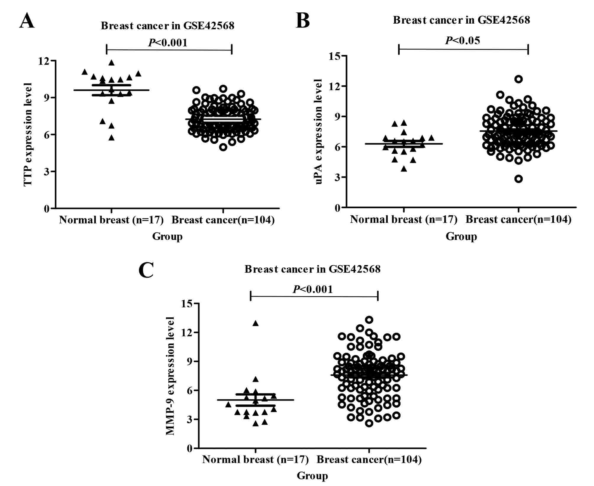
TTP is downregulated, whereas uPA and
MMP-9 are upregulated in human breast cancer compared with normal
breast in the GEO database
In breast cancer microarray datasets from the GEO
database, a microarray dataset (GSE42568) was identified in which
TTP mRNA levels were significantly downregulated in breast cancer
compared with in normal breast tissue (Fig. 1A). In the same microarray dataset, uPA
and MMP-9 mRNA levels were identified to be significantly
upregulated in breast cancer compared with in normal breast tissue
(Fig. 1B and C).
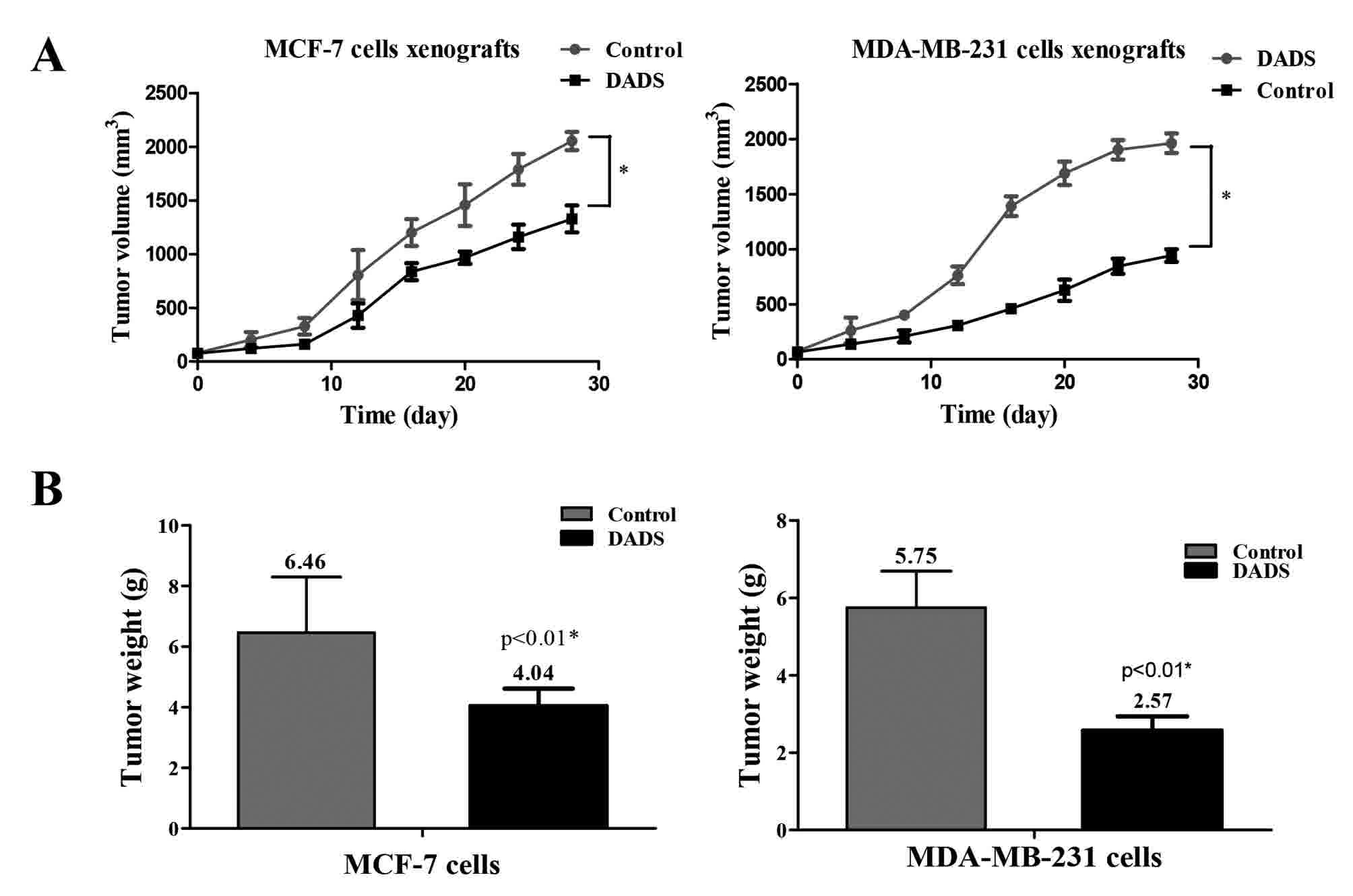
Effect of DADS on the progression of
breast cancer in vivo
Compared with the control group, the mean volume and
weight of the tumors from mice in the DADS-treated group were
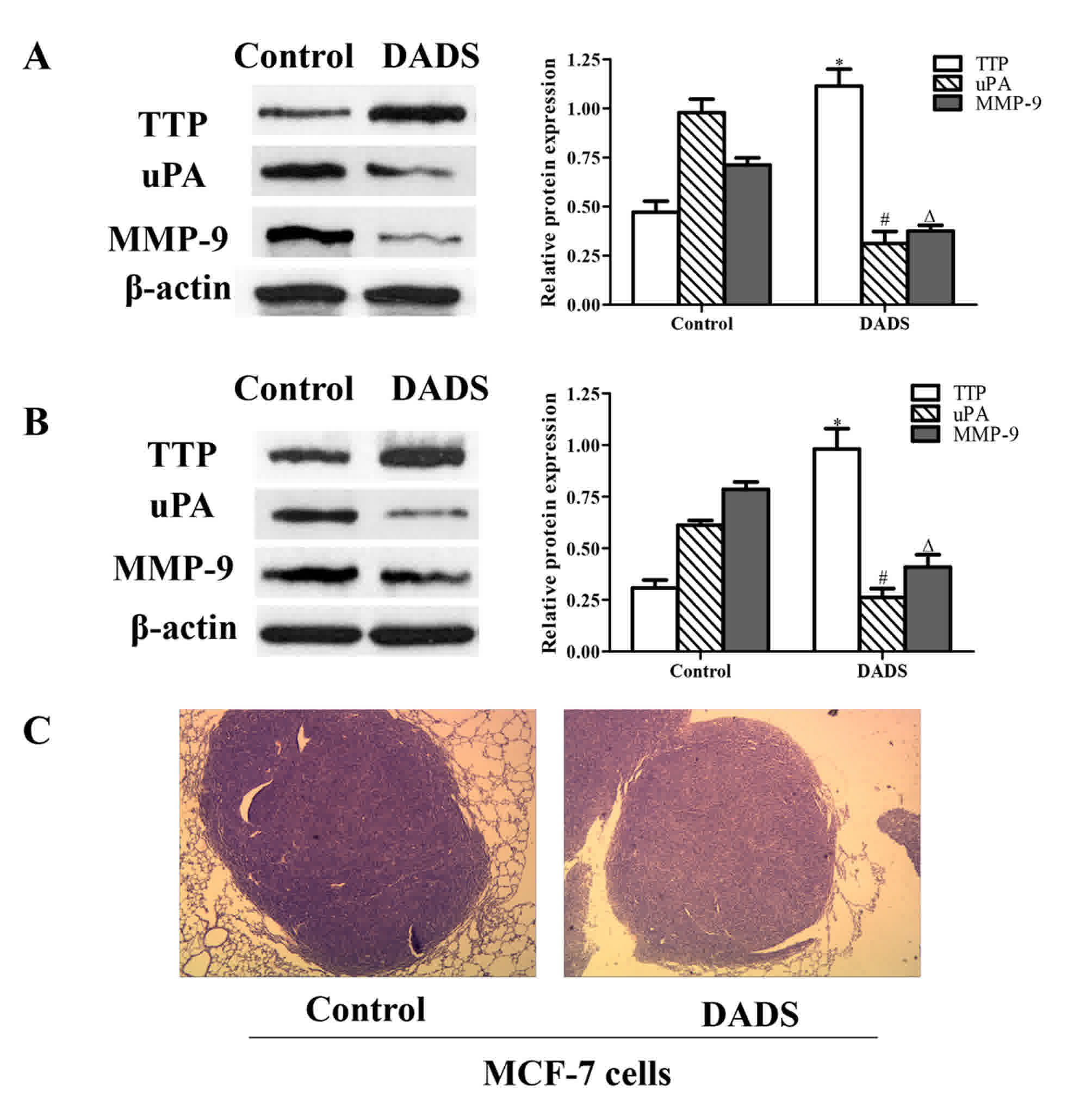
significantly decreased (Fig. 2).
Protein was extracted from the xenografts, and western blot
analysis was performed to detect the expression of uPA, MMP-9 and
TTP. uPA and MMP-9 protein expression levels were significantly
downregulated, whereas TTP expression was significantly upregulated
in the DADS-treated group compared with the corresponding control
groups, suggesting that DADS induced the expression of TTP in
breast cancer xenografts (Fig. 3A and
B). Furthermore, H&E staining revealed that MCF-7-derived
tumors in the DADS-treated group were markedly smaller compared
with the control group (Fig. 3C).
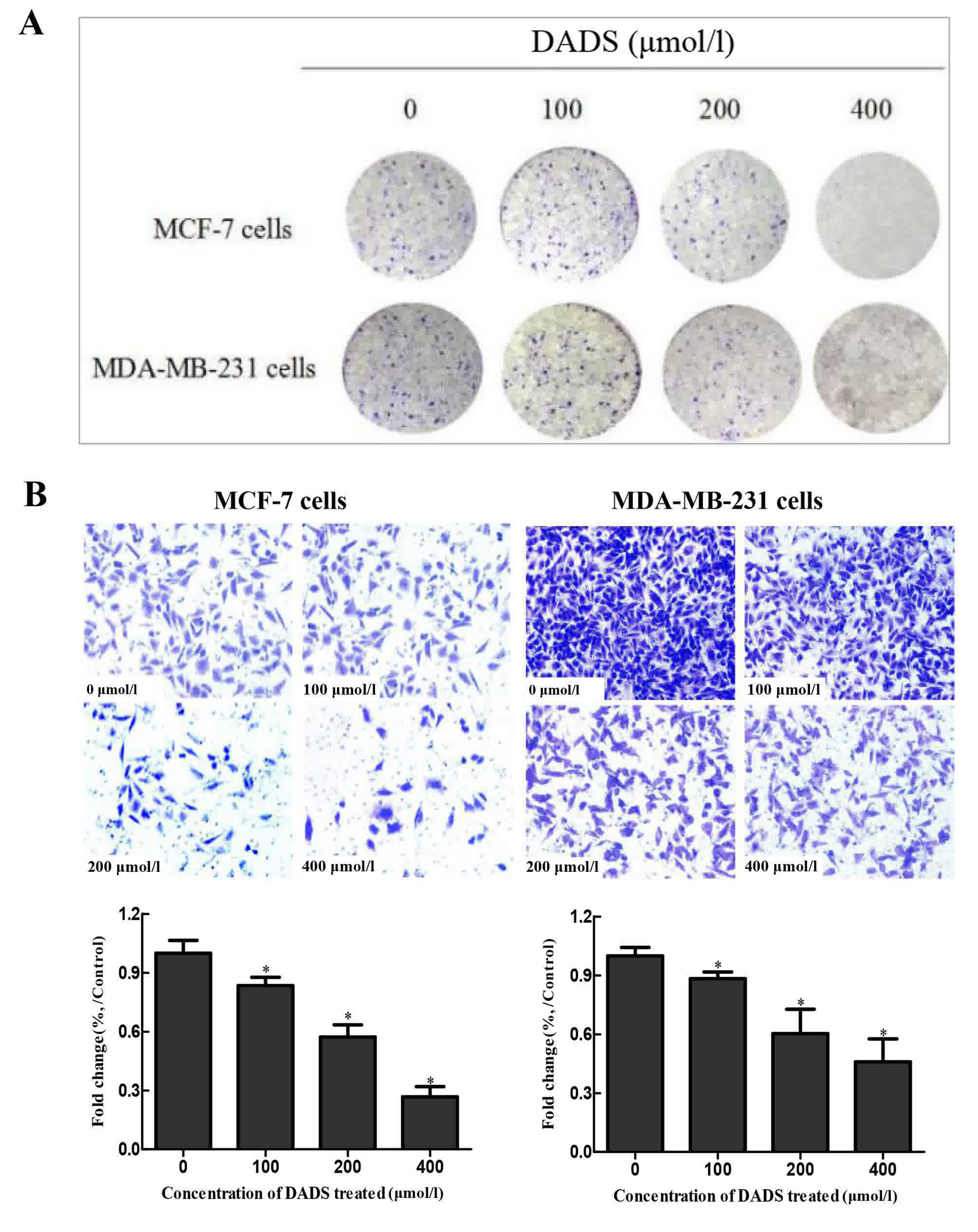
Effect of DADS on the proliferation,
invasion and migration of breast cancer cells in vitro
To examine the effect of DADS on cellular
proliferation, a tablet cloning assay was performed on MCF-7 and
MDA-MB-231 cells treated with DADS. It was demonstrated that the
clone formation of cells was markedly decreased in a dose-dependent
manner following DADS treatment (Fig.
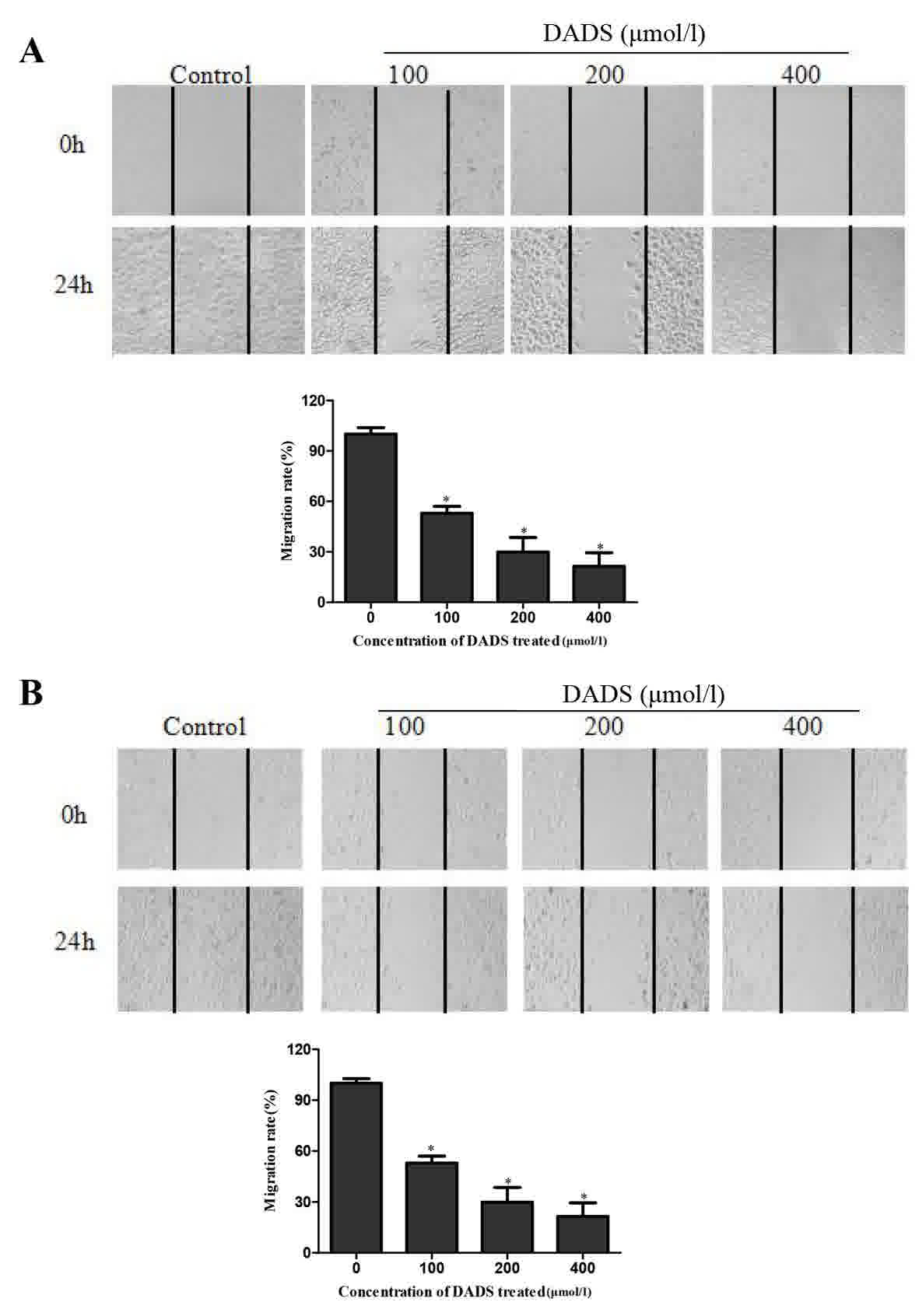
4A). In addition, invasion and migration rates of MCF-7 and
MDA-MB-231 human breast cancer cells were significantly decreased
following DADS treatment, as determined using the Transwell
(Fig. 4B) and wound healing assays
(Fig. 5) compared with the untreated
control groups.
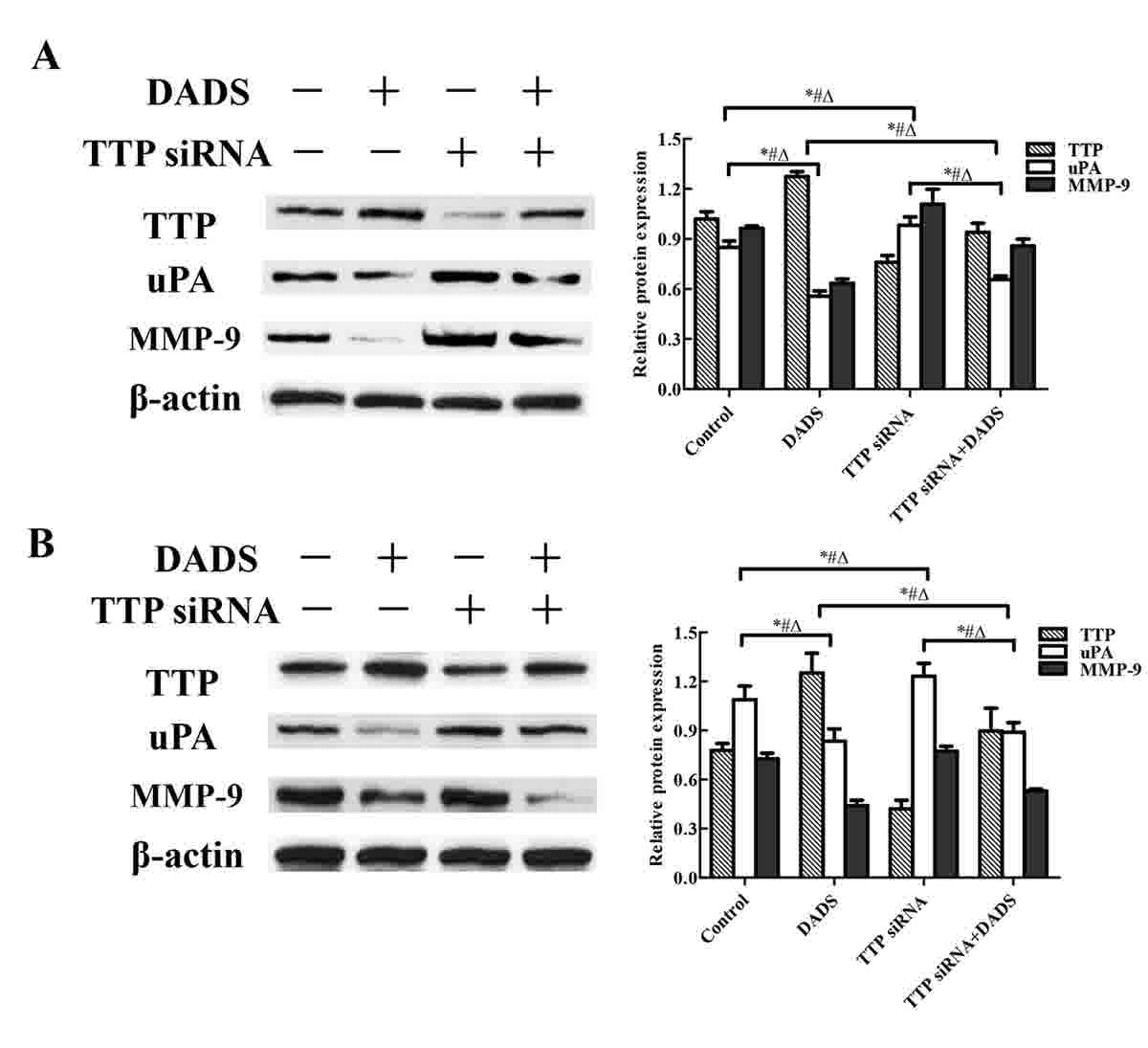
Effect of DADS on the expression of
TTP, uPA and MMP-9 in breast cancer cells
Western blotting was used to examine the expression
of TTP and proteins associated with invasion, including uPA and
MMP-9 in breast cancer cells. As presented in Fig. 6, DADS treatment significantly affected
the expression of TTP, uPA and MMP-9 in a dose- and time-dependent
manner (P<0.05).
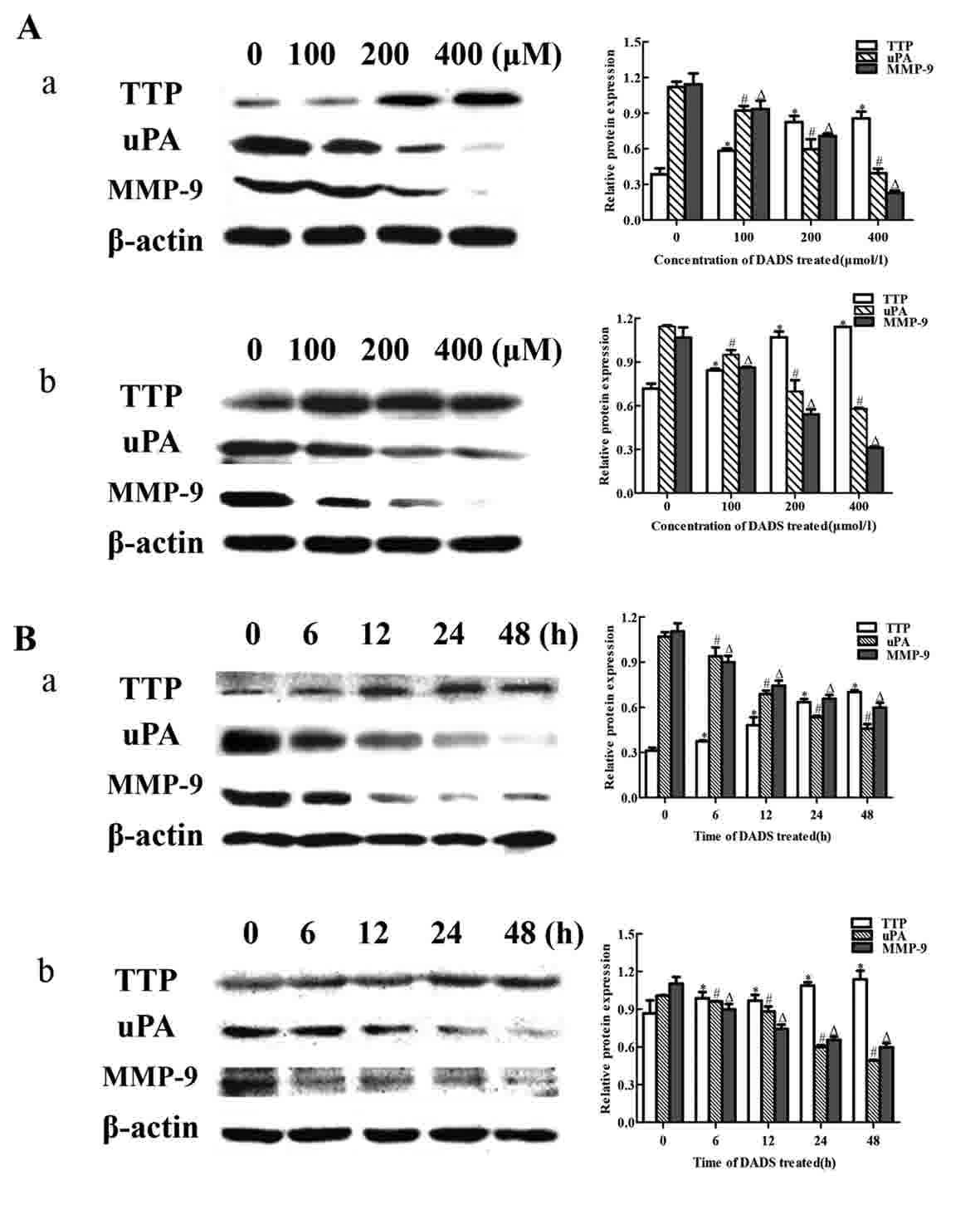 | Figure 6.DADS downregulates the expression of
uPA and MMP-9, and upregulates TTP expression in a dose- and
time-dependent manner in breast cancer cells. Western blot analysis
of (A) cells treated with different concentrations of DADS (0, 100,
200 and 400 µM) for 24 h and (B) cells treated with 200 µmol/l DADS
for different periods of time (0, 6, 12, 24 and 48 h). (a) MCF-7
cells. (b) MDA-MB-231 cells. *#ΔP<0.05 vs. 0 µmol/l
DADS or 0 h group; n=3. Data are presented as the mean ± standard
deviation. DADS, diallyl disulfide; MMP, matrix metalloproteinase;
uPA, urokinase-type plasminogen activator; TTP,
tristetraprolin. |
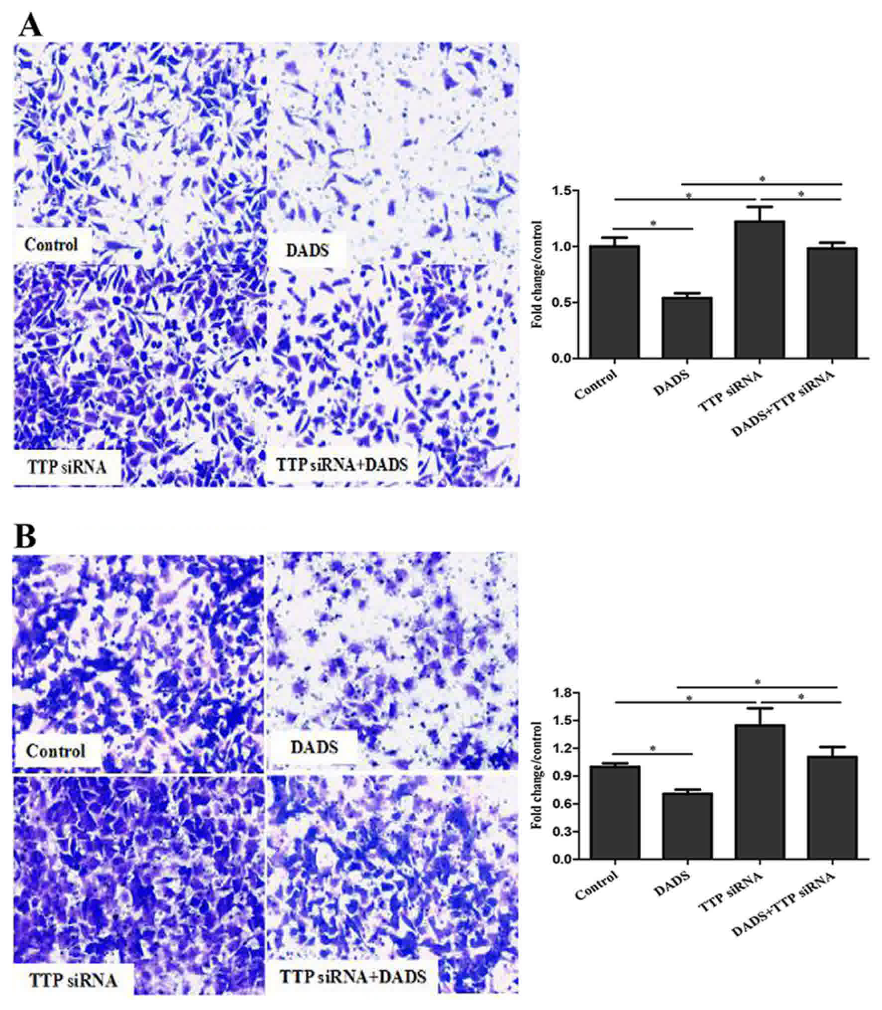
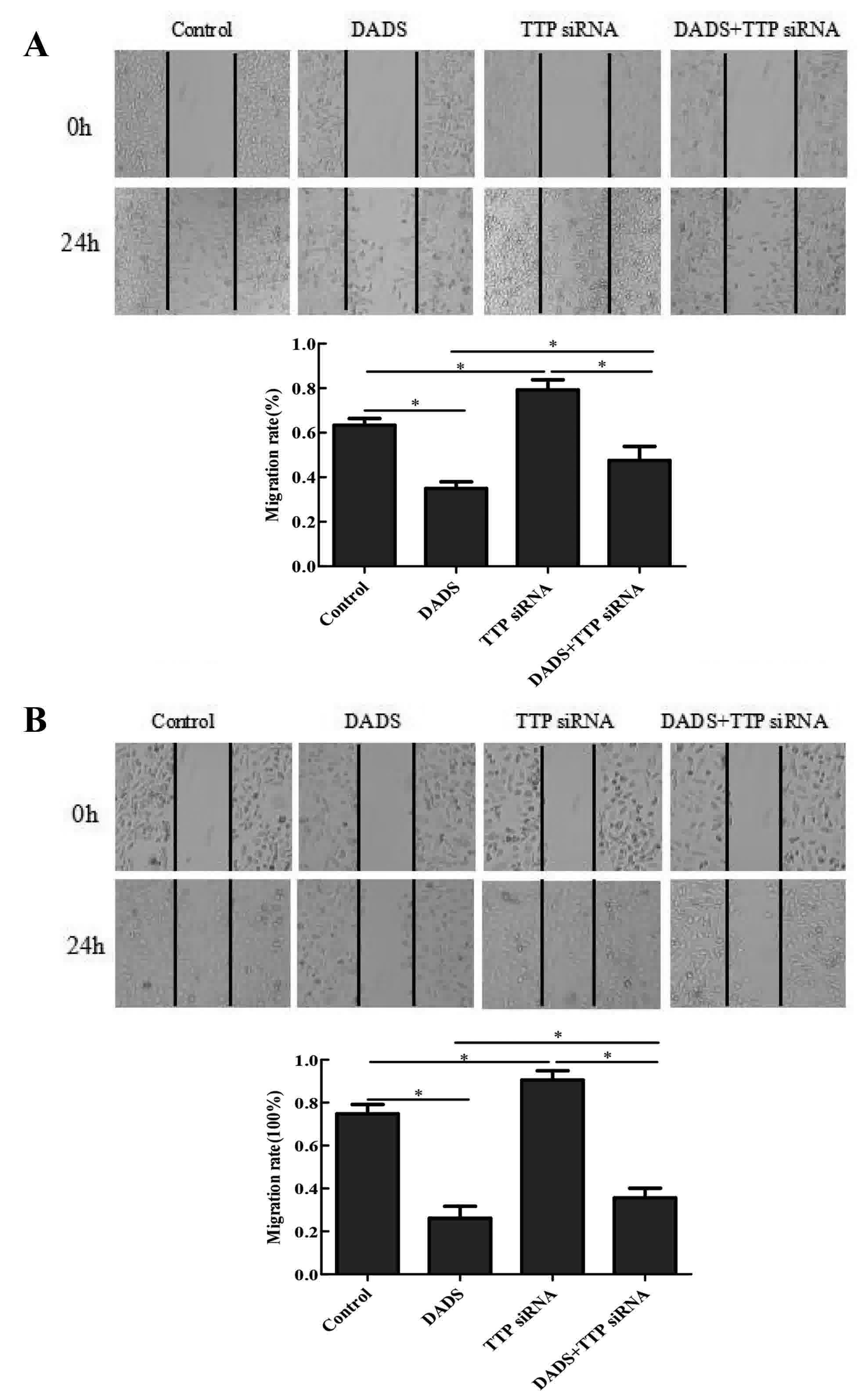
Effect of knocking down TTP on the
progression of breast cancer
Following transfection of MCF-7 and MDA-MB-231 cells
with TTP siRNA for 24 h, western blot analysis revealed that TTP
siRNA significantly decreased the effect that DADS exhibited on
TTP, uPA and MMP-9 expression (Fig.
7). The Transwell assay demonstrated that TTP siRNA
significantly reversed the anti-invasive effect of DADS in breast
cancer cells (Fig. 8). Furthermore,
the wound healing assay revealed that TTP siRNA counteracted the
suppressive effect of DADS on breast cancer cell migration
(Fig. 9). All the aforementioned
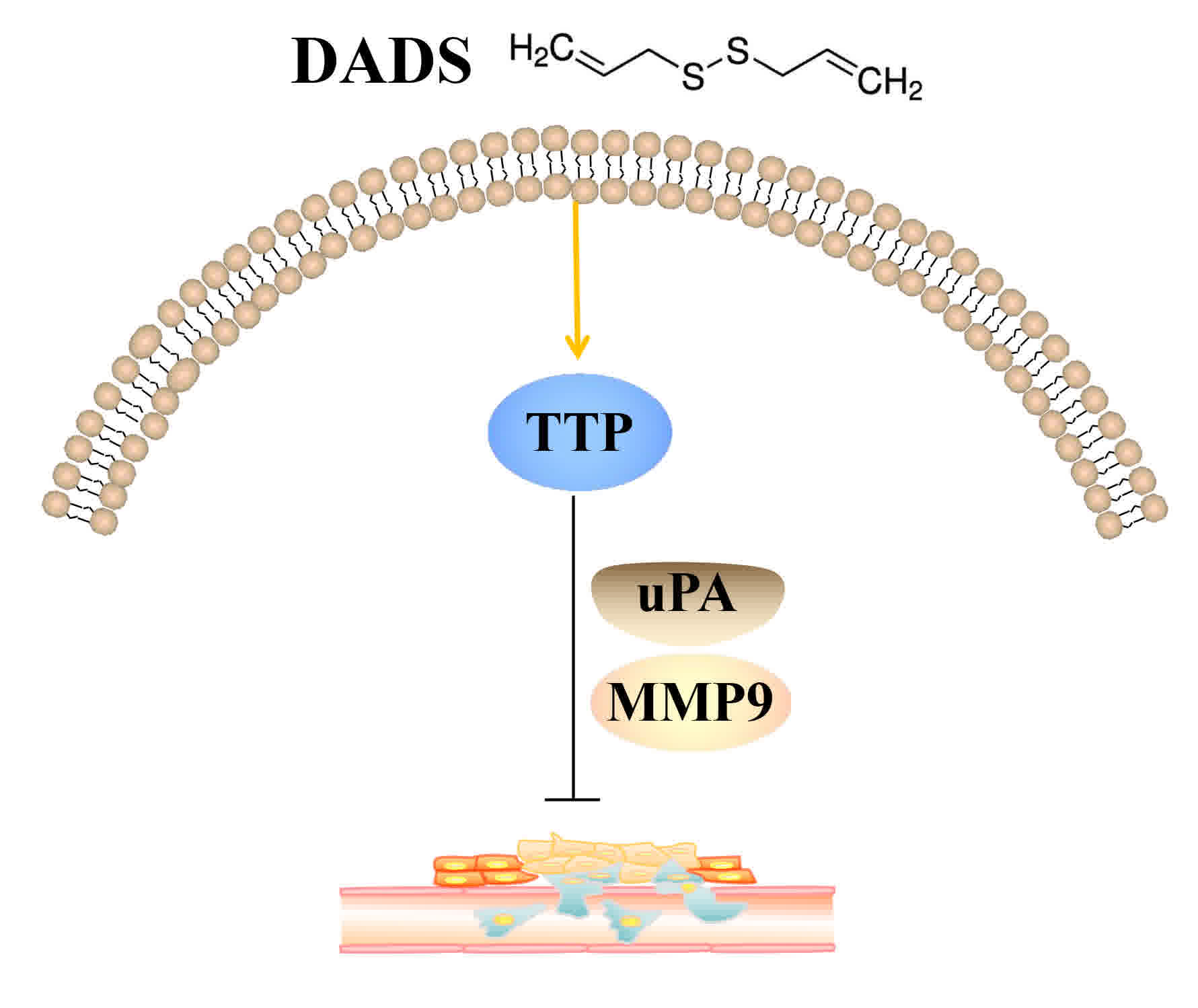
results indicate that DADS may inhibit the proliferation, invasion,
and migration of breast cancer cells by upregulating TTP while
downregulating the expression of uPA and MMP9 (Fig. 10) and TTP may be a novel target of
DADS in the progress of breast cancer.
Discussion
Breast cancer is the most commonly diagnosed type of
cancer and the primary cause of cancer-associated mortalities in
women worldwide (20). Over the last
two decades, with the development of early diagnostic methods and
more effective treatments, the mortality rate of patients with
breast cancer has declined (21).
However, metastatic breast cancer still lacks effective treatment
and remains the primary cause of breast cancer-associated mortality
(3). Therefore, full understanding
the underlying mechanism of metastasis is required in order to
establish methods that are able to effectively inhibit breast
cancer invasion and metastasis.
DADS, one of the major organosulfur compounds in
garlic, exhibits a wide variety of biological activities in
vivo and exhibits promising effects as a therapeutic agent in
various types of cancer. Certain anticancer mechanisms include
inhibiting the production of reactive oxygen species, inhibiting
cell cycle arrest and inducing apoptosis (4,22,23). However, the mechanism of action of
DADS in breast cancer remains unknown. Therefore, further study of
the function of DADS and how it inhibits the progress of breast
cancer cells is required.
In the present study, the antitumor activity of DADS
in breast cancer was evaluated using in vivo and in
vitro models. A mouse xenograft in vivo model was used,
which demonstrated that mice treated with DADS exhibited tumors
with significantly smaller weights and volumes compared with those
in the control group. Furthermore, DADS treatment significantly
inhibited the growth of breast cancer tumor xenografts by
upregulating TTP protein expression. Using tablet cloning,
Transwell and wound healing assays, it was revealed that DADS
significantly inhibited the cloning formation, invasion and
migration of MCF-7 and MDA-MB-231 cells compared with untreated
cells.
Cell invasion and metastasis are two important
biological characteristics of malignant tumor cells, but the
degradation of the extracellular matrix occurs first. As the tumor
prepares to metastasize, uPA protease is overexpressed in numerous
cancer cells, including in breast carcinoma, where it serves an
essential function in the metastatic process (24). uPA activates a cascade of proteases by
binding to its receptor, uPA receptor (uPAR), which ultimately
leads to the degradation of uPAR (25). MMPs degrade proteins that regulate
various cellular behaviors associated with cancer cell
differentiation, migration and invasion due to their proteolytic
nature. MMP-9 serves a key function in facilitating tumor
metastasis by degrading the extracellular matrix around the
vicinity of the tumor and destroying the basement membrane
integrity, leading to local invasion and distant metastasis of
tumor cells (26). Previous research
has demonstrated that uPA and MMP-9 are associated with the
regulation of invasion and metastasis in various types of malignant
tumor besides breast cancer, including prostate and colon cancer
(27–29). The results of the present study
revealed the function of DADS in inhibiting the progression of
breast cancer cells.
TTP, a tandem CCCH zinc-finger RNA-binding protein,
regulates the stability of several ARE-containing mRNAs. TTP serves
a fundamental function in various physiological and pathological
processes, including differentiation, reprogramming, metabolism,
proliferation, pluripotency and tumorigenesis (30). The loss of TTP has been reported in
several types of human cancer and is associated with poor prognosis
(30). Previous studies identified
that TTP expression is suppressed in numerous cancer cells compared
with in normal cell types (31). In
addition, a lack of TTP is associated with a variety of
cancer-associated processes. The regulation of TTP expression has
been identified to serve a function in several cancer types,
including colon and breast cancer (32,33).
However, its function in breast cancer has not been fully
investigated. TTP binds to the AREs within the 3′-untranslated
regions of uPA and uPAR, and the overexpression of TTP decreases
the expression of uPA and uPAR by enhancing the degradation of
their mRNAs (34). A previous study
suggested that TTP may be used as a promising therapeutic target to
treat human glioma (34). A previous
study identified that TTP regulates the degradation of growth
factors and proto-oncogenes (16). In
addition, a variety of inflammatory factors are able to enhance the
invasion and metastasis of tumor cells, including tumor necrosis
factor (TNF)-α, interleukin (IL)-1β and IL-6. TTP is known to
contribute to cytokine homeostasis by facilitating the degradation
of TNF-α mRNA, which is essential for reducing inflammation
(35).
TTP is a molecular marker of breast cancer that
regulates the proliferation, invasion and metastasis of breast
cancer cells (36). In the present
study, it was demonstrated that, following treatment with DADS, the
protein expression of TTP was significantly upregulated, but uPA
and MMP-9 expression levels were downregulated in a dose- and
time-dependent manner in MCF-7 and MDA-MB-231 cells. Furthermore,
silencing the expression of TTP using siRNA significantly increased
the expression of uPA and MMP-9 protein, and also significantly
enhanced the invasive and migratory capabilities of MCF-7 and
MDA-MB-231 cells. These results suggested that TTP may be a novel
target of DADS in inhibiting the progression of breast cancer.
However, there remains a lack of evidence that TTP is a target of
DADS, which are to be addressed in our further studies. This
prompts further investigation into the function of DADS in
uncontrollable inflammation and tumor growth. In conclusion, the
results of the present study suggest that DADS regulates TTP
expression and is a promising therapeutic target for breast cancer.
Further studies on TTP are required to provide a novel basis for
the targeted therapy of breast cancer, and to develop safe and
effective anticancer drugs.
Acknowledgements
Not applicable.
Funding
National Natural Science Foundation of China (grant
no. 81541163); the Open Fund Based on Innovation Platform of Hunan
Provincial Education Department (grant no. 15K111); High-level
talent research start-up fund of University of South China in 2017
(grant no. 24), Projects for Science Research of Hunan Provincial
Education Department (grant no. 16C1394); Hunan Provincial Bureau
of Traditional Chinese Medicine (grant no. 2014144); Hunan
Provincial Cooperative Innovation Center for Molecular Target New
Drug Study (grant no. 2014405); Innovation Program of College in
Cooperative Innovation Center for Molecular Target New Drug of
University of South China (grant no. 0223-0002-00028) and the
College Student's Innovative Project of University of South China
(grant no. 2016-133).
Availability of data and materials
The datasets used and/or analyzed during the current
study are available from the corresponding author on reasonable
request.
Authors' contributions
TX and XWL designed the study and wrote the paper;
XLH, XFX and WQX performed the experiments; SJZ and JT provided
reagents and mice, gave technical support and conceptual advice,
and reviewed and edited the manuscript. All authors read and
approved the manuscript.
Ethics approval and consent to
participate
All experimental procedures conformed to the Guide
for the Care and Use of Laboratory Animals published by the US
National Institutes of Health (NIH Publication No. 85–23, revised
1996) and was approved by the Institutional Animal Ethics Committee
of University of South China.
Consent for publication
Not applicable.
Competing interests
The authors declare that they have no competing
interests.
Glossary
Abbreviations
Abbreviations:
|
DADS
|
diallyl disulfide
|
|
MMP
|
matrix metalloproteinase
|
|
uPA
|
urokinase-type plasminogen
activator
|
|
AU
|
adenylate-uridylate
|
|
ARE
|
AU-rich element
|
|
TTP
|
tristetraprolin
|
|
siRNA
|
small interfering RNA
|
References
|
1
|
Niu HY, Niu CY, Wang JH, Zhang Y and He P:
Health-related quality of life in women with breast cancer: A
literature-based review of psychometric properties of breast
cancer-specific measures. Asian Pac J Cancer Prev. 15:3533–3536.
2014. View Article : Google Scholar : PubMed/NCBI
|
|
2
|
Lee Katie SY and Knobf MT: Primary breast
cancer decision-making among Chinese American Women: Satisfaction,
regret. Nurs Res. 64:391–401. 2015. View Article : Google Scholar : PubMed/NCBI
|
|
3
|
Shao S and Zhao X, Zhang X, Luo M, Zuo X,
Huang S, Wang Y, Gu S and Zhao X: Notch1 signaling regulates the
epithelial-mesenchymal transition and invasion of breast cancer in
a Slug-dependent manner. Mol Cancer. 14:282015. View Article : Google Scholar : PubMed/NCBI
|
|
4
|
Xiao X, Chen B, Liu X, Liu P, Zheng G, Ye
F, Tang H and Xie X: Diallyl disulfide suppresses SRC/Ras/ERK
signaling-mediated proliferation and metastasis in human breast
cancer by up-regulating miR-34a. PLoS One. 9:e1127202014.
View Article : Google Scholar : PubMed/NCBI
|
|
5
|
Su B, Su J, Zeng Y, Liu F, Xia H, Ma YH,
Zhou ZG, Zhang S, Yang BM, Wu YH, et al: Diallyl disulfide
suppresses epithelial-mesenchymal transition, invasion and
proliferation by downregulation of LIMK1 in gastric cancer.
Oncotarget. 7:10498–10512. 2016.PubMed/NCBI
|
|
6
|
Lai KC, Hsu SC, Kuo CL, Yang JS, Ma CY, Lu
HF, Tang NY, Hsia TC, Ho HC and Chung JG: Diallyl sulfide, diallyl
disulfide, and diallyl trisulfide inhibit migration and invasion in
human colon cancer colo 205 cells through the inhibition of matrix
metalloproteinase-2, −7, and −9 expressions. Environ Toxicol.
28:479–488. 2013. View Article : Google Scholar : PubMed/NCBI
|
|
7
|
Luo N, Zhao LC, Shi QQ, Feng ZQ, Chen DL
and Li J: Induction of apoptosis in human leukemic cell lines by
diallyl disulfide via modulation of EGFR/ERK/PKM2 signaling
pathways. Asian Pac J Cancer Prev. 16:3509–3515. 2015. View Article : Google Scholar : PubMed/NCBI
|
|
8
|
Huang J, Yang B, Xiang T, Peng W, Qiu Z,
Wan J, Zhang L, Li H, Li H and Ren G: Diallyl disulfide inhibits
growth and metastatic potential of human triple-negative breast
cancer cells through inactivation of the β-catenin signaling
pathway. Mol Nutr Food Res. 59:1063–1075. 2015. View Article : Google Scholar : PubMed/NCBI
|
|
9
|
Ciocci M, Iorio E, Carotenuto F, Khashoggi
HA, Nanni F and Melino S: H2S-releasing nanoemulsions: A new
formulation to inhibit tumor cells proliferation and improve tissue
repair. Oncotarget. 7:84338–84358. 2016. View Article : Google Scholar : PubMed/NCBI
|
|
10
|
Yi L, Shan J, Chen X, Li G, Li L, Tan H
and Su Q: Involvement of calreticulin in cell proliferation,
invasion and differentiation in diallyl disulfide-treated HL-60
cells. Oncol Lett. 12:1861–1867. 2016. View Article : Google Scholar : PubMed/NCBI
|
|
11
|
Su B, Su J, He H, Wu Y, Xia H, Zeng X, Dai
W, Ai X, Ling H, Jiang H and Su Q: Identification of potential
targets for diallyl disulfide in human gastric cancer MGC-803 cells
using proteomics approaches. Oncol Rep. 33:2484–2494. 2015.
View Article : Google Scholar : PubMed/NCBI
|
|
12
|
Sun Y, Wang X, Zhou Q, Lu Y, Zhang H, Chen
Q, Zhao M and Su S: Inhibitory effect of emodin on migration,
invasion and metastasis of human breast cancer MDA-MB-231 cells in
vitro and in vivo. Oncol Rep. 33:338–346. 2015. View Article : Google Scholar : PubMed/NCBI
|
|
13
|
Jacob A and Prekeris R: The regulation of
MMP targeting to invadopodia during cancer metastasis. Front Cell
Dev Biol. 3:42015. View Article : Google Scholar : PubMed/NCBI
|
|
14
|
Lampelj M, Arko D, Cas-Sikosek N, Kavalar
R, Ravnik M, Jezersek-Novakovic B, Dobnik S, Dovnik NF and Takac I:
Urokinase plasminogen activator (uPA) and plasminogen activator
inhibitor type-1 (PAI-1) in breast cancer-correlation with
traditional prognostic factors. Radiol Oncol. 49:357–364. 2015.
View Article : Google Scholar : PubMed/NCBI
|
|
15
|
Melanson BD, Bose R, Hamill JD, Marcellus
KA, Pan EF and McKay BC: The role of mRNA decay in p53 induced gene
expression. RNA. 17:2222–2234. 2011. View Article : Google Scholar : PubMed/NCBI
|
|
16
|
Brooks SA and Blackshear PJ:
Tristetraprolin (TTP): Interactions with mRNA and proteins, and
current thoughts on mechanisms of action. Biochim Biophys Acta.
1829:666–679. 2013. View Article : Google Scholar : PubMed/NCBI
|
|
17
|
Barrios-García T, Tecalco-Cruz A,
Gómez-Romero V, Reyes-Carmona S, Meneses-Morales I and León-Del-Río
A: Tristetraprolin represses estrogen receptor α transactivation in
breast cancer cells. J Biol Chem. 289:15554–15565. 2014. View Article : Google Scholar : PubMed/NCBI
|
|
18
|
Tran DDH, Koch A, Allister A, Saran S,
Ewald F, Koch M, Nashan B and Tamura T: Treatment with MAPKAP2
(MK2) inhibitor and DNA methylation inhibitor, 5-aza dC,
synergistically triggers apoptosis in hepatocellular carcinoma
(HCC) via tristetraprolin (TTP). Cell Signal. 28:1872–1880. 2016.
View Article : Google Scholar : PubMed/NCBI
|
|
19
|
Van Tubergen EA, Banerjee R, Liu M, Vander
Broek R, Light E, Kuo S, Feinberg SE, Willis AL, Wolf G, Carey T,
et al: Inactivation or loss of TTP promotes invasion in head and
neck cancer via transcript stabilization and secretion of MMP9,
MMP2, and IL-6. Clin Cancer Res. 19:1169–1179. 2013. View Article : Google Scholar : PubMed/NCBI
|
|
20
|
Redig AJ and McAllister SS: Breast cancer
as a systemic disease: A view of metastasis. J Intern Med.
274:113–126. 2013. View Article : Google Scholar : PubMed/NCBI
|
|
21
|
Warrier S, Tapia G, Goltsman D and Beith
J: An update in breast cancer screening and management. Womens
Health (Lond). 12:229–239. 2016. View Article : Google Scholar : PubMed/NCBI
|
|
22
|
Lei XY, Yao SQ, Zu XY, Huang ZX, Liu LJ,
Zhong M, Zhu BY, Tang SS and Liao DF: Apoptosis induced by diallyl
disulfide in human breast cancer cell line MCF-7. Acta Pharmacol
Sin. 29:1233–1239. 2008. View Article : Google Scholar : PubMed/NCBI
|
|
23
|
Wang G, Liu G, Ye Y, Fu Y and Zhang X:
Upregulation of miR-34a by diallyl disulfide suppresses invasion
and induces apoptosis in SGC-7901 cells through inhibition of the
PI3K/Akt signaling pathway. Oncol Lett. 11:2661–2667. 2016.
View Article : Google Scholar : PubMed/NCBI
|
|
24
|
Pei S, Yang X, Wang H, Zhang H, Zhou B,
Zhang D and Lin D: Plantamajoside, a potential anti-tumor herbal
medicine inhibits breast cancer growth and pulmonary metastasis by
decreasing the activity of matrix metalloproteinase-9 and −2. BMC
Cancer. 15:9652015. View Article : Google Scholar : PubMed/NCBI
|
|
25
|
Noh H, Hong S and Huang S: Role of
urokinase receptor in tumor progression and development.
Theranostics. 3:487–495. 2013. View Article : Google Scholar : PubMed/NCBI
|
|
26
|
Page-McCaw A, Ewald AJ and Werb Z: Matrix
metalloproteinases and the regulation of tissue remodelling. Nat
Rev Mol Cell Biol. 8:221–233. 2007. View
Article : Google Scholar : PubMed/NCBI
|
|
27
|
Moirangthem A, Bondhopadhyay B, Mukherjee
M, Bandyopadhyay A, Mukherjee N, Konar K, Bhattacharya S and Basu
A: Simultaneous knockdown of uPA and MMP9 can reduce breast cancer
progression by increasing cell-cell adhesion and modulating EMT
genes. Sci Rep. 6:219032016. View Article : Google Scholar : PubMed/NCBI
|
|
28
|
Shi C, Zhang N, Feng Y, Cao J, Chen X and
Liu B: Aspirin inhibits IKK-β-mediated prostate cancer cell
invasion by targeting matrix Metalloproteinase-9 and Urokinase-Type
plasminogen activator. Cell Physiol Biochem. 41:1313–1324. 2017.
View Article : Google Scholar : PubMed/NCBI
|
|
29
|
Tong W, Wang Q, Sun D and Suo J: Curcumin
suppresses colon cancer cell invasion via AMPK-induced inhibition
of NF-κB, uPA activator and MMP9. Oncol Lett. 12:4139–4146. 2016.
View Article : Google Scholar : PubMed/NCBI
|
|
30
|
Guo J, Qu H, Chen Y and Xia J: The role of
RNA-binding protein tristetraprolin in cancer and immunity. Med
Oncol. 34:1962017. View Article : Google Scholar : PubMed/NCBI
|
|
31
|
Griseri P, Bourcier C, Hieblot C,
Essafi-Benkhadir K, Chamorey E, Touriol C and Pagès G: A synonymous
polymorphism of the Tristetraprolin (TTP) gene, an AU-rich
mRNA-binding protein, affects translation efficiency and response
to Herceptin treatment in breast cancer patients. Hum Mol Genet.
20:4556–4568. 2011. View Article : Google Scholar : PubMed/NCBI
|
|
32
|
Sobolewski C, Sanduja S, Blanco FF, Hu L
and Dixon DA: Histone deacetylase inhibitors activate
tristetraprolin expression through induction of early growth
response protein 1 (EGR1) in colorectal cancer cells. Biomolecules.
5:2035–2055. 2015. View Article : Google Scholar : PubMed/NCBI
|
|
33
|
Pandiri I, Chen Y, Joe Y, Kim HJ, Park J,
Chung HT and Park JW: Tristetraprolin mediates the
anti-proliferative effects of metformin in breast cancer cells.
Breast Cancer Res Treat. 156:57–64. 2016. View Article : Google Scholar : PubMed/NCBI
|
|
34
|
Ryu J, Yoon NA, Lee YK, Jeong JY, Kang S,
Seong H, Choi J, Park N, Kim N, Cho WJ, et al: Tristetraprolin
inhibits the growth of human glioma cells through downregulation of
urokinase plasminogen activator/urokinase plasminogen activator
receptor mRNAs. Mol Cells. 38:156–162. 2015.PubMed/NCBI
|
|
35
|
Prabhala P and Ammit AJ: Tristetraprolin
and its role in regulation of airway inflammation. Mol Pharmacol.
87:629–638. 2015. View Article : Google Scholar : PubMed/NCBI
|
|
36
|
Gebeshuber CA, Zatloukal K and Martinez J:
miR-29a suppresses tristetraprolin, which is a regulator of
epithelial polarity and metastasis. EMBO Rep. 10:400–405. 2009.
View Article : Google Scholar : PubMed/NCBI
|