Introduction
Gastric cancer is the fourth most common malignancy
and accounts for >740,000 cancer-associated mortalities/year
globally (1,2). Despite great improvements made in
therapeutic methods in recent years, the prognosis is still
unsatisfactory (3). Novel
combinations of conventional chemotherapies, including the SPIRITS
trial (S1 plus cisplatin vs. S1) demonstrated improved overall
survival in patients treated with S1 plus cisplatin (13.0 months)
compared with those treated with S1 alone (11.0 months) (4). Additionally, other combinations of
cytotoxic agents, including docetaxel, irinotecan, capecitabine and
oxaliplatin, have also been reported to prolong survival. However,
therapeutic efficacy is still limited by two major factors: Drug
resistance and side effects (5–7).
Human epithelial growth factor receptor 2 (HER2) is
over-expressed in a significant proportion of gastric cancers
(6–23%) (8). It is associated with
tumor invasion, metastasis, chemoresistance and poor prognosis
(9). Trastuzumab, as a recombinant
humanized monoclonal antibody that targets the extracellular domain
IV of HER2, is one of the most promising targets in human
malignancy in recent years (10). In
trial investigating the use of trastuzumab for gastric cancer, the
addition of trastuzumab (Herceptin®) to chemotherapy
significantly improved overall survival (13.8 months, 95% CI 12–16)
compared with chemotherapy alone (11.1 months, 95% CI 10–13) in
patients with HER2-positive gastric cancer (11), which indicated that combined
chemotherapy with trastuzumab may be a novel treatment for patients
with HER2-positive advanced gastric cancer. However, the majority
of patients with gastric cancer still develop acquired resistance
to trastuzumab (12). To achieve
improved benefits for HER2-targeted therapy, the development of
novel drug delivery systems that may decrease the dosage and
periods of molecular targeted therapy are urgently required.
Nanoparticle (NP)-based therapeutics offer an
innovative method to overcome the limitations of current agents
(13). NPs possess unique properties
that enable them to be used as imaging probes, which may be traced
via magnetic resonance imaging (MRI) and therapeutic agents at the
same time (14); however, they can be
further loaded to deliver specific drugs (15–16) or
target specific molecules (17,18).
Superparamagnetic iron oxide (SPIO), known to be a highly efficient
T2 contrast agent for MRI, is an ideal small molecular
probe for medical use (19). Although
SPIO exploit an enhanced permeability and retention effect (EPR)
for tumor uptake, EPR is still inefficient with relatively low
concentrations of NPs reaching tumors (20). Vascular heterogeneity commonly exists
in large tumors, particularly metastases, and leads to
unpredictable rates of NP extravasation as well as decreased
perfusion and overall uptake (21,22).
Therefore, the development of active targeting NPs is required.
However, reports on SPIO with a combined feature of targeted
intracellular drug release and imaging function are rare.
In the present study, a dual-functioning NPs
conjugate, Au-Fe3O4, for HER2 targeted
oxaliplatin delivery and intracellular drug release triggered via
pH, was developed. In order to illustrate its targeting and
therapeutic potential, cell culture and animal experiments of
oxaliplatin-Au-Fe3O4-Herceptin NPs against
human gastric cancer cell line SGC-7901 were conducted. It was
demonstrated in the present study that selective targeting of
HER2-positive gastric cancer cells using
oxaliplatin-Au-Fe3O4-Herceptin NPs may
increase the efficacy and decrease the side effects of oxaliplatin
chemotherapy.
Materials and methods
Preparation of
Au-Fe3O4 NPs modified with poly(ethylene
glycol) (PEG)
For typical synthesis of 8 nm Au NPs, a precursor
solution containing hexane (10 ml), oleylamine (10 ml) and 0.1 g of
HAuCl4.4H2O (Aladdin Shanghai
Biochemical Technology Co., Ltd., Shanghai, China) was prepared and
magnetically stirred at 15°C under low flow N2 (15°C, 15
MPar). Following 10 min, a premixed solution (at 15°C) containing
tetrabutylammonium bromide (0.5 mmol), hexane (1 mmol) and
oleylamine (1 mmol) was injected into the precursor solution; the
solution changed color to deep purple within 5 sec. The mixture was
incubated at 15°C for 1 h prior to the precipitation by absolute
ethyl alcohol. The precipitated Au NPs were collected by
centrifugation (11,688 × g, 8 min), dissolved in absolute ethyl
alcohol and detected via transmission electron microscope (TEM;
JEOL 1230; JEOL, Ltd., Tokyo, Japan) at 80–200 kV.
Au NPs (0.48 mmol), octadecene (20 mmol), oleic acid
(8 mmol) and oleylamine (8 mmol) were then mixed prior to adding
iron acetylacetonate (6 mmol) and dodecanediol (20 mmol) into the
solution at 180°C for 2 h followed by 300°C for 1 h. Following
cooling to room temperature, Au-Fe3O4 NPs
were collected by centrifugation (16,501 × g, 10 min) and detected
via TEM at 80–200 kV.
Surface modification of
Au-Fe3O4 NPs
For Au-Fe3O4 modification, 50
ml α,ω-Bis{2-[(3-carboxy-1-oxopropyl)amino]ethyl}PEG
(Mr=2,000; Sigma-Aldrich; Merck KGaA, Darmstadt,
Germany), 1.0 mg of N-hydroxysuccinimide (NHS; Sigma-Aldrich; Merck
KGaA), 1.25 g of dicyclohexylcarbodiimide (Aladdin Shanghai
Biochemical Technology Co., Ltd., Shanghai, China) and 1.5 g of
dopamine hydrochloride were dissolved in a mixture of
CHCl3 (20 mmol), dimethylformamide (8 mmol) and
anhydrous Na2CO3 (6 mmol). The solution was
stirred at 37°C for 2 h, Au-Fe3O4 NPs (5 mg)
were added, and the resulting solution was stirred overnight at
37°C under a N2 flow. The modified NPs were precipitated
by adding cyclohexane (25 mmol), collected by centrifugation at
16,501 × g, dried under N2 flow at 40°C (23). Surfactants and other salts were
removed via dialysis (molecular mass cut off, 14,000 kDa; Spectrum
Laboratories, Inc., Rancho Dominguez, CA, USA) for 24 h in PBS or
water. The final iron concentration of the particles was determined
by inductively coupled plasma mass spectrometry (ICP-MS; Element
XR, Thermo Fisher Scientific, Inc., Waltham, MA, USA), which is a
type of mass spectrometry capable of detecting metals and several
non-metals at concentrations as low as one part in 1015
(part/quadrillion) on non-interfered low-background isotopes. The
ICP multi-element standard solution IV (1,000 mg/kg, Merck KGaA,
Darmstadt, Germany) was used as background equivalent concentration
in the ICP-MS measurement. The nitric acid (HNO3) and hydrochloric
acid (HCl) used were ultra-pure 100 grade (Merck KGaA). High
purified water was obtained by a Milli-Q system (Merck KGaA). The
operating conditions of ICP-MS are listed in Table I.
 | Table I.Operating conditions of ICP-MS. |
Table I.
Operating conditions of ICP-MS.
| Parameter | Setting |
|---|
| Radio frequency
power | 1300 W |
| Plasma gas flow
rate | 16 l/min |
| Auxiliary gas flow
rate | 0.85 l/min |
| Carrier gas flow
rate | 1.12 l/min |
| Nebulizer | Micromist nebulizer
with uptake rate of 200 l/min |
| Spray chamber | Isomist spray
chamber |
| Sampling cone | 1.0 mm |
| Skimmer cone | 0.7 mm |
| Sampling depth | 2.6 mm |
| Replicate of
mesurement | 5 times |
| Measured element
and resolution mode (m/Δm) | 57Fe
(4000), 195Pt (4000) |
Conjugation of Herceptin and
oxaliplatin to Au-Fe3O4 NPs
To conjugate the anti-HER2 antibody Herceptin,
Au-Fe3O4 NPs in methyl ester sulfonate
(Aladdin Shanghai Biochemical Technology Co., Ltd.) were mixed with
1-ethyl-3-(3-dimethylaminopropyl) carbodiimide (EDC; Sigma-Aldrich;
Merck KGaA) for 15 min at 4°C. Sulfo-NHS (Sigma-Aldrich; Merck
KGaA) was added into the solution, which was then subjected to
PD-10 column (GE Healthcare Life Sciences, Uppsala, Sweden)
filtering to remove excessive EDC and sulfo-NHS, according to the
manufacturer's protocol. Herceptin (Genentech Inc., San Francisco,
CA, USA) was added into the conjugate following the alteration of
pH to 7.4. The antibody-conjugated NPs were separated from unbound
Herceptin and Au-Fe3O4 NPs using 300 K
ultra-filtration (Optima MAX-TL, Beckman Coulter, Inc., Brea, CA,
USA). The peptide bond of Au-Fe3O4-Herceptin
was recorded by infrared (IR) spectroscopy on a Nicolet™ iS10
spectrometer (Thermo Fisher Scientific, Inc.) in the range between
2,500–400 cm−1 and a resolution of 2 cm−1.
The Herceptin mass spectrum was detected by matrix-assisted
laser-desorption ionization/time-of-flight (TOF)/TOF using
Ultraflex III TOF/TOF MS (Bruker-Michrom, Inc., Auburn, CA, USA).
MALDI-TOF MS measurements were taken using reflectron positive-ion
mode. Acceleration was performed at 25 kV. Laser power was set as
high as possible allowing baseline separation of isotopic peaks.
Sample spectra were acquired by summing 25,000 laser shots at a
frequency of 2,000 Hz, using a window from m/z 2,000 to 20,000
(Table II). To confirm the
structures of the peaks, tandem mass spectrometry (MALDI-TOF/TOF
MS/MS) was performed using laser-induced disassociation. Data
acquisition was performed with the FlexImaging 3.0 software
(Bruker-Michrom, Inc.).
| Table II.Operating conditions of MALDI-TOF
MS. |
Table II.
Operating conditions of MALDI-TOF
MS.
| Parameters | Settings |
|---|
| Mode | Reflectron
positive-ion mode |
| Laser | 200 Hz smartbeam-I
laser |
| Delay | 80 ns |
| Ion source | 1 voltage, 25 kV; 2
voltage, 23.4 kV |
| Lens voltage | 6 kV |
| Mass range | 2,000–20,000
kDa |
| Software | FlexImaging 3.0
software (Bruker-Michrom, Inc.) |
For oxaliplatin-binding ligand synthesis, ethyl
bromoacetate (8 mmol) and KI (3 mmol) were added to a solution of
cystamine dihydrochloride (1.125 g, 5 mmol) in 100 ml acetone and
10 ml Et3N, stirred for 6 h at room temperature and the
insoluble solid was removed via filtration. The filtrated crude
products was dried and purified on a Biotage flash chromatography
(Isolera™, Biotage Inc. Uppsala, Sweden) with SiliaFLASH™
cartridges (SiliCycle Inc. Quebec City, Canada) according to the
manufacturer's protocol (24). An
appropriate solvent mixture (petrol/EtOAc, 3:1) was used as eluent,
obtaining a yield of ~80% (Table
III). The carboxylic groups were deprotected in methanol
solution (0.496 g, 1 mmol), 5 ml of 1 M NaOH aqueous solution was
added, stirred for 30 min at room temperature and a small amount of
water was added if any precipitate remained. Following 24 h of
stirring, 20 ml distilled water was added, the solution was
acidified to pH 3.0 using 1 M HCl and the resulting precipitate was
collected by centrifugation (16,501 × g) at 37°C and washed with
EtOH/H2O (1:1). The detection of ligands of
antibody-coupled Au-Fe3O4 NPs was preceded by
nuclear magnetic resonance (NMR; Bruker Advance 2B/400 MHz; Bruker
Corporation).
| Table III.Chromatographic conditions. |
Table III.
Chromatographic conditions.
| Parameters | Settings |
|---|
| Column | SiliaFLASH™ |
| Sample volume | 8 ml |
| Band length | 6 mm |
| Application
rate | 10 s/µl |
| Scanner | Camag TLC scanner
III |
| Scanning speed | 20 mm/s |
| Data
resolution | 100 µm/step |
| Measurement
mode | Absorption |
| Flow rate | 10 ml/min |
| Pressure | 8 psi |
The antibody-coupled Au-Fe3O4
NPs (1 mg) were mixed with oxaliplatin binding ligand solution
(described previously) for 6 h at room temperature, uncoupled
ligands were removed using a PD-10 column. Oxaliplatin (20 mg/ml;
Eloxatin; Sanofi S.A., Paris, France) was added to the NP solution,
stirred overnight at room temperature in the dark and unconjugated
oxaliplatin was removed via low speed centrifugation (12,000 × g)
followed by purification on a PD-10 column. The amount of
oxaliplatin bound to the NPs was determined via ICP-MS under the
same experimental conditions as previously stated (Element XR,
Thermo Fisher Scientific, Inc.).
Oxaliplatin release assay
Oxaliplatin-Au-Fe3O4-Herceptin
NPs (50 µg/2 ml) were placed into Slide-A-Lyzer™ Dialysis Cassettes
(molecular mass cut off, 2,000 kDa; Thermo Fisher Scientific,
Inc.), which was in 30 ml PBS at room temperature. At the time
intervals of 1, 2, 4, 8 and 19 h, 1 ml PBS was sampled. The
platinum concentration was determined via Element XR ICP-MS (Thermo
Fisher Scientific, Inc.) additional details can be found in
Table I.
Cell lines and animals
Stomach adenocarcinoma cell line SGC-7901 was
purchased from American Type Culture Collection (Manassas, VA,
USA). SGC-7901 cells were cultured in RPMI-1640 medium (Hangzhou
Gino Biomedical Technology Co., Ltd., Hangzhou, China) supplemented
with 10% fetal bovine serum (FBS; Hangzhou Sijiqing Biological
Engineering Materials, Co., Ltd., Hangzhou, China) and 1%
penicillin/streptomycin (Genom Biologic). Cells were incubated in a
standard humidified incubator in 5% CO2 at 37°C and
passaged every 3–5 days using trypsin-EDTA (Genom Biologic). A
total of 50, 4–6-week-old female BALB/c nu/nu mice (18–22 g,
Shanghai Institutes for Biological Sciences, Shanghai, China) used
in the present study were housed 5 to a cage under pathogen-free
conditions and maintained on a daily 12/12 h light/dark cycle at
Zhejiang University Laboratory Animal Center (Hangzhou, China), a
fully accredited Association for Accreditation of Laboratory Animal
Care facility. Animals were housed in a sterile environment, fed
sterilized food and water ad libitum. All animal studies
were carried out in strict accordance with the guidelines for the
welfare and use of animals in cancer research of the Committee of
the National Cancer Research Institute. The Zhejiang University
Animal Research Committee approved the present study protocol. All
surgery was performed under sodium pentobarbital anesthesia (40
mg/kg), and all efforts were made to minimize suffering (25). Animal health and behavior was
monitored every 12 h, and were euthanized when the mice display
early markers associated with mortality or a poor prognosis,
including abdominal distension, emaciation or cachexy.
Cell viability assay (cytotoxicity
assay)
The cytotoxicity of NPs on SGC-7901 was determined
using MTS assays (26). Briefly,
SGC-7901 cells (4×10³ cells/well) were seeded into
96-well plates and incubated overnight at 37°C.
Au-Fe3O4 NPs with different Fe concentrations
(0, 50, 100, 150, 200 and 250 mg/l) were added to the designed
wells and incubated at 37°C for 24, 48 and 72 h. Following
incubation at 37°C with MTS solution
(CellTiter96®AQueous One Solution Cell
Proliferation Assay, Promega Corporation, Madison, WI, USA) for 2
h, the absorbance was measured using a TU-1901 Ultraviolet-Visible
Spectrophotometer (Beijing Purkinje General Instrument, Co, Ltd.,
Beijing, China) at 570 nm. The same method was used to measure the
cytotoxicity of Au-Fe3O4-Herceptin and
oxaliplatin-Au-Fe3O4-Herceptin at Fe
concentrations of 0, 30, 50, 80 and 100 mg/l.
Cellular internalization of NPs
Oxaliplatin-Au-Fe3O4-Herceptin
NPs with 0.01 mg/ml Fe were added to SGC-7901 cells until they were
70% confluent, cultured with RPMI-1640 medium (10% FBS, 1%
penicillin) and incubated for 2 h at 37°C. Cells
(1×10⁵/ml) were plated on sterile glass slides were
first fixed with 2.5% glutaraldehyde (Aladdin Shanghai Biochemical
Technology Co., Ltd.) in phosphate buffer (pH 7.0, Aladdin Shanghai
Biochemical Technology Co., Ltd.) for 4 h and then with 1%
OsO4 (Aladdin Shanghai Biochemical Technology Co., Ltd.)
in phosphate buffer (pH 7.0) for 1 h at room temperature. The
specimens were placed in a 1:1 mixture of absolute acetone and
Spurr's resin (AGAR Scientific, Ltd., Stansted, UK) for 1 h at room
temperature, then placed in capsules contained embedding medium
(Sigma-Aldrich; Merck KGaA) and heated at 70°C for 9 h. The
specimen sections of cells were stained with uranyl acetate and
alkaline lead citrate for 15 min consecutively and observed under
TEM of Model H-7650 (Hitachi, Ltd., Tokyo, Japan) at 80 kV.
Xenograft tumor model and in vivo
imaging by MRI
A total of 20 4–6-week-old female BALB/c nu/nu mice
weighing 18–22 g were used for tumor implantation. Sub-confluent
SGC-7901 cells were harvested and re-suspended in PBS to a final
concentration of 1.0×10⁸/ml. Tumor cell suspension was
subcutaneously injected into the flank of each mouse. After 4
weeks, when the subcutaneous mass reached a diameter of 1.0 cm,
prior to the development of signs of distress,
oxaliplatin-Au-Fe3O4-Herceptin NPs complex
was injected into the mouse model via the caudal vein at an
equivalent iron concentration (0.5 mg/kg). Following 24 h,
relaxation time measurements (T1 and T2) of
an aqueous solution of
oxaliplatin-Au-Fe3O4-Herceptin and NPs
complex in vivo were acquired using a clinical magnetic
resonance scanner (Signa EXCITE 3.0T HD; GE Healthcare Life
Sciences) without contrast. An 11-cm circular coil was used for all
the MRI studies. With regards to the T2-weighted
sequence, a repetition time of 750 msec and an echo time of 50–300
msec were employed. To alleviate animal suffering, all mice were
euthanized by the end of MRI studies via carbon dioxide
asphyxiation followed by cervical dislocation, as the majority of
mice become moribund within days of this time point. Subcutaneous
tumor masses were harvested and immediately immersed in 10% neutral
buffered formalin (pH 6.8–7.2, Thermo Fisher Scientific, Inc.) for
2 h at room temperature and then paraffin embedded and sliced (4
µm).. Iron staining was performed using an iron stain kit
(Sigma-Aldrich; Merck KGaA) according to the manufacturer's
protocol. Images (magnification, ×200) were evaluated with a light
microscope (Leica DMi1, Leica Microsystems GmbH, Wetzlar,
Germany).
Statistical analysis
Statistical analysis was performed with Student's
t-test and one-way analysis of variance using SPSS 10.0 software
(SPSS, Inc., Chicago, IL, USA). Bonferroni correction was applied
for multiply comparisons dividing the significance level by the
number of tested variables. All experiments were performed at least
in triplicates and are expressed as the mean ± standard deviation
(SD). P<0.05 was considered to indicate a statistically
significant difference.
Results and Discussion
Synthesis and characterization of
Au-Fe3O4 NPs
The 8–20 nm Au-Fe3O4 NPs were
synthesized by decomposing iron pentacarbonyl on the surfaces of Au
NPs in the presence of oleic acid and oleylamine. TEM was used to
characterize the synthesis of the dumbbell-like
Au-Fe3O4 NPs. Au NPs were observed as uniform
spheres with an average size of 8 nm, based on the size
(diameter)-distribution of ~100 NPs. The Au NPs within the
Au-Fe3O4 NP complexes appeared to be black
under TEM images (Fig. 1A) due to the
heavy atom effect (27).
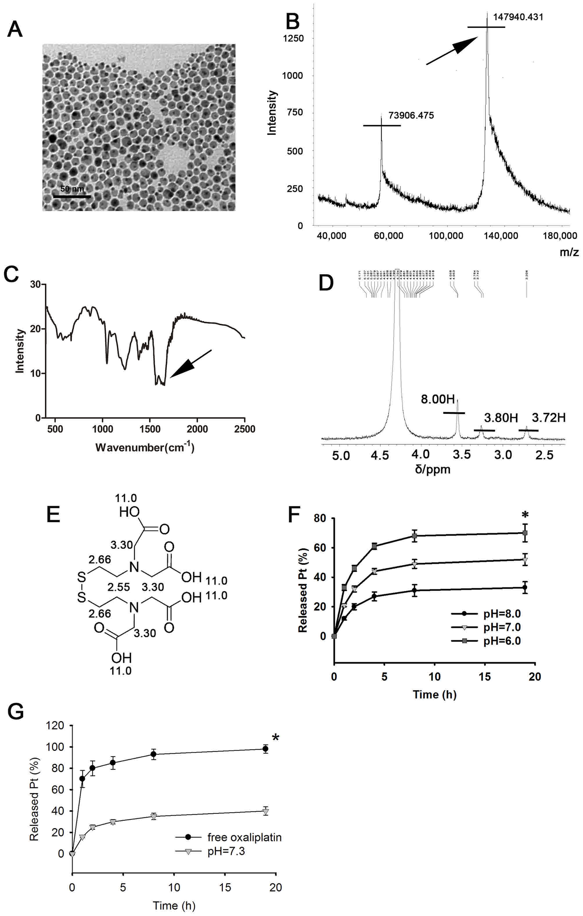 | Figure 1.Synthesis and characterization of
oxaliplatin-Au-Fe3O4-Herceptin NPs. (A)
Transmission electron microscope images of dumbbell-shaped
structure of 8–20 nm Au-Fe3O4 NPs, formed of
8 nm Au NPs (black) and 20 nm Fe3O4 NPs
(grey). (B) Mass spectrometry analysis of
Au-Fe3O4-Herceptin NPs. A specific peak for
human epithelial growth factor receptor antibody, Herceptin
(Mr=148), at m/z ~148,000 was detected by
matrix-assisted laser-desorption ionization-TOF-TOF. (C) Infrared
spectrum of Au-Fe3O4-Herceptin NPs. Specific
double peaks of carbonyl bands at 1580 and 1650 cm−1
(arrow) representing the amide bonds linking the polyethylene
glycol to the silane. (D) NMR of the oxaliplatin-binding ligand
(D2O, 300 MHz) revealed peaks at δ 3.51 (8.00 H, s,
3-H), 2.97–3.02 (3.80 H, t, 2-H) and 2.72–2.76 (3.72 H, t, 1-H).
(E) The NMR analog of oxaliplatin-binding ligand. (F) The released
oxaliplatin level from pH=6.0 group was significantly increased
compared with pH=8.0 group. (G) The released oxaliplatin level at
pH=7.3 after 4 h of incubation was significantly compared with the
free oxaliplatin group. *P<0.01.*P<0.01 vs. pH=8.0. NMR,
Nuclear Magnetic Resonance; NPs, nanoparticles; TOF, time of
flight; Pt, oxiplatin. |
Xu et al (28)
reported the structure of Au-Fe3O4 NP in
2009. Compared with conventional single-component Au or
Fe3O4 NPs, Au-Fe3O4 NPs
possess unique advantages. First, the structure contains magnetic
(Fe3O4) and optically active plasmonic (Au)
units and can therefore be stably detected optically and
magnetically, compared with the fast signal loss observed in common
fluorescent labeling. Secondly, the presence of
Fe3O4 and Au surfaces facilitate the
attachment of different chemical functional groups, enabling a
variety of target-specific imaging and delivery applications.
Thirdly, the size of NPs can be controlled to optimize their
magnetic and optical properties, and as small particles are only
capable of accommodating a few DNA strands, proteins, antibodies or
therapeutic molecules, kinetic cell targeting and drug release
assays can be performed (23).
Due to the high surface area-to-volume ratio, NPs
have a tendency to aggregate and absorb plasma proteins upon
intravenous injection, leading to rapid clearance by the
reticuloendothelial system (RES) (29). The size of the
Au-Fe3O4 NPs in the present study is suitable
for in vivo applications, as it has been demonstrated that
NPs between 10–150 nm in diameter can effectively escape rapid
clearance by the RES. NPs are commonly protected with a polymer
coating to improve their dispersity and stability (30). In the present study, PEG was used to
align the oil phase of the NPs to the water phase, dopamine was
reacted with the oleic acid on the surface of the
Fe3O4 NPs, then the amino group of dopamine
was reacted with the carboxyl group of PEG under catalysis of the
EDC-sulfo-NHS system. This ensured that PEG was only connected to
the Fe3O4 NPs. The ζ-potential of the
dumbbell-shaped Au-Fe3O4 NPs prior to surface
modification was 7.1±1.2 mV. Following being loaded with PEG, the
dumbbell-like Au-Fe3O4 NPs possessed a
ζ-potential of 13.8±1.6 mV. It is notable that the surface charge
density of NPs is an important parameter that determines their
colloidal stability and cellular interactions (31).
Fe3O4 surface
functionalization and multifunctional probe loading
Herceptin is a recombinant, DNA-derived, humanized
monoclonal antibody glycoprotein that selectively targets the
extracellular domain of HER2 (32).
Herceptin contains 1,328 amino acids and has a Mr of
~148 (33). Herceptin has been
approved for the clinical treatment of HER2-overexpressing breast
cancer, metastatic gastric cancer and gastro-esophageal junction
adenocarcinoma (34,35). In the present study, the EDC-sulfo-NHS
system was used to activate the carboxyl groups of
Au-Fe3O4 NPs modified with PEG, as
aforementioned. Removal of excess EDC and sulfo-NHS prior to
addition of Herceptin was used to avoid self-crosslinking of the
antibody. Following activation using the EDC-sulfo-NHS system,
Herceptin was conjugated to the modified
Au-Fe3O4 NPs through PEG
(Mr=2,000) and dopamine via a condensation reaction with
the formation of a peptide bond. Mass spectrometry analysis of
oxaliplatin-Au-Fe3O4-Herceptin NPs revealed a
specific peak for Herceptin at m/z ~148,000 (Fig. 1B), which was not detected in
Au-Fe3O4 NPs. IR spectrum analysis of
oxaliplatin-Au-Fe3O4-Herceptin NPs revealed
the presence of a peptide bond, indicated by specific double split
peaks between 1,580–1,650 cm−1 (Fig. 1C).
Oxaliplatin has long been used as a chemotherapeutic
agent (36); however, it does not
specifically target tumor cells and can be taken-up by any rapidly
growing cells, leading to toxic side effects (37). The present study proposed that the
conjugation of oxaliplatin onto
Au-Fe3O4-Herceptin NPs, which can actively
accumulate in HER2 positive tumor tissues, may greatly decrease the
side effects of oxaliplatin. The Au particle of the
Au-Fe3O4-Herceptin NPs was coated with thiol
(HS)-PEG-NH2 with the thiol for oxaliplatin binding. NMR results of
the ligand (D2O, 300 MHz) revealed three peaks at δ 3.51
(8.00 H, s, 3-H), 2.97–3.02 (3.80 H, t, 2-H) and 2.72–2.76 (3.72 H,
t, 1-H), as presented in Fig. 1D. The
NMR of the analog is depicted in Fig.
1E. Under certain conditions, the disulfide bond of the ligand
was broken. One sulfur atom of the ligand was connected to an Au NP
and another to oxaliplatin. In the present study, according to the
weight percentage of Pt/Au (13.2%), giving a Pt/Au atom ratio of
13:100, ~2,084 Pt units were bound to each Au NP.
The results of drug release profiles in vitro
at different pH values indicated that
oxaliplatin-Au-Fe3O4-Herceptin NPs released
13, 21 and 32% of oxaliplatin in the first h and a total of 25, 43
and 58% by 4 h at pH 8.0, 7.0 and 6.0, respectively (P=0.003)
(Fig. 1F). It appears that
oxaliplatin became more prone to detach from the conjugated NPs in
lower pH conditions, and that Pt release is pH dependent. When
human serum has a pH of ~7.35, only ~25% of oxaliplatin will be
released from the conjugated NPs by 4 h of incubation (28), which delayed the oxaliplatin release
by ~4 times compared with free oxaliplatin (80% release, Fig. 1G). However, following uptake by target
cells, particularly in the endosomal/lysosomal compartments, which
have a pH of ~5.0, >60% of oxaliplatin was released from
oxaliplatin-Au-Fe3O4-Herceptin NPs (28). This pH dependent release character
could reduce the systemic toxicity effect to a great extent and is
ideal for the selective targeting therapy in vivo.
Cellular uptake and cytotoxicity of
oxaliplatin-Au-Fe3O4-Herceptin NPs in
SGC-7901 cells in vitro
The SGC-7901 cell line is known to overexpress HER2
and is an ideal cell line to assess the efficacy of HER2 targeted
therapy (38,39).
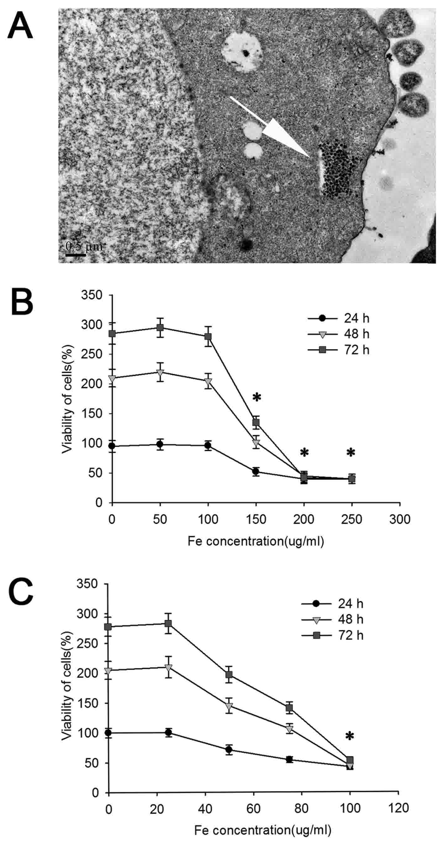
Oxaliplatin-Au-Fe3O4-Herceptin NPs (0.01
mg/ml Fe) were incubated with SGC-7901 cells for 2 h. TEM image
analysis revealed the presence of NPs in the endosome/lysosome,
which indicated the successful uptake of NPs through endocytosis
(Fig. 2A).
The cytotoxicity of NPs depends on their
concentration and the surfactants used (28). In the present study, the water-soluble
Au-Fe3O4 NPs were tested over a concentration
range of 0–250 mg Fe/l. The MTS assay (Fig. 2B) revealed that the
Au-Fe3O4 NPs did not induce appreciable cell
viability (~100%) for ≤100 µg/ml Fe; however, cell viability
decreased in a concentration-dependent manner at a Fe
concentrations >100 µg/ml, with only half of the cells viable at
a Fe concentration of 150 µg/ml (P=0.005). Therefore, within the
safe zone (0–100 µg/ml), Au-Fe3O4 did not
inhibit cell growth and the cytotoxicity of the NPs itself to the
tumor cells may be negligible.
The cytotoxicity assays were further performed to
assess the effect of
oxaliplatin-Au-Fe3O4-Herceptin NPs on the
viability of HER2-positive SGC-7901 cells in vitro (Fig. 2C). The control experiments
demonstrated that the cytotoxicity of
oxaliplatin-Au-Fe3O4-Herceptin NPs was
markedly increased compared with Au-Fe3O4 NPs
(P=0.005). The oxaliplatin-Au-Fe3O4-Herceptin
NPs had a half-maximal inhibitory concentration toward SGC-7901
cells of 75 µg/ml (Fe concentration). The toxicity was considered
to be as a result of two sources, one from Herceptin, the other
from oxaliplatin released by hydrolysis.
A variety of specific antigens, including HER2, are
frequently overexpressed on the surface of tumor cells. These
antigens provide specific targets, which could be selectively bound
by monoclonal antibodies. Therefore, when linked to a monoclonal
antibody, NP conjugates may enable target-specific delivery via
high affinity antibody-antigen interactions and receptor-mediated
endocytosis (40–42). A number of cationic NPs have been
indicated to enter cells by transiently generating holes in the
cell membrane (43). However, most of
the uptake of NPs into mammalian cells and macrophages usually
occurs via endocytosis (44,45). In the present study, TEM indicated
that the platinum-tethered NPs synthesized in the present study
were internalized by SGC-7901 cells via endocytosis. Therefore,
conjugation of Herceptin to Au-Fe3O4 NPs is
an effective way for targeted internalization.
Imaging of the selective uptake and
accumulation of
oxaliplatin-Au-Fe3O4-Herceptin NPs in
HER2-expressing gastric cancer cells in vivo
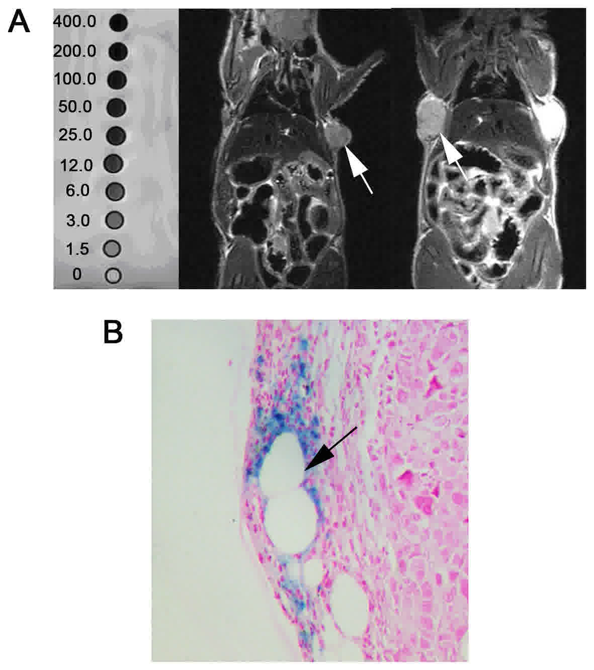
The specific targeting efficiency of
oxaliplatin-Au-Fe3O4-Herceptin NPs with
various iron concentrations towards HER2-positive tumor cells was
investigated in vivo using a xenograft model of SGC-7901 and
T2-weighted MRI without contrast. As depicted in
Fig. 3A, the T2 relaxation
time decreased with the increase of the iron concentration in
T2-weighted MRI (Fe concentration of 0.0, 1.5, 3.0, 6.0,
12.0, 25.0, 50.0, 100.0, 200.0, 400.0 µg/ml).
For the purpose of acquiring adequate NPs
accumulation at site and a safe dosage for the mice, in the present
study, a dose of 0.35 mg/kg Fe was selected for the in vivo
experiments based on the in vitro cytotoxicity assays. A
total of 24 h following caudal vein injection, the mice were sodium
pentobarbital anesthetized and received MRI examination. In
T2-weighted imaging, the signal intensity of
subcutaneous mass was significantly decreased in the
oxaliplatin-Au-Fe3O4-Herceptin NPs group,
compared with the group treated with Au-Fe3O4
NPs alone, which demonstrated no detectable intensity alteration.
It indicated that the HER2-labeled NPs exhibit increased
specificity in their attachment to SGC-7901 cells.
Following the sacrifice, subcutaneous tumor masses
were paraffin embedded and stained with Prussian blue.
Fe4[Fe(CN)6]3 sediment (blue) was
then observed around the tumor feeding vascular wall, which
indicated oxaliplatin-Au-Fe3O4-Herceptin NPs
aggregation. These results demonstrated that the
oxaliplatin-Au-Fe3O4-Herceptin NPs could
specifically accumulate in the xenograft tumor mass with HER2
expression via blood flow. The mice were re-imaged by MRI two days
following the first scan and no marked signal loss was observed.
The study for long term tracking of the NPs in vivo remains
underway.
Conclusion
In the present study, a dual-functioning NPs
conjugate Au-Fe3O4 for HER2 targeted
oxaliplatin delivery and intracellular drug release triggered via
pH was developed. In order to illustrate its targeting and
therapeutic potential, cell culture and animal experiments of
oxaliplatin-Au-Fe3O4-Herceptin NPs against
human gastric cancer cell line SGC-7901 were conducted. It was
demonstrated that selective targeting of HER2-positive gastric
cancer cells using
oxaliplatin-Au-Fe3O4-Herceptin NPs may
increase the efficacy and decrease the side effects of oxaliplatin
chemotherapy. Oxaliplatin-Au-Fe3O4-Herceptin
NPs with high stability in aqueous solution, HER2 targeting, pH
dependent drug release and MRI detectable capability were
successfully developed in the present study. Evaluation of
oxaliplatin-Au-Fe3O4-Herceptin NPs in
vivo by HER2 positive gastric cancer cell line SGC-7901
revealed that NPs demonstrated selective uptake and accumulated in
HER2-expressing SGC-7901 xenograft tumor mass, which makes them a
promising multifunctional platform for simultaneous magnetic
traceable and HER2 targeted chemotherapy for gastric cancer.
Acknowledgements
The authors would like to thank Professor Shouheng
Sun of Brown University (Providence, RI, USA) for assisting with
the synthesis of Au-Fe3O4.
Funding
Zhejiang medical health science and technology plan
(grant nos. 2013KYA164 and 2015KYB171), Zhejiang Provincial Natural
Science Foundation (grant nos. LY15E010005, LQ15H190001 and
LY18H180005).
Availability of data and materials
The datasets used and/or analyzed during the
current study are available from the corresponding author on
reasonable request.
Authors' contributions
DL managed the experiment design, data collection,
animal experiment, MRI analysis and manuscript preparation; XWL and
CLC assisted the preparation of Au-Fe3O4 NPs
and the surface modification. CL, CBZ and WDZ managed the cell
culture and assisted the establishment of xenograft tumor model and
in vivo imaging. JGZ assisted the animal experiment and MRI
analysis. JF and KC in charge of the preparation of
Au-Fe3O4 NPs, surface modification and
ligation of HER2 and oxaliplatin. LC managed the experiment design
and manuscript preparation. All authors have approved this
manuscript.
Ethics approval and consent to
participate
All animal studies were carried out in strict
accordance with the guidelines for the welfare and use of animals
in cancer research of the Committee of the National Cancer Research
Institute. The Zhejiang University Animal Research Committee
approved the present study protocol.
Consent for publication
Not applicable.
Competing interests
The authors declare that they have no competing
interests.
References
|
1
|
Ferlay J, Soerjomataram I, Dikshit R, Eser
S, Mathers C, Rebelo M, Parkin DM, Forman D and Bray F: Cancer
incidence and mortality worldwide: Sources, methods and major
patterns in GLOBOCAN 2012. Int J Cancer. 136:E359–E386. 2015.
View Article : Google Scholar : PubMed/NCBI
|
|
2
|
Sato K, Choyke PL and Kobayashi H:
Photoimmunotherapy of gastric cancer peritoneal carcinomatosis in a
mouse model. PLoS One. 9:e1132762014. View Article : Google Scholar : PubMed/NCBI
|
|
3
|
Wagner AD, Unverzagt S, Grothe W, Kleber
G, Grothey A, Haerting J and Fleig WE: Chemotherapy for advanced
gastric cancer. Cochrane Database Syst Rev. 17:CD0040642010.
|
|
4
|
Koizumi W, Narahara H, Hara T, Takagane A,
Akiya T, Takagi M, Miyashita K, Nishizaki T, Kobayashi O, Takiyama
W, et al: S-1 plus cisplatin versus S-1 alone for first-line
treatment of advanced gastric cancer (SPIRITS trial): A phase III
trial. Lancet Oncol. 9:215–221. 2008. View Article : Google Scholar : PubMed/NCBI
|
|
5
|
Van Cutsem E, Moiseyenko VM, Tjulandin S,
Majlis A, Constenla M, Boni C, Rodrigues A, Fodor M, Chao Y, Voznyi
E, et al: Phase III study of docetaxel and cisplatin plus
fluorouracil compared with cisplatin and fluorouracil as first-line
therapy for advanced gastric cancer: A report of the V325 Study
Group. J Clin Oncol. 24:4991–4997. 2006. View Article : Google Scholar : PubMed/NCBI
|
|
6
|
Dank M, Zaluski J, Barone C, Valvere V,
Yalcin S, Peschel C, Wenczl M, Goker E, Cisar L, Wang K and Bugat
R: Randomized phase III study comparing irinotecan combined with
5-fluorouracil and folinic acid to cisplatin combined with
5-fluorouracil in chemotherapy naive patients with advanced
adenocarcinoma of the stomach or esophagogastric junction. Ann
Oncol. 19:1450–1457. 2008. View Article : Google Scholar : PubMed/NCBI
|
|
7
|
Cunningham D, Starling N, Rao S, Iveson T,
Nicolson M, Coxon F, Middleton G, Daniel F, Oates J and Norman AR:
Upper Gastrointestinal Clinical Studies Group of the National
Cancer Research Institute of the United Kingdom: Capecitabine and
oxaliplatin for advanced esophagogastric cancer. N Engl J Med.
358:36–46. 2008. View Article : Google Scholar : PubMed/NCBI
|
|
8
|
Tai W, Mahato R and Cheng K: The role of
HER2 in cancer therapy and targeted drug delivery. J Control
Release. 146:264–275. 2010. View Article : Google Scholar : PubMed/NCBI
|
|
9
|
Lorenzen S and Lordick F: How will human
epidermal growth factor receptor 2-neu data impact clinical
management of gastric cancer? Curr Opin Oncol. 23:396–402. 2011.
View Article : Google Scholar : PubMed/NCBI
|
|
10
|
Fornaro L, Lucchesi M, Caparello C, Vasile
E, Caponi S, Ginocchi L, Masi G and Falcone A: Anti-HER agents in
gastric cancer: From bench to bedside. Nat Rev Gastroenterol
Hepatol. 8:369–383. 2011. View Article : Google Scholar : PubMed/NCBI
|
|
11
|
Cid Pazo RA and Antón A: Advanced
HER2-positive gastric cancer: Current and future targeted
therapies. Crit Rev Oncol Hematol. 85:350–362. 2013. View Article : Google Scholar : PubMed/NCBI
|
|
12
|
Shimoyama S: Unraveling trastuzumab and
lapatinib inefficiency in gastric cancer: Future steps (review).
Mol Clin Oncol. 2:175–181. 2014. View Article : Google Scholar : PubMed/NCBI
|
|
13
|
Tyagi P and Santos JL: Macromolecule
nanotherapeutics: Approaches and challenges. Drug Discov Today. Jan
8–2018.(Epub ahead of print). View Article : Google Scholar : PubMed/NCBI
|
|
14
|
Jain TK, Morales MA, Sahoo SK,
Leslie-Pelecky DL and Labhasetwar V: Iron oxide nanoparticles for
sustained delivery of anticancer agents. Mol Pharm. 2:194–205.
2005. View Article : Google Scholar : PubMed/NCBI
|
|
15
|
Sun C, Lee JS and Zhang M: Magnetic
nanoparticles in MR imaging and drug delivery. Adv Drug Deliv Rev.
60:1252–1265. 2008. View Article : Google Scholar : PubMed/NCBI
|
|
16
|
Kettering M, Zorn H, Bremer-Streck S,
Oehring H, Zeisberger M, Bergemann C, Hergt R, Halbhuber KJ, Kaiser
WA and Hilger I: Characterization of iron oxide nanoparticles
adsorbed with cisplatin for biomedical applications. Phys Med Biol.
54:5109–5121. 2009. View Article : Google Scholar : PubMed/NCBI
|
|
17
|
Yang L, Mao H, Cao Z, Wang YA, Peng X,
Wang X, Sajja HK, Wang L, Duan H, Ni C, et al: Molecular imaging of
pancreatic cancer in an animal model using targeted multifunctional
nanoparticles. Gastroenterology. 136:1514–1525.e2. 2009. View Article : Google Scholar : PubMed/NCBI
|
|
18
|
Davis ME, Chen ZG and Shin DM:
Nanoparticle therapeutics: An emerging treatment modality for
cancer. Nat Rev Drug Discov. 7:771–782. 2008. View Article : Google Scholar : PubMed/NCBI
|
|
19
|
Tong L, Zhao M, Zhu S and Chen J:
Synthesis and application of superparamagnetic iron oxide
nanoparticles in targeted therapy and imaging of cancer. Front Med.
5:379–387. 2011. View Article : Google Scholar : PubMed/NCBI
|
|
20
|
Jain RK and Stylianopoulos T: Delivering
nanomedicine to solid tumors. Nat Rev Clin Oncol. 7:653–664. 2010.
View Article : Google Scholar : PubMed/NCBI
|
|
21
|
Fang J, Nakamura H and Maeda H: The EPR
effect: Unique features of tumor blood vessels for drug delivery,
factors involved, and limitations and augmentation of the effect.
Adv Drug Deliv Rev. 63:136–151. 2011. View Article : Google Scholar : PubMed/NCBI
|
|
22
|
Minchinton AI and Tannock IF: Drug
penetration in solid tumours. Nat Rev Cancer. 6:583–592. 2006.
View Article : Google Scholar : PubMed/NCBI
|
|
23
|
Xu C, Xie J, Ho D, Wang C, Kohler N, Walsh
EG, Morgan JR, Chin YE and Sun S: Au-Fe3O4 dumbbell nanoparticles
as dual-functional probes. Angew Chem Int Ed Engl. 47:173–176.
2008. View Article : Google Scholar : PubMed/NCBI
|
|
24
|
Stevens WC Jr and Hill DC: General methods
for flash chromatography using disposable columns. Mol Divers.
13:247–252. 2009. View Article : Google Scholar : PubMed/NCBI
|
|
25
|
Workman P, Aboagye EO, Balkwill F, Balmain
A, Bruder G, Chaplin DJ, Double JA, Everitt J, Farningham DA,
Glennie MJ, et al: Guidelines for the welfare and use of animals in
cancer research. Br J Cancer. 102:1555–1577. 2010. View Article : Google Scholar : PubMed/NCBI
|
|
26
|
Gauduchon J, Gouilleux F, Maillard S,
Marsaud V, Renoir JM and Sola B: 4-Hydroxytamoxifen inhibits
proliferation of multiple myeloma cells in vitro through
down-regulation of c-Myc, up-regulation of p27Kip1, and modulation
of Bcl-2 family members. Clin Cancer Res. 11:2345–2354. 2005.
View Article : Google Scholar : PubMed/NCBI
|
|
27
|
Imasaka T: Ultraviolet femtosecond laser
ionization mass spectrometry. Chem Rec. 8:23–32. 2008. View Article : Google Scholar : PubMed/NCBI
|
|
28
|
Xu C, Wang B and Sun S: Dumbbell-like
Au-Fe3O4 nanoparticles for target-specific platin delivery. J Am
Chem Soc. 131:4216–4217. 2009. View Article : Google Scholar : PubMed/NCBI
|
|
29
|
Cao Z, Zhu W, Wang W, Zhang C, Xu M, Liu
J, Feng ST, Jiang Q and Xie X: Stable cerasomes for simultaneous
drug delivery and magnetic resonance imaging. Int J Nanomedicine.
9:5103–5116. 2014. View Article : Google Scholar : PubMed/NCBI
|
|
30
|
Hu Y, Mignani S, Majoral JP, Shen M and
Shi X: Construction of iron oxide nanoparticle-based hybrid
platforms for tumor imaging and therapy. Chem Soc Rev.
47:1874–1900. 2018. View Article : Google Scholar : PubMed/NCBI
|
|
31
|
Kim J, Fox C, Peng S, Pusung M, Pectasides
E, Matthee E, Hong YS, Do IG, Jang J, Thorner AR, et al:
Preexisting oncogenic events impact trastuzumab sensitivity in
ERBB2-amplified gastroesophageal adenocarcinoma. J Clin Invest.
124:5145–5158. 2014. View Article : Google Scholar : PubMed/NCBI
|
|
32
|
Baselga J, Coleman RE, Cortés J and Janni
W: Advances in the management of HER2-positive early breast cancer.
Crit Rev Oncol Hematol. 119:113–122. 2017. View Article : Google Scholar : PubMed/NCBI
|
|
33
|
Schneider GF, Subr V, Ulbrich K and Decher
G: Multifunctional cytotoxic stealth nanoparticles. A model
approach with potential for cancer therapy. Nano Lett. 9:636–642.
2009. View Article : Google Scholar : PubMed/NCBI
|
|
34
|
Won E, Janjigian YJ and Ilson DH: HER2
directed therapy for gastric/esophageal cancers. Curr Treat Options
Oncol. 15:395–404. 2014. View Article : Google Scholar : PubMed/NCBI
|
|
35
|
Figueroa-Magalhães MC, Jelovac D, Connolly
RM and Wolff AC: Treatment of HER2-positive breast cancer. Breast.
23:128–136. 2014. View Article : Google Scholar : PubMed/NCBI
|
|
36
|
Riddell IA: Cisplatin and Oxaliplatin: Our
current understanding of their actions. Met Ions Life Sci. 18:pii.
2018.doi: 10.1515/9783110470734-007. PubMed/NCBI
|
|
37
|
Montagnani F, Turrisi G, Marinozzi C,
Aliberti C and Fiorentini G: Effectiveness and safety of
oxaliplatin compared to cisplatin for advanced, unresectable
gastric cancer: A systematic review and meta-analysis. Gastric
Cancer. 14:50–55. 2011. View Article : Google Scholar : PubMed/NCBI
|
|
38
|
Liu K, Chen H, You Q, Shi H and Wang Z:
The siRNA cocktail targeting VEGF and HER2 inhibition on the
proliferation and induced apoptosis of gastric cancer cell. Mol
Cell Biochem. 386:117–124. 2014. View Article : Google Scholar : PubMed/NCBI
|
|
39
|
Wang T, Zhao J, Ren JL, Zhang L, Wen WH,
Zhang R, Qin WW, Jia LT, Yao LB, Zhang YQ, et al: Recombinant
immunoproapoptotic proteins with furin site can translocate and
kill HER2-positive cancer cells. Cancer Res. 67:11830–11839. 2007.
View Article : Google Scholar : PubMed/NCBI
|
|
40
|
Wang WJ, Lei YY, Mei JH and Wang CL:
Recent progress in HER2 associated breast cancer. Asian Pac J
Cancer Prev. 16:2591–2600. 2015. View Article : Google Scholar : PubMed/NCBI
|
|
41
|
Mar N, Vredenburgh JJ and Wasser JS:
Targeting HER2 in the treatment of non-small cell lung cancer. Lung
Cancer. 87:220–225. 2015. View Article : Google Scholar : PubMed/NCBI
|
|
42
|
Iqbal N and Iqbal N: Human epidermal
growth factor receptor 2 (HER2) in cancers: Overexpression and
therapeutic implications. Mol Biol Int. 2014:8527482014. View Article : Google Scholar : PubMed/NCBI
|
|
43
|
Verma A, Uzun O, Hu Y, Hu Y, Han HS,
Watson N, Chen S, Irvine DJ and Stellacci F:
Surface-structure-regulated cell-membrane penetration by
monolayer-protected nanoparticles. Nat Mater. 7:588–595. 2008.
View Article : Google Scholar : PubMed/NCBI
|
|
44
|
López-Castro JD, Maraloiu AV, Delgado JJ,
Calvino JJ, Blanchin MG, Gálvez N and Domínguez-Vera JM: From
synthetic to natural nanoparticles: Monitoring the biodegradation
of SPIO (P904) into ferritin by electron microscopy. Nanoscale.
3:4597–4599. 2011. View Article : Google Scholar : PubMed/NCBI
|
|
45
|
Lunov O, Zablotskii V, Syrovets T, Röcker
C, Tron K, Nienhaus GU and Simmet T: Modeling receptor-mediated
endocytosis of polymer-functionalized iron oxide nanoparticles by
human macrophages. Biomaterials. 32:547–555. 2011. View Article : Google Scholar : PubMed/NCBI
|