Introduction
Gastric cancer is one of the most prevalent types of
cancer globally, and is the second most frequent cause of
cancer-associated mortality worldwide, with a particularly high
incidence in East Asia, including China (1). Despite significant advances in
treatment, including drugs and surgical technologies, the overall
5-year survival rate in China remains low (40%), as the majority of
gastric cancer cases are diagnosed at stage III or IV with a high
rate of lymph node metastasis (50–75%) (1). Therefore, it is of clinical significance
to further elucidate the pathogenesis of gastric cancer, in
addition to identifying novel prognostic markers and therapeutic
strategies.
Human matrix metalloproteinases (MMPs) are a family
of 23 structurally-associated enzymes that remodel and degrade the
extracellular matrix (ECM). Based on substrate specificity, they
are classified into collagenases (MMP-1, −8 and −13), gelatinases
(MMP-2 and −9), stromelysins (MMP-7 and −26), membrane-type MMPs
(MMP-14, −15, −16, −17, −24 and −25) and other MMPs (MMP-19, −20,
−21, −23 and −28) (2,3). MMPs are able to act on non-ECM
components to mediate the release and activation of soluble
factors, including growth factors and cytokines, from the ECM;
these enzymes are frequently overexpressed in various forms of
human cancer and are associated with malignancy (2,3). The role
of MMPs in cancer invasion and metastasis has been the subject of
various studies due to their ECM-degrading capacity (4–10). In
gastric cancer, previous studies have linked the overexpression of
MMP-2, −21, −9, −3, −7, −28 and −13 with tumor aggressiveness
(4–8).
High expression levels of MMP-3, −7, −2, −9 and −21 have been
suggested to be independent prognostic markers of poor overall
survival in patients with gastric cancer (4–8).
Additionally, MMP-7 and MMP-28 have been reported to promote
gastric cancer cell invasion and migration (9,10).
However, the association between the expression levels of MMP-21
and MMP-28 and pathological parameters has yet to be elucidated in
gastric cancer.
In the current study, the protein expression levels
of MMP-21 and MMP-28 in a large cohort of 436 gastric cancer cases
were investigated, in order to examine their potential association
with the clinical features and overall survival of patients with
gastric cancer who had not received neo-adjuvant chemotherapy.
Materials and methods
Patients and tissue microarrays
The study protocol was approved by The Ethics
Committee of Zhejiang Provincial People's Hospital (Hangzhou,
China) and written informed consent was gained from all
participants. A total of 436 formalin-fixed paraffin-embedded
(FFPE) gastric carcinoma tissue specimens and 92 non-cancerous
tissue specimens were retrospectively collected at the Department
of Gastrointestinal Surgery of Zhejiang Provincial People's
Hospital between 1 January 2003 and 31 December 2008. The patients
with gastric cancer ranged in age from 17 to 91 years old (median,
60 years old) and had received no radiotherapy or chemotherapy
prior to undergoing surgery. The histomorphology of all primary
tumor specimens (obtained through surgical resection) was examined
using hematoxylin and eosin staining at the Department of Pathology
of Zhejiang Provincial People's Hospital. Clinical parameters,
including gender, age, differentiation status, lymph node
metastasis and tumor-node-metastasis (TNM) stage were obtained from
the Department of Pathology's record system of Zhejiang Provincial
People's Hospital and are presented in Table I. All cases were classified according
to the World Health Organization (WHO) criteria for the
pathological classification of tumors (11). During the follow-up period (≤5 years),
overall survival was determined as the time of diagnosis to the
date of mortality or the last follow-up. Follow-up information for
all patients was updated every 3 months by telephone, visits and
questionnaires. Mortality of the patients was verified with the
family and by review of public records.
 | Table I.Clinicopathological characteristics of
MMP-21- and MMP-28-positive patients with gastric cancer. |
Table I.
Clinicopathological characteristics of
MMP-21- and MMP-28-positive patients with gastric cancer.
| Clinicopathological
characteristics | No. of patients | MMP-21 positive, no.
of patients (%) | P-value | MMP-28 positive, no.
of patients (%) | P-value |
|---|
| Sex |
|
| 0.474 |
| 0.824 |
| Male | 311 | 96 (30.9) |
| 106 (34.1) |
|
|
Female | 125 | 43 (34.4) |
| 44 (35.2) |
|
| Tumor diameter
(cm) |
|
| <0.001 |
| <0.001 |
|
<5 | 256 | 52 (20.3) |
| 62 (24.2) |
|
| ≥5 | 180 | 87 (48.3) |
| 88 (48.9) |
|
| Lauren type |
|
| 0.277 |
| 0.560 |
|
Intestinal | 264 | 79 (29.9) |
| 88 (33.3) |
|
|
Diffuse | 172 | 60 (34.9) |
| 62 (36.0) |
|
| Differentiation |
|
| 0.405 |
| 0.117 |
| Well | 13 | 3 (23.1) |
| 1 (7.7) |
|
|
Moderate | 128 | 36 (28.1) |
| 42 (32.8) |
|
| Poor | 293 | 100 (34.1) |
| 107 (36.5) |
|
|
Undifferentiated | 2 | 0 (0) |
| 0 (0) |
|
| Histology type |
|
| 0.196 |
| 0.512 |
| Papillary
adenocarcinoma | 16 | 8 (50.0) |
| 5 (31.3) |
|
| Tubular
adenocarcinoma | 326 | 96 (29.4) |
| 107 (32.8) |
|
|
Mucinous adenocarcinoma | 29 | 10 (34.5) |
| 13 (44.8) |
|
|
Signet-ring cell
carcinoma | 65 | 25 (38.5) |
| 25 (38.5) |
|
| Infiltration
depth |
|
| <0.001 |
| <0.001 |
| T1 | 57 | 2 (3.5) |
| 5 (8.8) |
|
| T2 | 109 | 21 (19.3) |
| 21 (19.3) |
|
| T3 | 244 | 100 (41.0) |
| 108 (44.3) |
|
| T4 | 26 | 16 (61.5) |
| 16 (61.5) |
|
| Lymph node
metastasis |
|
| <0.001 |
| <0.001 |
|
Negative | 166 | 18 (10.8) |
| 19 (11.4) |
|
|
Positive | 270 | 121 (44.8) |
| 131 (48.5) |
|
| Vascular
invasion |
|
| <0.001 |
| <0.001 |
|
Negative | 183 | 22 (12.0) |
| 23 (12.6) |
|
|
Positive | 253 | 117 (46.2) |
| 127 (50.2) |
|
| Distance
metastasis |
|
| <0.001 |
| <0.001 |
|
Negative | 375 | 99 (26.4) |
| 106 (28.3) |
|
|
Positive | 61 | 40 (65.6) |
| 44 (72.1) |
|
| TNM stage |
|
| <0.001 |
| <0.001 |
| I | 90 | 5 (5.6) |
| 6 (6.7) |
|
| II | 104 | 14 (13.5) |
| 15 (14.4) |
|
|
III | 173 | 72 (41.6) |
| 79 (45.7) |
|
| IV | 69 | 48 (69.6) |
| 50 (72.5) |
|
Tissue microarray (TMA) blocks containing a total of
528 cases (436 cancer tissue samples and 92 non-cancerous tissue
samples) were constructed by reviewing the core area of tumor
region from the hematoxylin and eosin-stained slides by two
independent pathologists and selecting one representative FFPE
archival block for each case. Core tissue biopsies (2 mm in
diameter) were obtained from individual FFPE blocks (donor blocks)
and arranged in recipient paraffin blocks (TMA blocks) using a
trephine.
Immunohistochemistry and staining
evaluation
The protein expression levels of MMP-21 and MMP-28
in the normal gastric mucosa and in gastric carcinoma tissues were
evaluated using immunohistochemistry. Tissue sections (4-µm-thick)
were obtained from the TMAs, deparaffinized in xylene and hydrated
using an ethanol-deionized water series (100, 95, 80 and 60%
ethanol and water). The sections were submerged in EDTA antigenic
retrieval buffer (0.1 M, pH 7.5) and microwaved 20 min for antigen
retrieval, then followed by treatment with 3%
H2O2 for 15 min to inhibit endogenous
peroxidase activity. Tissue sections were blocked by incubation
with 1% bovine serum albumin (cat no. B2064; Sigma-Aldrich; Merck
KGaA, Darmstadt, Germany) to prevent nonspecific binding at room
temperature for 20 min. Tissue sections were washed three times in
phosphate buffered saline (PBS; 0.1 M, pH 7.5) three times and
incubated with rabbit monoclonal antibodies against MMP-21 (cat no.
ab52817) and MMP-28 (cat no. ab155507) at a dilution of 1:250 (both
Abcam, Cambridge, USA) overnight at 4°C. Normal goat serum (cat no.
50062Z; Thermo Fisher Scientific, Inc., Waltham, MA, USA) was used
as a negative control. The tissue sections were subsequently
incubated with a horseradish peroxidase-conjugated goat anti-rabbit
secondary antibody (1:1,000 diluted in PBS; cat no. ZDR-5306;
OriGene Technologies, Inc., Rockville, MD, USA) for 20 min at room
temperature. Finally, 3,3′-diaminobenzidine was used to visualize
the signal development and the tissue sections were counterstained
with hematoxylin.
Immunohistochemical evaluation was performed
independently by two pathologists who were blinded to the clinical
data. The immunoreactivity levels of each case were estimated under
a light microscope by assessing the average signal intensity (on a
scale of 0–3) and the proportion of cells exhibiting positive
staining (0, <5%; 1, 5–25%; 2, 26–50%; 3, 51–75%; 4, 76–100%).
The intensity and proportion scores were then multiplied to obtain
a composite score, with 0–3 defined as negative and 4–12 defined as
positive, as described previously (12).
Statistical analysis
All statistical analyses were conducted using SPSS
software (version 11.0; SPSS, Inc., Chicago, IL, USA). Categorical
data were evaluated using χ2 or Fisher's exact tests.
Survival curves were produced using the Kaplan-Meier estimator
method and the log-rank test was used to analyze variation between
curves. Multivariate analysis using the Cox proportional hazards
regression model was performed to assess the prognostic value of
MMP-21 and MMP-28 protein expression. Correlation coefficients
between protein expression levels and clinicopathological findings
were estimated using the Pearson's correlation coefficient method.
The data was presented as the mean ± standard deviation. P<0.05
was considered to indicate a statistically significant
difference.
Results
Clinicopathological
characteristics
The clinicopathological characteristics of the
patients are presented in Table I. A
total of 436 patients with gastric cancer (311 males, 125 females)
were included in the present study. Based on the WHO classification
criteria for gastric carcinoma, patient diagnoses included 16 cases
of papillary adenocarcinoma, 326 cases of tubular adenocarcinoma,
29 cases of mucinous adenocarcinoma and 65 cases of signet ring
cell carcinoma. Of these, 13 cases were well differentiated, 128
cases were moderately differentiated, 293 cases were poorly
differentiated, and 2 cases were undifferentiated. According to the
TNM staging criteria, 90 cases were TNM stage I, 104 were stage II,
173 were stage III and 69 were stage IV. In total, 253 cases
presented with vessel invasion and 183 cases presented without
vessel invasion; 270 cases exhibited lymph node metastasis and 166
cases were without lymph node metastasis; 61 cases had distant
metastasis and 375 cases did not exhibit distant metastasis.
Association between MMP-21/MMP-28
expression and the clinicopathological characteristics of patients
with gastric cancer
Tumor tissue specimens from a total of 436 patients
with positive staining for MMP-21 and MMP-28 exhibited yellow-brown
granules in the cytoplasm of gastric cancer cells, along with
negative staining in normal gastric epithelial cells (Figs. 1 and 2).
Positive staining that was detected in the gastric lamina propria
cells was nonspecific and was therefore excluded from the staining
evaluation. The MMP-21 and MMP-28 positive detection rates were
31.9% (139/436) and 34.4% (150/436), respectively (data not shown),
in the gastric carcinoma tissue specimens. All normal gastric
tissue samples were determined to be negative for MMP-21 and
MMP-28.
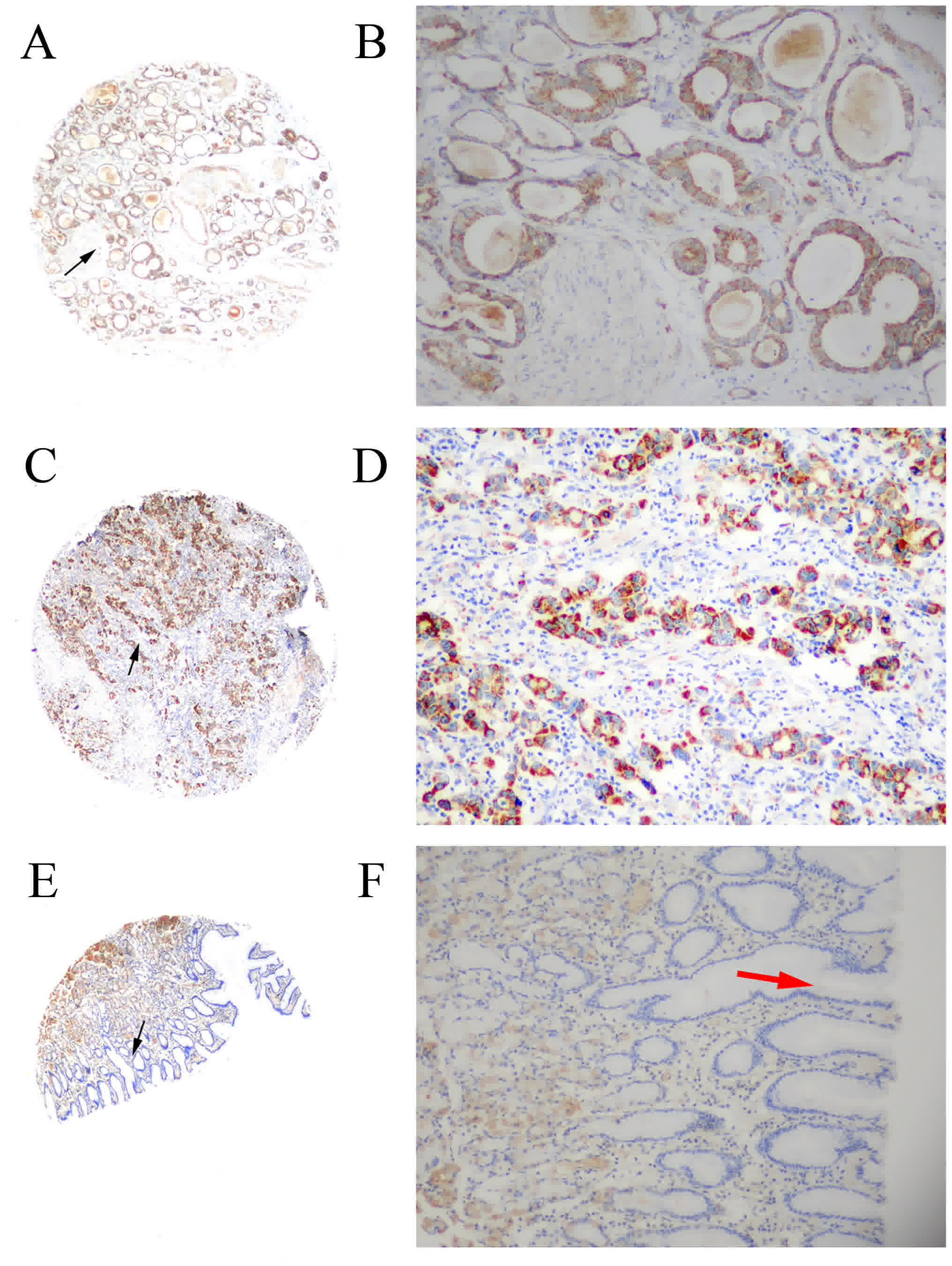 | Figure 1.Immunohistochemical analysis of MMP-21
protein expression levels in gastric cancer tissue and
non-cancerous human gastric mucosa using tissue microarray
analysis. (A) MMP-21 staining in moderately differentiated
adenocarcinoma (magnification, ×40). (B) Magnified image
(magnification, ×200) of the area indicated by black arrow in the
previous image. Immunostaining of MMP-21 produced yellow-brown
granules, primarily in the cytoplasm. (C) MMP-21 staining in poorly
differentiated adenocarcinoma (magnification, ×40). (D) Magnified
image (magnification, ×200) of the area indicated by black arrow in
the previous image. Immunostaining of MMP-21 produced yellow-brown
granules, primarily in the cytoplasm. (E) MMP-21 staining in
non-cancerous human gastric mucosa (magnification, 40×). Negative
staining was observed in normal gastric epithelial cells and a
positive signal, considered as nonspecific staining, was detected
in gastric basal cells. (F) Magnified image (magnification, ×200)
of the area indicated by black arrow in the previous image. The red
arrow indicates negative staining in normal gastric epithelial
cells. MMP, matrix metalloproteinase. |
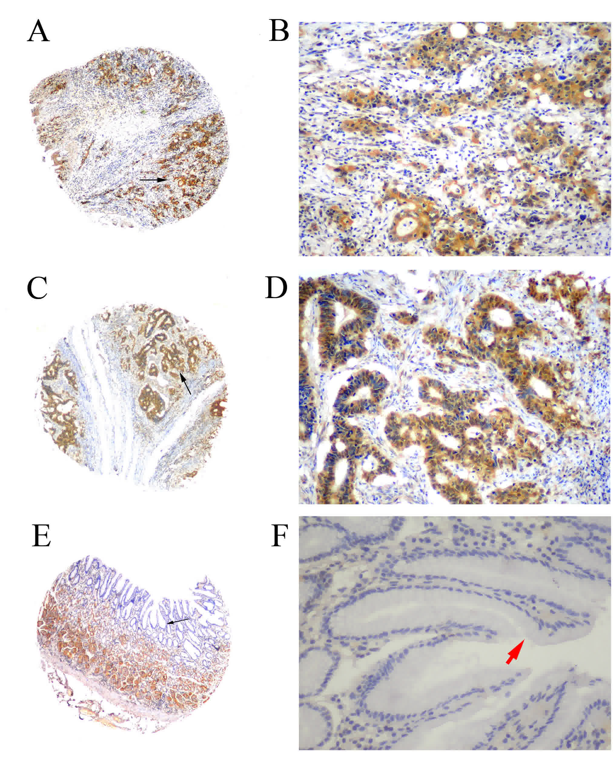 | Figure 2.Immunohistochemical analysis of MMP-28
expression in gastric cancer tissue and non-cancerous human gastric
mucosa using tissue microarray analysis. (A) MMP-28 staining in
poorly differentiated adenocarcinoma (magnification, ×40). (B)
Magnified image (magnification, ×200) of the area indicated by
black arrow in the previous image. Immunostaining of MMP-28
produced yellow-brown granules, primarily in the cytoplasm. (C)
MMP-28 staining in moderately differentiated adenocarcinoma
(magnification, ×40). (D) Magnified image (magnification, ×200) of
the area indicated by black arrow in the previous image.
Immunostaining of MMP-28 produced yellow-brown granules, primarily
in the cytoplasm. (E) MMP-28 staining in non-cancerous human
gastric mucosa (magnification, ×40). Negative staining was observed
in normal gastric epithelial cells and a positive signal,
considered as nonspecific staining, was detected in gastric basal
cells. (F) Magnified image (magnification, ×200) of the area
indicated by black arrow in the previous image. The red arrow
indicates negative staining in the cytoplasm of normal gastric
epithelial cells. MMP, matrix metalloproteinase. |
The positive staining of MMP-21 and MMP-28 was
significantly correlated with tumor diameter, depth of invasion,
vessel invasion, lymph node and distant metastasis and TNM stage
(P<0.05; Table I). MMP-21 and
MMP-28 staining did not correlate significantly with gender, Lauren
type (11), differentiation or
histology type (P>0.05; Table
I).
Results for the association between MMP-21/MMP-28
expression and the individual clinicopathological characteristics
of patients with gastric cancer are presented in Table I. The rate of positive MMP-21 staining
in patients with lymph node metastasis (44.8%; 121/270) was
significantly higher compared with those without lymph node
metastasis (10.8%; 18/166) (P<0.05). The MMP-21 expression rate
in patients with distant metastasis (65.6%; 40/61) was also
significantly higher, compared with those without distant
metastasis (26.4%; 99/375) (P<0.05). Patients with stage III or
IV gastric cancer exhibited significantly higher MMP-21 positivity
(41.6 and 69.6%, respectively) compared with those with stage I or
II gastric cancer (5.6 and 13.5%, respectively) (P<0.05).
Similarly, patients with T3 and T4 tumors exhibited a significantly
higher level of MMP-21 expression (41 and 61.5%, respectively),
compared with those with T1 and T2 stage tumors (3.5 and 19.3%,
respectively) (P<0.05).
In addition, the MMP-21 detection rate was 48.3%
(87/180) in gastric carcinoma specimens of a tumor diameter ≥5 cm,
which was significantly higher compared with specimens of a tumor
diameter <5 cm (20.3%; 51/256) (P<0.05). The MMP-28 positive
expression rate was significantly higher in gastric carcinoma
specimens of tumor diameter ≥5 cm (48.9%; 88/180) compared with
specimens of tumor diameter <5 cm (24.2%; 62/256) (P<0.05).
The frequency of MMP-28-positive tissue samples from patients with
lymph node metastasis (48.5%; 131/270) was significantly increased
compared with tissue specimens without lymph node metastasis
(12.6%; 23/183) (P<0.001). The detection rate of MMP-28 was
50.2% (127/253) and 72.1% (44/61) in tissue specimens with vascular
invasion and distant metastasis, respectively, which was
significantly higher compared with tissue specimens without
vascular invasion (12.6%; 23/183) or distant metastasis (28.3%;
106/375) (both P<0.05). MMP-28 was detected in 6.7% (6/90) of
TNM stage I tissues and 14.4% (15/104) of stage II tissue samples,
which was significantly lower compared with stage III (45.7%;
79/173) and IV (72.5%; 50/69) tissue samples (both P<0.001).
MMP-28-positivity was significantly positively correlated with the
infiltration depth of gastric cancer and the MMP-28 positive rate
was gradually increased with T stage (T1, 8.8%; T2, 19.3%; T3,
44.3%; T4, 61.5%; P<0.001). There was no significant association
between MMP-21 and MMP-28 expression levels and other
clinicopathological parameters. Cox multivariate analysis revealed
that Lauren classification (P=0.028), TNM stage (P=0.002), MMP-21
(P<0.001) and MMP-28 (P=0.001) expression levels were
significant independent prognostic factors for GC survival
(Table II). These results indicate
that MMP-21 and MMP-28 are independent predictors of survival in
patients with gastric cancer.
| Table II.Multivariate Cox's proportional
hazard analysis of overall survival with different
clinicopathological characteristics. |
Table II.
Multivariate Cox's proportional
hazard analysis of overall survival with different
clinicopathological characteristics.
|
| 95% confidence
interval |
|
|
|
|
|
|---|
|
|
|
|
|
|
|
|
|---|
| Clinicopathological
characteristic | Lower bound | Upper bound | Wald value | B-value | Standard error | Odds ratio | P-value |
|---|
| Lauren
classification | 1.048 | 2.304 | 4.800 | 0.441 | 0.201 | 1.550 | 0.028a |
| TNM stage | 1.248 | 2.671 | 9.631 | 0.602 | 0.194 | 1.826 | 0.002a |
| MMP-21 | 1.796 | 4.057 | 22.823 | 0.993 | 0.208 | 2.700 | <0.001a |
| MMP-28 | 1.294 | 2.778 | 10.790 | 0.640 | 0.195 | 1.896 | 0.001a |
MMP-21 and MMP-28 overexpression is
associated with poor prognosis
In total, 260 gastric cancer cases were negative for
MMP-21 and MMP-28 expression, whereas 113 gastric cancer cases
exhibited positive expression for MMP-21 and MMP-28 simultaneously,
with a significant correlation between positive MMP-21 and MMP-28
expression (r=0.675; P<0.001) (data not shown).
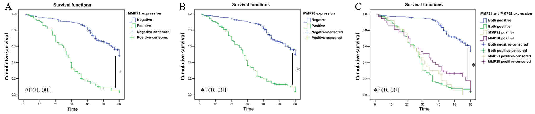
In the present cohort of patients (n=436), the mean
survival time in MMP-21-positive patients was significantly shorter
compared with that of MMP-21-negative patients (27.95±1.19 vs.
50.42±0.86 months; P<0.001; data not shown) and the 5-year
survival rate of MMP-21-positive patients (7.9%; 11/139) was
significantly lower compared with MMP-21-negative patients (56.2%;
167/297) (P<0.001; Fig. 3A). The
mean survival time of MMP-28-positive patients was also
significantly shorter compared with that of MMP-28-negative
patients (29.06±1.27 vs. 50.73±0.85 months; P<0.001; data not
shown) and the 5-year survival rate of MMP-28-positive patients
(9.3%; 14/150) was significantly lower, compared with that of
MMP-28-negative patients (57.3%; 164/286) (P<0.001; Fig. 3B). In the present cohort of patients
(n=436), the mean survival time of MMP-28- and MMP-21-positive
patients was significantly shorter compared with that of MMP-21-
and MMP-28-negative patients (27.65±1.31 vs. 52.87±0.77 months;
P<0.001; data not shown). The 5-year survival time and rate of
MMP-28- and MMP-21-positive patients was significantly lower
compared with that of MMP-28- and MMP-21-negative patients
(27.66±1.31 vs. 52.88±0.77 months, P<0.001; 8% vs. 62.3%,
P<0.001, respectively; Fig.
3C).
Discussion
Surgical resection is the most effective treatment
for advanced and metastatic gastric cancer in ~60% of patients;
however, ~40% of cases exhibit recurrence following curative
surgery (1). Thus, predicting patient
prognosis is a challenge in the management of gastric cancer and
there is a requirement for sensitive novel prognostic markers.
Several MMPs have been reported to serve a role in
inflammation, mucosal wound healing and cancer progression
(2). MMP-28 and MMP-21 are the most
recently cloned human MMPs, and are important in cancer progression
(4,10). MMP-21 is a 57 kDa proprotein
convertase-activated MMP, and is a newly identified member of the
MMP family (13). It is encoded by a
gene containing only seven exons, whereas other MMP genes comprise
ten exons (10,13). MMP-21 is widely expressed in a variety
of human malignancies, including Merkel cell carcinoma, pancreatic
adenocarcinoma, colon cancer, breast cancer, squamous cell
carcinoma and hepatocellular carcinoma (13–20).
MMP-28 is a 59 kDa protein, containing a signal sequence,
propeptide, zinc-binding catalytic domain and haemopexin-like
C-terminal domain (21). Within the
propeptide is a furin activation sequence, as MMP-28 is activated
by furin (21,22). The highest expression levels of MMP-28
mRNA are observed in the basal and suprabasal keratinocytes of the
skin, the testis and the lung (23).
MMP-28 is also expressed in several forms of carcinoma, and is
associated with proliferative cells in epithelial wound healing
(23). In oral squamous cell
carcinoma and esophageal carcinoma cells, downregulation of MMP-28
expression leads to a reduction in anchorage-independent growth
(23,24). In lung cancer, a previous study has
suggested that MMP-28 induces epithelial-mesenchymal transition
(EMT) via the activation of transforming growth factor β (25).
Numerous transcription factors, including
specificity protein (sp)1, transcription factor 4, paired box
protein, notch, retinoic acid receptor, mothers against
decapentaplegic homolog 3 and activator protein 1, have been
reported to bind to the promoter region of the MMP-21 gene
(26). The MMP-28 promoter region
contains a conserved GT-box that binds to sp1 and sp3, and is
essential for the expression of this gene (27,28).
GT-boxes and the transcription factors described above are required
for the expression of numerous genes involved in the regulation of
cell growth, cell cycle progression and cancer progression
(28). Unlike other MMPs, which are
primarily expressed in vivo by stromal cells, the expression
of MMP-21 and MMP-28 is primarily restricted to epithelial and
tumor cells, suggesting that MMP-21 and MMP-28 may regulate a wide
range of cellular functions during cancer progression (10,28).
Therefore, the present study investigated the expression levels and
prognostic value of MMP-21 and MMP-28 in gastric cancer.
The results of the present study demonstrated
significantly elevated protein expression levels of MMP-21 and
MMP-28 in a cohort of 436 patients with gastric cancer, with a 31.9
and 34.4% positive detection rate, respectively. MMP-21 and MMP-28
expression was significantly associated with the depth of tumor
invasion, lymph node metastasis, vascular invasion and distant
metastasis, suggesting that the role of MMP-21 and MMP-28 in the
breakdown of the ECM is important for the invasion and metastasis
of gastric cancer. The expression of MMP-21 and MMP-28 was observed
to significantly increase from stage I to stage IV gastric cancer,
further indicating a role for these enzymes in gastric cancer
progression. However, neither type of MMP was associated with
gender, age, Lauren type, histological type or tumor
differentiation status. Additionally, Kaplan-Meier survival
analysis revealed that patients with higher levels of MMP-21 and
MMP-28 expression had a poorer overall survival. However, the
underlying factors, including the upstream or downstream genes or
which pathways involve MMP21 and MMP28 remains unclear. It may be
that MMP-21 and MMP-28 are associated with ECM-degrading capacity
and EMT induction by regulating the expression of a number of
different transcription factors, which may degrade or denature
collagens, including type IV, V, VII, IX and X collagens.
The current study identified that MMP-21 and MMP-28
expression levels were significantly correlated with poorer patient
outcomes and are independent prognostic factors for gastric cancer.
The expression of these enzymes may be used as a prognostic marker
in these patients, in addition to the TNM staging system, in order
to enable the identification of patients with a high risk of
disease recurrence or metastasis, who are candidates for aggressive
adjuvant chemotherapy.
The current study demonstrated that MMP-21 and
MMP-28 expression levels were significantly associated with tumor
invasion, metastasis and poor prognosis in patients with gastric
cancer. Although prospective studies are required to further
determine the prognostic value of MMP-21 and MMP-28 in malignant
tumors, the results of the current study support their role as
independent prognostic factors in gastric cancer. In addition, the
results of the present study indicate that MMP-21 and MMP-28 are
novel therapeutic targets for the treatment and prevention of
gastric cancer metastasis.
Acknowledgements
The authors would like to thank ICU specialty nurse
Yajuan Yu from Department of Intensive Care Units (ICU), Zhejiang
Provincial Hospital of Tradition Chinese Medicine (TCM), Xiasha
Campus for help and support our research.
Funding
The present study was supported by the Medicine and
Health Research Foundation of Zhejiang Province (grant nos.,
2014KYA160 and 2013KYB022), The National Natural Science Foundation
of China (grant no., 81502090), Zhejiang Provincial Natural Science
Foundation of China (grant no., LY14H160039).
Availability of data and materials
The datasets generated and analyzed in the present
study are included in this published article.
Authors' contributions
XH and ZZ designed and wrote the paper, JZ and QP
collected the samples and produced the TMA arrays, WY performed the
IHC staining and YW analyzed the data.
Ethics and consent to participate
The study protocol was approved by The Ethics
Committee of Zhejiang Provincial People's Hospital (Hangzhou,
China) and the written informed consent was gained from all
participants.
Consent for publication
All patients provided written informed consent for
the publication of their data.
Competing interests
The authors declare that they have no competing
interests.
References
|
1
|
Yabusaki H, Nashimoto A, Matsuki A and
Aizawa M: Significance of surgical treatment in multimodal therapy
for stage IV highly advanced gastric cancer.
Hepatogastroenterology. 60:377–381. 2013.PubMed/NCBI
|
|
2
|
Curran S and Murray GI: Matrix
metalloproteinases: Molecular aspects of their roles in tumour
invasion and metastasis. Eur J Cancer. 36:1621–1630. 2000.
View Article : Google Scholar : PubMed/NCBI
|
|
3
|
Stetler-Stevenson WG: Type IV collagenases
in tumor invasion and metastasis. Cancer Metastasis Rev. 9:289–303.
1990. View Article : Google Scholar : PubMed/NCBI
|
|
4
|
Wu T, Li Y, Lu J, Qiao Q, Bao G, Wang N,
He X and Du X: Increased MMP-21 expression is associated with poor
overall survival of patients with gastric cancer. Med Oncol.
30:3232013. View Article : Google Scholar : PubMed/NCBI
|
|
5
|
Ye Y, Zhou X, Li X, Tang Y, Sun Y and Fang
J: Inhibition of epidermal growth factor receptor signaling
prohibits metastasis of gastric cancer via downregulation of MMP7
and MMP13. Tumor Biol. 35:10891–10896. 2014. View Article : Google Scholar
|
|
6
|
Łukaszewicz-Zając M, Mroczko B,
Guzińska-Ustymowicz K, Pryczynicz A, Gryko M, Kemona A, Kędra B and
Szmitkowski M: Matrix metalloproteinase 2 (MMP-2) and their tissue
inhibitor 2 (TIMP-2) in gastric cancer patients. Adv Med Sci.
58:235–243. 2013. View Article : Google Scholar : PubMed/NCBI
|
|
7
|
Liu HQ, Song S, Wang JH and Zhang SL:
Expression of MMP-3 and TIMP-3 in gastric cancer tissue and its
clinical significance. Oncol Lett. 2:1319–1322. 2011. View Article : Google Scholar : PubMed/NCBI
|
|
8
|
Koskensalo S, Mrena J, Wiksten JP,
Nordling S, Kokkola A, Hagström J and Haglund C: MMP-7
overexpression is an independent prognostic marker in gastric
cancer. Tumor Biol. 31:149–155. 2010. View Article : Google Scholar
|
|
9
|
Chen J, Liu X, Jiao H, Peng L, Huo Z, Yang
W, Shen Q, Li T and Liu Q: Prognostic and clinical significance of
STAT3 and MMP9 in patients with gastric cancer: A meta-analysis of
a Chinese cohort. Int J Clin Exp Med. 8:546–557. 2015.PubMed/NCBI
|
|
10
|
Jian P, Yanfang T, Zhuan Z, Jian W,
Xueming Z and Jian N: MMP28 (epilysin) as a novel promoter of
invasion and metastasis in gastric cancer. BMC Cancer. 11:2002011.
View Article : Google Scholar : PubMed/NCBI
|
|
11
|
Hu B, El Hajj N, Sittler S, Lammert N,
Barnes R and Meloni-Ehrig A: Gastric cancer: Classification,
histology and application of molecular pathology. J Gastrointest
Oncol. 3:251–261. 2012.PubMed/NCBI
|
|
12
|
Zhao ZS, Wang YY, Chu YQ, Ye ZY and Tao
HQ: SPARC is associated with gastric cancer progression and poor
survival of patients. Clin Cancer Res. 16:260–268. 2010. View Article : Google Scholar : PubMed/NCBI
|
|
13
|
Huang Y, Li W, Chu D, Zheng J, Ji G, Li M,
Zhang H, Wang W, Du J and Li J: Overexpression of matrix
metalloproteinase-21 is associated with poor overall survival of
patients with colorectal cancer. J Gastrointest Surg. 15:1188–1194.
2011. View Article : Google Scholar : PubMed/NCBI
|
|
14
|
Suomela S, Koljonen V, Skoog T, Kukko H,
Böhling T and Saarialho-Kere U: Expression of MMP-10, MMP-21,
MMP-26, and MMP-28 in merkel cell carcinoma. Virchows Arch.
455:495–503. 2009. View Article : Google Scholar : PubMed/NCBI
|
|
15
|
Bister V, Skoog T, Virolainen S, Kiviluoto
T, Puolakkainen P and Saarialho-Kere U: Increased expression of
matrix metalloproteinases-21 and −26 and TIMP-4 in pancreatic
adenocarcinoma. Mod Pathol. 20:1128–1140. 2007. View Article : Google Scholar : PubMed/NCBI
|
|
16
|
Shagisultanova EI, Novikova IA, Sidorenko
YS, Marchenko GN, Strongin AY and Malkhosyan SR: The matrix
metalloproteinase-21 gene 572C/T polymorphism and the risk of
breast cancer. Anticancer Res. 24:199–201. 2004.PubMed/NCBI
|
|
17
|
Skoog T, Elomaa O, Pasonen-Seppänen SM,
Forsberg S, Ahokas K, Jeskanen L, Pärssinen J, Tiala I, Rollman O,
Lohi J and Saarialho-Kere U: Matrix metalloproteinase-21 expression
is associated with keratinocyte differentiation and upregulated by
retinoic acid in HaCaT cells. J Invest Dermatol. 129:119–130. 2009.
View Article : Google Scholar : PubMed/NCBI
|
|
18
|
Ahokas K, Lohi J, Illman SA, Llano E,
Elomaa O, Impola U, Karjalainen-Lindsberg ML and Saarialho-Kere U:
Matrix metalloproteinase-21 is expressed epithelially during
development and in cancer and is up-regulated by transforming
growth factor-β1 in keratinocytes. Lab Invest. 83:1887–1899. 2003.
View Article : Google Scholar : PubMed/NCBI
|
|
19
|
Ahokas K, Lohi J, Lohi H, Elomaa O,
Karjalainen-Lindsberg ML, Kere J and Saarialho-Kere U: Matrix
metalloproteinase-21, the human orthologue for XMMP, is expressed
during fetal development and in cancer. Gene. 301:31–41. 2002.
View Article : Google Scholar : PubMed/NCBI
|
|
20
|
Boyd S, Virolainen S, Pärssinen J, Skoog
T, van Hogerlinden M, Latonen L, Kyllönen L, Toftgard R and
Saarialho-Kere U: MMP-10 (Stromelysin-2) and MMP-21 in human and
murine squamous cell cancer. Exp Dermatol. 18:1044–1052. 2009.
View Article : Google Scholar : PubMed/NCBI
|
|
21
|
Pavlaki M, Zucker S, Dufour A, Calabrese
N, Bahou W and Cao J: Furin functions as a nonproteolytic chaperone
for matrix metalloproteinase-28: MMP-28 propeptide sequence
requirement. Biochem Res Int. 2011:6303192011. View Article : Google Scholar : PubMed/NCBI
|
|
22
|
Rodgers UR, Kevorkian L, Surridge AK,
Waters JG, Swingler TE, Culley K, Illman S, Lohi J, Parker AE and
Clark IM: Expression and function of matrix metalloproteinase
(MMP)-28. Matrix Biol. 28:263–272. 2009. View Article : Google Scholar : PubMed/NCBI
|
|
23
|
Marchenko GN and Strongin AY: MMP-28, a
new human matrix metalloproteinase with an unusual cysteine-switch
sequence is widely expressed in tumors. Gene. 265:87–93. 2001.
View Article : Google Scholar : PubMed/NCBI
|
|
24
|
Saarialho-Kere U, Kerkelä E, Jahkola T,
Suomela S, Keski-Oja J and Lohi J: Epilysin (MMP-28) expression is
associated with cell proliferation during epithelial repair. J
Invest Dermatol. 119:14–21. 2002. View Article : Google Scholar : PubMed/NCBI
|
|
25
|
Illman SA, Lehti K, Keski-Oja J and Lohi
J: Epilysin (MMP-28) induces TGF-beta mediated epithelial to
mesenchymal transition in lung carcinoma cells. J Cell Sci.
119:3856–3865. 2006. View Article : Google Scholar : PubMed/NCBI
|
|
26
|
Marchenko GN, Marchenko ND and Strongin
AY: The structure and regulation of the human and mouse matrix
metalloproteinase-21 gene and protein. Biochem J. 372:503–515.
2003. View Article : Google Scholar : PubMed/NCBI
|
|
27
|
Illman SA, Keski-Oja J and Lohi J:
Promoter characterization of the human and mouse epilysin (MMP-28)
genes. Gene. 275:185–194. 2001. View Article : Google Scholar : PubMed/NCBI
|
|
28
|
Swingler TE, Kevorkian L, Culley KL,
Illman SA, Young DA, Parker AE, Lohi J and Clark IM: MMP28 gene
expression is regulated by Sp1 transcription factor acetylation.
Biochem J. 427:391–400. 2010. View Article : Google Scholar : PubMed/NCBI
|