Introduction
Gastric cancer is an important health concern with
an age-adjusted incidence rate of 9.7/100,000 males and 4.8/100,000
females in the United States (1),
however, this incidence rate is higher in Japan, Finland, Iceland,
Brazil, Korea and China (2). Notable
risk factors include diet (high salt, low animal fat, high complex
carbohydrates, nitrites and red or processed meat), mucosal
atrophy, chronic gastritis, smoking and Helicobacter pylori
infection (3,4). At presentation, only 40% of patients
with gastric cancer are curable, and the 10-year cancer-associated
survival rate is 51% when the cardia is not involved (5). Treatment typically consists of a
combination of surgery and chemotherapy (6,7).
Macrophages from the peripheral blood that
infiltrate tumor tissues are named tumor-associated macrophages
(TAMs). TAMs are an important part of solid tumors and have an
essential role in tumor progression (8–10). Noy
et al (11) have hypothesized
that the greater the number of macrophages in the tumor, the more
efficient their anti-tumor effect. However, previous studies have
also identified that the presence of TAMs is associated with a poor
prognosis in a number of malignancies (12–14).
Certain characteristics of the TAMs may have a functional role in
this effect on tumors.
Tunica Interna endothelial cell kinase (Tie2) is a
receptor tyrosine kinase expressed on endothelial cells and
hematopoietic stem cells (15).
Tie2-expressing macrophages (TEMs) are a subgroup of TAMs, which
were initially identified in a mouse breast cancer model (16) and are characterized by high expression
levels of the pro-angiogenic receptor Tie2. Venneri et al
(17) also identified TEMs in the
peripheral blood, where they accounted for 2–7% of the blood
mononuclear cells from healthy donors (17). TEMs were primarily located in the
hypoxic regions of tumors and may be involved in tumor
angiogenesis, thus promoting tumor progression and metastasis
(18). Previous studies have
demonstrated that the degree of TEM infiltration into tumor hypoxic
regions may be an adverse prognostic factor for patients with
cancer (14,19); however, a small number of studies
(14,20) focused on the effects of Tie2 on tumor
recurrence and disease-free survival.
Therefore, the aim of the present study was to
assess the prognostic impact of Tie2 expression in TEMs identified
in patients with gastric cancer. The results of the present study
indicate Tie2 to be a novel prognostic marker for these patients or
a potential target for therapy.
Materials and methods
Patient characteristics
From January 2009 to December 2009, 76 newly
diagnosed patients (51 males and 26 females aged 28 to 86 years)
with gastric cancer who underwent surgical tumor resection at the
Department of Surgery and Center of Minimally Invasive
Gastrointestinal Surgery, Southwest Hospital, Third Military
Medical University (Chongqing, China) by the same gastrointestinal
surgery team were enrolled in the present study. Histopathological
diagnosis was performed by an experienced pathologist according to
the criteria of the American Joint Commission on Cancer (21). The exclusion criteria included a
history of previously treated cancer and preoperative chemotherapy
or radiotherapy. All patients received adjuvant oxaliplatin and
capecitabine chemotherapy. The current study was approved by the
Institutional Review Board of the Southwest Hospital, Third
Military Medical University and written informed consent was
obtained from all patients.
Data collection
Detailed clinicopathological data was collected from
the medical records of each patient, including sex, age, tumor
location, tumor diameter and the extent of tumor resection.
Follow-up was censored in December 2013 and the collection of
subsequent treatment, recurrence and survival status data was
completed by this date. Progression-free survival (PFS) was defined
as the time interval from diagnosis to first tumor progression,
recurrence or metastasis. Overall survival (OS) was measured from
diagnosis to the date of mortality or to December 2013.
Immunohistochemistry
Gastric cancer tissues were collected during surgery
and fixed in 10% formalin for 24 h at 18°C, prior to paraffin
embedding. Paraffin sections (4 µm thick) of gastric cancer tissues
were mounted on silanized slides, dewaxed at 56°C for 30 min,
deparaffinized with xylene (slices were placed in >99% xylene I
and xylene II for 5 min at a time) and rehydrated using ethanol
(100, 100, 95, 85, 75 and 75% ethanol, 3 min at room temperature)
and washed with PBS for 3 min. Peroxidase activity was quenched
with 3% hydrogen peroxidase at room temperature for 15 min and
non-specific background staining was eliminated using blocking
buffer (2% goat serum, 0.2% Triton X-100 and 0.1% bovine serum
albumin in PBS) at room temperature for 1 h. Primary antibodies
against Tie2 (dilution, 1:50; cat no. ab24859; Abcam, Cambridge,
UK) or carbonic anhydrase IX (CAIX; dilution, 1:400; cat no.
ab107257; Abcam) were applied overnight at 4°C. Following washing
three times in PBS for 3 min, immunodetection was performed using a
labeled polymer horseradish peroxidase mouse antibody (cat no.
SC-51948; dilution, 1:100; Santa Cruz Biotechnology, Inc., Dallas,
TX, USA) incubated for 10 min at room temperature. Slides were
subsequently washed with dH2O, visualized with
3,3′-diaminobenzidine for 10 min and counterstained with Mayers
hematoxylin. The negative controls were obtained by omitting the
primary antibodies.
Slides were evaluated using an Eclipse TE2000-S
microscope (Nikon Corporation, Tokyo, Japan) by two independent
investigators who were blinded to the clinical outcomes. Images
were captured under ×400 magnification using Image-Pro Plus v5.0
software (Media Cybernetics, Inc., Rockville, MD, USA). Tie2 and
CAIX expression levels in cancer cells were graded as follows:
-(≤10% of positive cells), +(20%-50% of positive cells), ++(≥50% of
positive cells).
Immunofluorescent staining
Paraffin sections were dewaxed by heating to 56°C
for 30 min, deparaffinized using xylene I and xylene II for 5 min
at a time and rehydrated using ethanol (100, 100, 95, 85, 75 and
75% ethanol, 3 min at room temperature) and washed with PBS for 3
min. Peroxidase activity was quenched using 3% hydrogen peroxidase
at room temperature for 15 min and non-specific background staining
was eliminated using a blocking buffer (2% goat serum, 0.2% Triton
X-100 and 0.1% bovine serum albumin in PBS) at room temperature for
1 h. Primary antibodies against Tie2 (dilution, 1:50; cat no.
SC-324; Santa Cruz Biotechnology, Inc.) or CD68 (dilution, 1:50;
cat no. ab955; Abcam) were applied overnight at 4°C. Following
washing three times with PBS for 10 min, the secondary antibodies
were added; donkey anti-Rabbit IgG (Alexa Fluor 647, dilution,
1:50; cat no. A31573, or donkey anti-Mouse IgG Alexa Fluor 488,
dilution, 1:50; cat no. A21202s; both Invitrogen; Thermo Fisher
Scientific, Inc., Waltham, MA, USA) at 37°C for 60 min. Then they
were washed 5 times with PBS, for 10 min each time. DAPI was then
added at 37°C for 5 min. This was followed by washing 3 times with
PBS, for 10 min each time. The slices were then sealed using
glycerin. Slides were viewed at a magnification of ×400 using a
fluorescence microscope (Eclipse TE2000-S Nikon Corporation) using
Image-Pro Plus v5.0 software (Media Cybernetics, Inc.).
Statistical analysis
Continuous data are presented as the mean ± standard
deviation and were analyzed using the Student's t test. Categorical
data are presented as frequencies and were analyzed using the
Fisher's exact test. Pearsons chi-square test was used to compare
the recurrence rate of gastric cancer up to 3 years following
surgery. Survival rates were analyzed using the Kaplan-Meier method
and the log-rank test. Data analysis was performed using SPSS
v.13.0 (SPSS, Inc., Chicago, IL, USA). P<0.05 was considered to
indicate a statistically significant difference.
Results
Characteristics of the patients
Table I presents the
characteristics of all enrolled patients. A total of 51 male and 25
female patients were enrolled in the present study. The median age
was 56 years (range, 28–86). The tumors were located in the gastric
antrum in 42 patients, in the body of the stomach in 16 patients
and in the cardia in 18 patients. The tumor histological grade was
I in 10 patients, II in 21 patients and III in 45 patients. The
tumor-node-metastasis (TNM) stage (22) was I in 9 patients, II in 13 patients,
III in 41 patients and IV in 13 patients. The median follow-up was
52.3 months (range, 48–60) and no patient failed to complete
follow-up.
 | Table I.Clinicopathological characteristics of
the patients with gastric cancer (n=76). |
Table I.
Clinicopathological characteristics of
the patients with gastric cancer (n=76).
| Characteristic | Tie2-positive, n
(%) | P-value |
|---|
| Age, years |
| NS |
|
<60 | 32/50 (64.0) |
|
| ≥60 | 19/26 (73.1) |
|
| Gender |
| NS |
| Male | 34/51 (66.7) |
|
|
Female | 17/25 (68.0) |
|
| Tumor
sizeb |
|
<0.001a |
| T1 | 0/2 (0.0) |
|
| T2 | 3/13 (23.1) |
|
| T3 | 40/53 (75.5) |
|
| T4 | 8/8 (100.0) |
|
| Lymph node
metastasesc |
|
<0.001a |
| N0 | 4/17 (23.5) |
|
| N1 | 18/28 (64.3) |
|
| N2 | 28/30 (93.3) |
|
| N3 | 1/1 (100.0) |
|
| TNM stage |
|
<0.001a |
| I | 0/9 (0.0) |
|
| II | 7/13 (53.9) |
|
|
III | 31/41 (75.6) |
|
| IV | 13/13 (100.0) |
|
| Histological
grade |
| NS |
| I | 9/10 (90.0) |
|
| II | 12/21 (57.1) |
|
|
III | 30/45 (66.7) |
|
| Tumor location |
| NS |
| Gastric
antrum | 26/42 (61.9) |
|
| Body of
the stomach | 10/16 (62.5) |
|
|
Cardia | 15/18 (83.3) |
|
Associations between
clinicopathological characteristics and TEM recruitment in the
gastric cancer stroma
The associations between certain clinicopathological
characteristics and TEM recruitment in gastric cancer stroma are
presented in Table I. The positive
rate of TEM recruitment in gastric cancer stroma was 67.2%. TEM
recruitment increased with TNM stage: 0, 53.9, 75.6 and 100% in
stages I, II, III and IV, respectively (P<0.001). Tumor size and
lymph node involvement were significantly associated with the
presence of TEMs in the tumor stroma (P<0.001). Age, gender,
histological grade and tumor location were not significantly
associated with the presence of TEMs in the tumor stroma.
Association between
clinicopathological characteristics and the expression of CAIX and
Tie2 in gastric cancer
CAIX and Tie2 expression levels in gastric cancer
cells are presented in Table II and
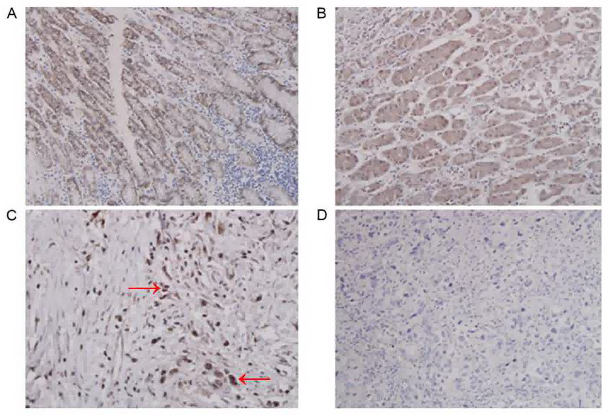
Fig. 1. The expression of CAIX and
Tie2 in gastric cancer was positive in 66/76 patients (86.8%) for
CAIX and in 65/76 patients (85.5%) for Tie2. There was no
significant difference between the two markers, irrespective of how
the patients were grouped (tumor size, lymph node involvement, TNM
stage or histological grade).
| Table II.CAIX and Tie2 expression in gastric
cancer (n=76). |
Table II.
CAIX and Tie2 expression in gastric
cancer (n=76).
|
| CAIX, n (%) | Tie2, n (%) |
|
|---|
|
|
|
|
|
|---|
|
Characteristics | − | + | ++ | − | + | ++ | P-value |
|---|
| Tumor
sizea |
|
|
|
|
|
| NS |
| T1 | 2 (100.0) | 0 | 0 | 2 (100.0) | 0 | 0 |
|
| T2 | 3 (25.0) | 8 (66.7) | 1 (8.3) | 3 (25.0) | 9 (75.0) | 0 |
|
| T3 | 5 (9.4) | 13 (24.5) | 35 (66.0) | 5 (9.4) | 24 (45.3) | 24 (45.3) |
|
| T4 | 0 | 0 | 9 (100.0) | 0 | 2 (22.2) | 7 (77.8) |
|
| Lymph
nodesb |
|
|
|
|
|
| NS |
| N0 | 9 (50.0) | 6 (33.3) | 3 (16.7) | 10 (55.6) | 6 (33.3) | 2 (11.1) |
|
| N1 | 1 (3.6) | 15 (53.6) | 12 (42.9) | 1 (3.6) | 21 (75.0) | 6 (21.4) |
|
| N2 | 0 | 2 (8.0) | 23 (92.0) | 0 | 8 (32.0) | 17 (68.0) |
|
| N3 | 0 | 0 | 5 (100.0) | 0 | 0 | 5 (100.0) |
|
| TNM stage |
|
|
|
|
|
| NS |
| I | 5 (55.5) | 4 (44.4) | 0 | 5 (55.5) | 4 (44.4) | 0 |
|
| II | 4 (33.3) | 6 (50.0) | 2 (16.7) | 4 (33.3) | 6 (50.0) | 2 (16.7) |
|
|
III | 1 (2.4) | 11 (26.2) | 30 (71.4) | 1 (2.4) | 24 (57.1) | 17 (40.5) |
|
| IV | 0 | 0 | 13 (100.0) | 0 | 1 (7.7) | 12 (92.3) |
|
| Histological
grade |
|
|
|
|
|
| NS |
| I | 2 (20.0) | 2 (20.0) | 6 (60.0) | 2 (20.0) | 2 (20.0) | 6 (60.0) |
|
| II | 3 (14.3) | 9 (42.9) | 9 (42.9) | 4 (19.1) | 11 (52.4) | 6 (28.6) |
|
|
III | 5 (11.1) | 14 (31.1) | 26 (57.8) | 5 (11.1) | 19 (42.2) | 21 (46.7) |
|
Tie2 expression level and its
association with hypoxia
Table II demonstrates
that Tie2 protein expression was increased in the hypoxic regions
of gastric tumors. Comparing the expression of Tie2 and CAIX in
gastric cancer tissues, it was identified that the expression of
Tie2 in gastric cancer tissues was consistent with the expression
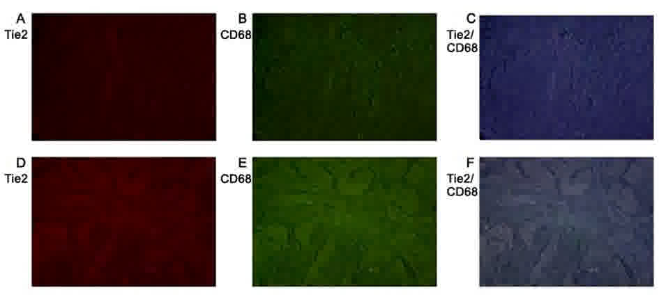
of hypoxia marker CAIX in gastric cancer tissues. Fig. 2 identifies that Tie2 and CD68 (a
macrophage-specific marker) expression colocalizes in gastric
cancer tissues. TEMs are a subset of mononuclear macrophages that
infiltrate tumor tissues.
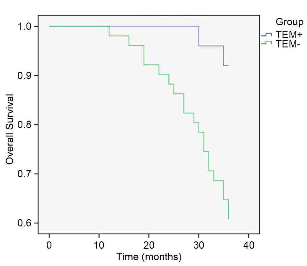
Recurrence of gastric cancer
The 1-, 2- and 3-year recurrence rates are presented
in Table III. The recurrence rates
of the TEM-positive group were 31.4, 56.9 and 66.7%, respectively,
compared with 8, 28 and 48%, respectively, for the TEM-negative
group (P<0.05). Tie2 patients with positive gastric cancer
experienced a significantly lower incidence of tumor recurrence
within three years after surgery than those with negative Tie2
expression. (P<0.05; Fig. 3).
| Table III.Tumor recurrence rate of gastric
cancer patients within three years of surgery. |
Table III.
Tumor recurrence rate of gastric
cancer patients within three years of surgery.
| Recurrence
date | TEM-negative, n
(%) | TEM-positive, n
(%) | P-value |
|---|
| 1-year | 2/25 (8.0) | 16/51 (31.4) | 0.024a |
| 2-year | 7/25 (28.0) | 29/51 (56.9) | 0.018a |
| 3-year | 12/25 (48.0) | 34/51 (66.7) | 0.118 |
Discussion
The aim of the present study was to assess the
prognostic impact of Tie2 expression in TEMs identified in patients
with gastric cancer. These results indicated that Tie2 and CD68
expression colocalizes in gastric cancer tissues. Tie2 positivity
increased with TNM stage. Tumor size and lymph node involvement
were significantly associated with the presence of TEM recruitment
in the tumor stroma. It was identified that there was no
significant difference in tumor stage between Tie2 and CAIX
expression, irrespective of how the patients were grouped (tumor
size, lymph node involvement, TNM stage or histological grade).
Tie2 protein expression was increased in the hypoxic regions of the
gastric tumors. The 1-, 2- and 3-year recurrence rates were
increased in the TEM-positive group, as compared with in the
TEM-negative group. In the TEM-negative group, the recurrence rate
of tumor recurrence within 3 years after surgery was significantly
higher than the TEM-positive group.
CAIX expression levels reflect the state of tumor
hypoxia, and its expression in solid tumors is an established
marker of poor prognosis (23–26). In
the present study, CAIX was used to locate the hypoxic areas in
gastric cancer tissues. However, there was no significant
difference in the association between CAIX and prognostic markers
compared with Tie2 and prognostic markers, suggesting that CAIX and
Tie2 are associated with tumor hypoxia. In addition, western blot
analysis demonstrated that Tie2 protein levels are increased in
hypoxic tumor tissues.
In the present study, Tie2 immunohistochemistry was
used to reflect the infiltration of tumors by TEMs, as TEMs are the
only cells known to express Tie2 (17). Positive Tie2 expression was associated
with tumor size, lymph node involvement and TNM stage, which are
established prognosis markers for gastric cancer (5). Indeed, previous studies have identified
that the degree of TEM infiltration of tumor hypoxic regions may be
an adverse prognostic factor in patients with breast cancer
(14,20); the results of the present study
suggest that this may also apply to gastric cancer.
In a study by Reed et al (27), high rates of disease recurrence have
been associated with the abundance of lymphatic channels and
vascularization within the gastric wall, which may provide
opportunity for cancer cells to migrate. Tie2 is a receptor
involved in angiogenesis (19). The
results of the present study demonstrated that the number of
patients with Tie2-positive gastric cancer was increased alongside
an increase in the rate of regional lymph node invasion, which is
concordant with the observations of Reed et al (27).
Following surgery, the gastric cancer recurrence
rate was increased in the Tie2-positive group and Tie2 protein
expression was higher in hypoxic gastric cancer tissues compared
with in normoxic gastric cancer tissues. The hypoxic
microenvironment of a tumor serves two roles. Firstly, hypoxia may
lead to tumor necrosis but the microenvironment surrounding the
tumor necrosis also induces angiogenesis, thus promoting tumor
progression and metastasis (28–33). A
number of previous studies have established that TEM infiltration
in the tumor tissue promotes tumor angiogenesis in the
microenvironment and, therefore, contribute to tumor progression
and the metastasis of tumor cells (10,30,31,33,34).
Furthermore, with the development of the tumor, the hypoxic state
of the tumor microenvironment induces the macrophage phenotype to
change from M1 to M2, resulting in an modification from immune
surveillance and cytotoxicity to immune escape, which promotes
tumor progression and metastasis (35). These previous studies support the
results of the present study, demonstrating that Tie2-positive TAMs
located in the stroma of gastric cancer have an affect on tumor
recurrence and patient survival.
The present study has limitations. The sample size
was small and from a single institution. In addition, a
comprehensive panel of prognostic markers was not assessed. Further
investigation is required to fully assess the role of Tie2 in the
prognosis of gastric cancer, including in vitro experiments
using TEMs in hypoxic and normoxic environments.
In conclusion, Tie2 positive expression
(representing infiltrated TEMs) in gastric tumors may be associated
with poorer survival. Tie2 has the potential to be used as a
prognostic marker for gastric cancer and may be a novel therapeutic
target.
Acknowledgements
The authors would like to thank Dr. Ariel Yang from
the Abramson Family Cancer Research Institute, University of
Pennsylvania (Philadelphia, USA) for his valuable help proofreading
the present study and Dr. Dong Yi from the Department of
Statistics, The Third Military Medical University (Chongquing,
China) for assistance with the statistical methods.
Funding
This study was supported by the National Natural
Science Foundation of China (grant no. 30901426).
Availability of data and materials
The datasets generated and analyzed in the present
study are included in this published article.
Authors' contributions
WJY, YXH, XY, and XLF performed the research. YXH
and PWY designed the research. WJY, YS, HLY, PY and HLD analyzed
the data. WJY and YXH wrote the paper.
Ethics approval and consent to
participate
The current study was approved by the Institutional
Review Board of the Southwest Hospital, Third Military Medical
University and written informed consent was obtained from all
patients.
Consent for publication
Written informed consent was obtained from all
patients.
Competing interests
The authors declare that they have no competing
interests.
References
|
1
|
Kohler BA, Ward E, McCarthy BJ, Schymura
MJ, Ries LA, Eheman C, Jemal A, Anderson RN, Ajani UA and Edwards
BK: Annual report to the nation on the status of cancer, 1975–2007,
featuring tumors of the brain and other nervous system. J Natl
Cancer Inst. 103:714–736. 2011. View Article : Google Scholar : PubMed/NCBI
|
|
2
|
Bertuccio P, Chatenoud L, Levi F, Praud D,
Ferlay J, Negri E, Malvezzi M and La Vecchia C: Recent patterns in
gastric cancer: A global overview. Int J Cancer. 125:666–673. 2009.
View Article : Google Scholar : PubMed/NCBI
|
|
3
|
Zhu H, Yang X, Zhang C, Zhu C, Tao G, Zhao
L, Tang S, Shu Z, Cai J, Dai S, et al: Red and processed meat
intake is associated with higher gastric cancer risk: A
meta-analysis of epidemiological observational studies. PLoS One.
8:e709552013. View Article : Google Scholar : PubMed/NCBI
|
|
4
|
Doll R, Peto R, Boreham J and Sutherland
I: Mortality from cancer in relation to smoking: 50 years
observations on British doctors. Br J Cancer. 92:426–429. 2005.
View Article : Google Scholar : PubMed/NCBI
|
|
5
|
Marrelli D, Morgagni P, de Manzoni G,
Coniglio A, Marchet A, Saragoni L, Tiberio G and Roviello F:
Italian Research Group for Gastric C (IRGGC): Prognostic value of
the 7th AJCC/UICC TNM classification of noncardia gastric cancer:
Analysis of a large series from specialized Western centers. Ann
Surg. 255:486–491. 2012. View Article : Google Scholar : PubMed/NCBI
|
|
6
|
Wagner AD, Unverzagt S, Grothe W, Kleber
G, Grothey A, Haerting J and Fleig WE: Chemotherapy for advanced
gastric cancer. Cochrane Database Syst Rev: CD004064. 2010.
View Article : Google Scholar
|
|
7
|
Gertler R, Rosenberg R, Feith M, Schuster
T and Friess H: Pouch vs. no pouch following total gastrectomy:
Meta-analysis and systematic review. Am J Gastroenterol.
104:2838–2851. 2009. View Article : Google Scholar : PubMed/NCBI
|
|
8
|
Pollard JW: Tumour-educated macrophages
promote tumour progression and metastasis. Nat Rev Cancer. 4:71–78.
2004. View
Article : Google Scholar : PubMed/NCBI
|
|
9
|
Balkwill F, Charles KA and Mantovani A:
Smoldering and polarized inflammation in the initiation and
promotion of malignant disease. Cancer Cell. 7:211–217. 2005.
View Article : Google Scholar : PubMed/NCBI
|
|
10
|
Lewis CE and Pollard JW: Distinct role of
macrophages in different tumor microenvironments. Cancer Res.
66:605–612. 2006. View Article : Google Scholar : PubMed/NCBI
|
|
11
|
Noy R and Pollard JW: Tumor-associated
macrophages: From mechanisms to therapy. Immunity. 41:49–61. 2014.
View Article : Google Scholar : PubMed/NCBI
|
|
12
|
van Netten JP, George EJ, Ashmead BJ,
Fletcher C, Thornton IG and Coy P: Macrophage-tumour cell
associations in breast cancer. Lancet. 342:872–873. 1993.
View Article : Google Scholar : PubMed/NCBI
|
|
13
|
Takanami I, Takeuchi K and Kodaira S:
Tumor-associated macrophage infiltration in pulmonary
adenocarcinoma: Association with angiogenesis and poor prognosis.
Oncology. 57:138–142. 1999. View Article : Google Scholar : PubMed/NCBI
|
|
14
|
Tsutsui S, Yasuda K, Suzuki K, Tahara K,
Higashi H and Era S: Macrophage infiltration and its prognostic
implications in breast cancer: The relationship with VEGF
expression and microvessel density. Oncol Rep. 14:425–431.
2005.PubMed/NCBI
|
|
15
|
Arai F, Hirao A, Ohmura M, Sato H,
Matsuoka S, Takubo K, Ito K, Koh GY and Suda T: Tie2/angiopoietin-1
signaling regulates hematopoietic stem cell quiescence in the bone
marrow niche. Cell. 118:149–161. 2004. View Article : Google Scholar : PubMed/NCBI
|
|
16
|
De Palma M, Venneri MA, Galli R, Sergi
Sergi L, Politi LS, Sampaolesi M and Naldini L: Tie2 identifies a
hematopoietic lineage of proangiogenic monocytes required for tumor
vessel formation and a mesenchymal population of pericyte
progenitors. Cancer Cell. 8:211–226. 2005. View Article : Google Scholar : PubMed/NCBI
|
|
17
|
Venneri MA, De Palma M, Ponzoni M, Pucci
F, Scielzo C, Zonari E, Mazzieri R, Doglioni C and Naldini L:
Identification of proangiogenic TIE2-expressing monocytes (TEMs) in
human peripheral blood and cancer. Blood. 109:5276–5285. 2007.
View Article : Google Scholar : PubMed/NCBI
|
|
18
|
Bingle L, Brown NJ and Lewis CE: The role
of tumour-associated macrophages in tumour progression:
Implications for new anticancer therapies. J Pathol. 196:254–265.
2002. View Article : Google Scholar : PubMed/NCBI
|
|
19
|
Medrek C, Ponten F, Jirstrom K and
Leandersson K: The presence of tumor associated macrophages in
tumor stroma as a prognostic marker for breast cancer patients. BMC
Cancer. 12:3062012. View Article : Google Scholar : PubMed/NCBI
|
|
20
|
Kübler K, Ayub TH, Weber SK, Zivanovic O,
Abramian A, Keyver-Paik MD, Mallmann MR, Kaiser C, Serçe NB, Kuhn W
and Rudlowski C: Prognostic significance of tumor-associated
macrophages in endometrial adenocarcinoma. Gynecol Oncol.
135:176–183. 2014. View Article : Google Scholar : PubMed/NCBI
|
|
21
|
Greene FL, Page DL, Fleming ID, et al:
AJCC cancer staging handbook. New York: Springer; 2001
|
|
22
|
Ajani JA, Beaii-saab T, Yang G, et al:
NCCN clinical practice guidelines in oncology[M]: gastric cancer.
2009.
|
|
23
|
Kaluz S, Kaluzova M and Stanbridge EJ: The
role of extracellular signal-regulated protein kinase in
transcriptional regulation of the hypoxia marker carbonic anhydrase
IX. J Cell Biochem. 97:207–216. 2006. View Article : Google Scholar : PubMed/NCBI
|
|
24
|
Trastour C, Benizri E, Ettore F, Ramaioli
A, Chamorey E, Pouysségur J and Berra E: HIF-1alpha and CA IX
staining in invasive breast carcinomas: Prognosis and treatment
outcome. Int J Cancer. 120:1451–1458. 2007. View Article : Google Scholar : PubMed/NCBI
|
|
25
|
Hussain SA, Ganesan R, Reynolds G, Gross
L, Stevens A, Pastorek J, Murray PG, Perunovic B, Anwar MS,
Billingham L, et al: Hypoxia-regulated carbonic anhydrase IX
expression is associated with poor survival in patients with
invasive breast cancer. Br J Cancer. 96:104–109. 2007. View Article : Google Scholar : PubMed/NCBI
|
|
26
|
Brahimi-Horn MC and Pouyssegur J: The
hypoxia-inducible factor and tumor progression along the angiogenic
pathway. Int Rev Cytol. 242:157–213. 2005. View Article : Google Scholar : PubMed/NCBI
|
|
27
|
Reed VK, Krishnan S, Mansfield PF, Bhosale
PR, Kim M, Das P, Janjan NA, Delclos ME, Lowy AM, Feig BW, et al:
Incidence, natural history, and patterns of locoregional recurrence
in gastric cancer patients treated with preoperative
chemoradiotherapy. Int J Radiat Oncol Biol Phys. 71:741–747. 2008.
View Article : Google Scholar : PubMed/NCBI
|
|
28
|
Hu B and Cheng SY: Angiopoietin-2:
Development of inhibitors for cancer therapy. Curr Oncol Rep.
11:111–116. 2009. View Article : Google Scholar : PubMed/NCBI
|
|
29
|
Etoh T, Inoue H, Tanaka S, Barnard GF,
Kitano S and Mori M: Angiopoietin-2 is related to tumor
angiogenesis in gastric carcinoma: Possible in vivo regulation via
induction of proteases. Cancer Res. 61:2145–2153. 2001.PubMed/NCBI
|
|
30
|
Murdoch C, Giannoudis A and Lewis CE:
Mechanisms regulating the recruitment of macrophages into hypoxic
areas of tumors and other ischemic tissues. Blood. 104:2224–2234.
2004. View Article : Google Scholar : PubMed/NCBI
|
|
31
|
Burke B, Giannoudis A, Corke KP, Gill D,
Wells M, Ziegler-Heitbrock L and Lewis CE: Hypoxia-induced gene
expression in human macrophages: Implications for ischemic tissues
and hypoxia-regulated gene therapy. Am J Pathol. 163:1233–1243.
2003. View Article : Google Scholar : PubMed/NCBI
|
|
32
|
Bingle L, Lewis CE, Corke KP, Reed MW and
Brown NJ: Macrophages promote angiogenesis in human breast tumour
spheroids in vivo. Br J Cancer. 94:101–107. 2006. View Article : Google Scholar : PubMed/NCBI
|
|
33
|
Murdoch C, Tazzyman S, Webster S and Lewis
CE: Expression of Tie-2 by human monocytes and their responses to
angiopoietin-2. J Immunol. 178:7405–7411. 2007. View Article : Google Scholar : PubMed/NCBI
|
|
34
|
Lewis CE, De Palma M and Naldini L:
Tie2-expressing monocytes and tumor angiogenesis: Regulation by
hypoxia and angiopoietin-2. Cancer Res. 67:8429–8432. 2007.
View Article : Google Scholar : PubMed/NCBI
|
|
35
|
Manzur M, Hamzah J and Ganss R: Modulation
of the ‘blood-tumor’ barrier improves immunotherapy. Cell Cycle.
7:2452–2455. 2008. View Article : Google Scholar : PubMed/NCBI
|