Introduction
Prostate Cancer (PCa) is the most common type of
cancer in men >40 years of age (1). Over the past decade, the age at
mortality and the rate of mortality caused by PCa have declined.
After lung cancer, PCa is the second leading cause of
cancer-associated mortality in men (2). In 2015, the American Cancer Society
announced that >220,000 men were diagnosed with PCa; it was
anticipated that 25,000 of these men would succumb as a result of
their cancer (3).
An increase in the serum level of prostate-specific
antigen (PSA) or an abnormal digital rectal examination result may
be warning signs for PCa. There is no general consensus on the
normal PSA level in men of different ages and therefore, its use
remains undetermined for an accurate and early diagnosis (4). One of the main difficulties in treating
PCa is the resistance of cancer cells to current therapy methods
(5).
MicroRNAs (miRNAs/miRs) are non-coding ribonucleic
acids, 18–25 nucleotides in length, that are highly conserved
across species. These molecular structures are involved in
controlling physiological and pathological cellular processes.
miRNAs primarily bind to the 3′-untranslated region (3′-UTR) of
target mRNA(s) through complementary base pairing, which will cause
degradation of mRNA or halt its translation (6–8).
As of yet, as demonstrated in the miRBase
(www.mirbase.org), >21,264 miRNAs have been
identified. With regards to cancer, miRNAs may be oncogenic or
tumor suppressors. Therefore, it is reasonable to assume that
cancer may be controlled by altering the expression of miRNAs
(9).
In recent decades, the role of miRNAs has been
studied in numerous types of human and animal cancer. The
expression of miRNAs has been studied in cancerous and healthy
cells, and different resulting profiles have revealed that miRNAs
may control certain cell cycle pathways, including apoptosis
(10–12). Defective apoptosis pathways may be
considered one of the most important features of cancer cells
(13). At present, two main pathways
for apoptosis have been discovered in cells. The extrinsic pathway
involves receptors [first apoptosis signal receptor (FAS),
complement receptor 4/5 (CR4/5), and tumor necrosis factor receptor
(TNFR)] on the cell surface, which, after binding with their
specific ligands (FAS ligand, TNF-related apoptosis-inducing ligand
and threose nucleic acid), oligomerize the FAS-associated death
domain (FADD) and activate apoptosis (14).
The second pathway is a mitochondrial pathway that
is regulated by B cell lymphoma 2 (BCL2) family proteins. BCL2
family proteins include pro-apoptotic [BCL2-associated X protein
(BAX) and BCL2 homologous killer protein (BAK)] and anti-apoptotic
[BCL2 and BCL2-like 1 (BCL2L1)] members. The balance between pro-
and anti-apoptotic proteins regulates the permeability of the
mitochondrial membrane (15–17). Although the exact mechanisms of
mitochondrial proteins remain clear, conformational changes in BAK
and BAX lead to pore formation in the outer mitochondrial membrane
and cytochrome C release (18), which
eventually leads to apoptosis. BCL2L1, which is a member of the
BCL2 protein group, serves a role in regulating mitochondrial
apoptosis through regulating the release of pro-apoptotic factors
from the mitochondria (15).
Mitochondrial apoptosis pathway stimulation is induced by DNA
damage or cytotoxic drugs that eventually cause cytochrome C to be
released from the mitochondria through the permeated membrane and
apoptosis to take place (16,17). The BCL2L1 gene is transcribed
to a few isoforms. The longest of these is translated to a
233-amino acid protein with anti-apoptotic function (15,19,20).
Considering the role of miRNAs in the suppression of
anti-apoptotic genes, it can be assumed that downregulation of
these miRNAs may be associated with the initiation or maintenance
of cancer (21).
The present study analyzed the association between
the expression of BCL2 and BCL2L1 anti-apoptosis
genes and the expression of the miRNAs that target them. Using
bioinformatics and literature review, a few miRNAs that had been
validated for having or were predicted to have inhibitory effects
on the BCL2 and BCL2L1 genes by targeting their
3′-UTR were nominated. Among them, 12 miRNAs were selected based on
their features, including being bound to more than one
anti-apoptotic gene, having more complementary base pairing
nucleotides in the seed region, being predicted by miRNA algorithm
tools and novelty. Finally, expression profiles of the 12 selected
miRNAs taken from the PCa tissue samples were examined and compared
with those taken from the benign prostatic hyperplasia (BPH) tissue
samples. Simultaneously, expression profiles of the target genes
(BCL2 and BCL2L1) were analyzed.
Materials and methods
miRNA selection
Four databases were used to identify 12 miRNAs. One
score was given for each target prediction by any of the databases
and the miRNAs with the 12 highest overall scores were selected.
miRWalk (http://zmf.umm.uni-heidelberg.de/apps/zmf/mirwalk2/)
provides a validated and predicted miRNA-target interaction
database of human, mouse and rat genes. It aggregates and compares
results from other miRNA-to-mRNA databases. miRanda (http://34.236.212.39/microrna/getDownloads.do) is an
online database for experimentally observed microRNA expression
patterns and predicted microRNA targets. miRDB (www.mirdb.org/) is also an online database for
predicted miRNA targets in animals. TargetScan (www.targetscan.org/) miRNA target predictor software
has context score contribution to include seed pairing stability
and target site.
Clinical samples
The present study was approved by the Pasteur
Institute of Iran Ethical Review Board. Written informed consent
was obtained from all participants prior to the clinical samples
being obtained. PCa tissues (n=30) were collected following radical
prostatectomy from untreated patients with PCa who were 48–80 years
old (mean, 65.66 years). Within 1 h of prostatectomy, the specimens
were dissected by a uropathologist (Hashemi Nejad Clinical Research
Developing Center, Tehran, Iran) and were stored at −80°C until RNA
extraction. Only samples containing >70% tumor cells were
included in the study. The Gleason score, pathological stage and
histological diagnosis were assessed according to the guidelines of
the Union for International Cancer Control 2002 (22). As a control group, RNA was prepared
from prostate tissue samples obtained from patients undergoing
radical surgery for BPH (n=30). The absence of cancer cells in the
prostate tissue was verified. In all cases, tissue samples were
randomly selected. Clinical samples were obtained from the
Department of Pathology, Hashemi Nejad Hospital (Tehran, Iran)
between 2014 and 2015 (Table I).
 | Table I.Patient demographics and
clinicopathological features. |
Table I.
Patient demographics and
clinicopathological features.
| Variable | No. | % |
|---|
| Age, years |
|
|
|
40-49 | 2 | 6.66 |
|
50–59 | 7 | 23.33 |
|
60–69 | 10 | 33.33 |
|
70–79 | 9 | 30 |
|
>80 | 2 | 6.66 |
|
Mean | 65.66 |
|
|
Median | 65.5 |
|
|
Maximum | 80 |
|
|
Minimum | 48 |
|
| TNM pT |
|
|
| 3a | 2 | 6.6 |
| 3b | 3 | 10 |
| 2b | 2 | 6.7 |
| 2c | 23 | 76.7 |
| TNM pN |
|
|
| 0 | 27 | 90 |
| 1 | 3 | 10 |
| TNM pM |
|
|
| x | 27 | 90 |
| 0 | 2 | 6.66 |
| 1 | 1 | 3.33 |
| Gleason score |
|
|
| 7 | 19 | 63.3 |
|
<7 | 8 | 26.7 |
|
>7 | 3 | 10 |
| PSA, ng/ml |
|
|
|
>10 | 21 | 70 |
|
4–10 | 9 | 30 |
|
<10 | 0 | 0 |
Cell lines
The human PCa PC3 and DU145 cell line, and human
umbilical vein endothelial cells (HUVECs) were purchased from the
Cell Bank of the Pasteur Institute of Iran (Tehran, Iran). Cells
were cultured in RPMI-1640 medium, supplemented with 10% fetal
bovine serum, 100 U/ml penicillin and 100 mg/ml streptomycin (all
Gibco; Thermo Fisher Scientific, Inc., Waltham MA, USA) at 37°C
with 5% CO2.
RNA extraction
Tissues
To begin with, 10–50 mg samples of frozen muscle in
pieces were homogenized in 1 ml 100% Triazole (Merck KGaA,
Darmstadt, Germany) in 15 ml centrifuge tubes. During the
homogenization process, the tubes were kept cold using liquid
nitrogen. The tubes were then left at room temperature for 5 min to
permit complete dissociation of nucleoprotein complexes.
Subsequently, 100 µl 1-Bromo-3-chloropropane was added to each
tube, vortexed for 10 sec and incubated at room temperature for 10
min. Following centrifugation at 20,817 × g -for 15 min at 4°C, the
mixture separated into 3 layers; the lower phenol-red chloroform
phase, the relatively turbid middle interphase and the colorless
upper aqueous phase. The latter was carefully transferred into
unused 1.5 ml tubes. Following the addition of an equal volume of
isopropanol and brief vortexing, the samples were stored at 4°C for
24 h, which was followed by centrifugation at 6,797 × g, for 1 h at
4°C. The precipitate RNA was visualized as a small palette.
Subsequently, the supernatant was carefully removed and the palette
was washed with 1 ml 75% ethanol and centrifuged at 6,797 × g for 5
min at 4°C. The supernatant was removed and the pellet was air
dried for 2–3 min and re-dissolved in 100 µl diethyl
pyrocarbonate-treated water. The quantity and quality of the
purified RNA samples were determined spectrophotometrically
(A260/280 >2.0; A260/230 >1.8), using a NanoDrop ND-2000
Spectrophotometer (Thermo Fisher Scientific, Inc.).
Cell lines
Total RNA was extracted from cells using a miRN easy
Mini kit (Qiagen GmbH, Hilden, Germany). In brief, cells were
seeded (to ~90% confluency, ~100,000 cells/well) onto 24-well
plates for 24 h, and were then disrupted by the addition of Qiazole
(Qiagen GmbH,), prior to the samples being separated to three
phases by the addition of chloroform (Merck KGaA). Ethanol (70%;
Merck KGaA) was added to the aqueous phase to precipitate RNA;
finally, according to the manufacturer's protocol, following
washing and centrifuging, total RNA was extracted.
cDNA synthesis
microRNA
Due to its specific structure, first strand
synthesis of cDNA was performed using an miScript II RT kit (Qiagen
GmbH, Hilden, Germany), which first adds a poly-A tail to the 3′
overhang of molecules and then incorporates a tagged sequence
upstream to the poly-T sequence for the universal reverse primer
used in subsequent reverse transcription-quantitative polymerase
chain reaction (RT-qPCR).
mRNA
A PrimeScript RT reagent kit (Takara Bio, Inc.,
Otsu, Japan) was used for reverse transcription of molecules,
according to the manufacturer's protocols.
RT-qPCR
microRNAs
RT-qPCR for the analysis of selected microRNAs was
performed in a Rotor-Gene 6000 (Corbett Life Science; Qiagen GmbH,
Hilden, Germany) with 7 µl qPCR Master mix 2×, 1 µl universal
reverse-primer sequence (as mentioned earlier), in addition to a
specific forward primer (Table II).
The SNORD47 (U47) and U6 microRNAs were used as reference genes.
The thermocycling conditions were as follows: 95°C for 15 min, 40
cycles of 94°C for 15 sec, 55°C for 30 sec and 70°C for 30 sec. The
quantitative cycle (CQ) was defined as a fractional cycle at which
the fluorescence of the sample passes through the defined
threshold. Analysis of the RT-qPCR data was performed using the
2−∆∆Cq method, which is defined as follows:
| Table II.Primers for 13 selective microRNAs
(forward)a and genes
(forward and reverse). |
Table II.
Primers for 13 selective microRNAs
(forward)a and genes
(forward and reverse).
| MicroRNA/gene | Sequence |
|---|
| 1301–3p |
5′-TTGCAGCTGCCTGGGAGTGACTTC-3′ |
| 193b-3p |
5′-AACTGGCCCTCAAAGTCCCGCT-3′ |
| 206 |
5′-TGGAATGTAAGGAAGTGTGTGG-3′ |
| 1266-5p |
5′-CCTCAGGGCTGTAGAACAGGGCT-3′ |
|
133b |
5′-TTTGGTCCCCTTCAACCAGCTA-3′ |
| 653-5p |
5′-GTGTTGAAACAATCTCTACTG-3′ |
| 143-3p |
5′-TGAGATGAAGCACTGTAGCTC-3′ |
| 219a-5p |
5′-TGATTGTCCAAACGCAATTCT-3′ |
| 185-5p |
5′-TGGAGAGAAAGGCAGTTCCTGA-3′ |
| 126-3p |
5′-TCGTACCGTGAGTAATAATGCG-3′ |
| 126-5p |
5′-CATTATTACTTTTGGTACGCG-3′ |
| 30c-2-3p |
5′-CTGGGAGAAGGCTGTTTACTCT-3′ |
| BCL2
forward |
5′-GGATCCAGGATAACGGAGGC-3′ |
| BCL2
reverse |
5′-GGCAGGCATGTTGACTTCAC-3′ |
| BCL2L1
forward |
5′-CCTTGGATCCAGGAGAACGGC-3′ |
| BCL2L1
reverse |
5′-GGGAGGGTAGAGTGGATGGTC-3′ |
| GAPDH
forward |
5′-AACGGGAAGCTTGTCATCAATGGAAA-3′ |
| GAPDH
reverse |
5′-GCATCAGCAGAGGGGGCAGAG-3′ |
Δ Cq (sample)=Cq (target microRNA sample)-Cq
(reference microRNA sample)
Δ Cq (control)=Cq (target microRNA control)-Cq
(reference microRNA control)
ΔΔ Cq=Δ Cq (sample)-Δ Cq (control)
Normalized target gene expression
level=2−ΔΔCq
mRNAs
First strand cDNAs were subjected to RT-qPCR using
SYBR® Premix Ex Taq™ (Tli RNase H Plus;
Takara Biotechnology Co., Ltd., Dalian, China). Reactions (20 ml)
were used containing forward and reverse primers for the BCL2,
BCL2-XL target genes and the GAPDH gene as the normalizer
(Table II). The thermocycling
conditions were as follows: 1 cycle at 95°C for 30 sec; 40 cycles
at 95°C for 5 sec and 60°C for 20 sec. Analysis of the RT-qPCR
results was performed as described earlier. All primers were
synthesized by TAG Copenhagen A/S (Copenhagen, Denmark).
Statistical analysis
The relative expressions of 12 miRNAs in PCa and BPH
tissue samples were compared using a Student's t-test with Welche's
correction. Data are presented as the mean ± standard deviation.
P<0.05 was considered to indicate a statistically significant
difference. GraphPad Prism 5 software (GraphPad Software, Inc., La
Jolla, CA, USA) was used for calculations and drawing graphs.
Results
miRNA selection
The results obtained from the four aforementioned
databases are presented in Table
III. A total of 12 miRNAs were selected based on the highest
scores. Furthermore, previously validated miRNAs that were
differentially downregulated in cancer cells were considered.
| Table III.Selection of microRNAs using 4
algorithmsa. |
Table III.
Selection of microRNAs using 4
algorithmsa.
| Name | Gene | miRanda | miRDB | miRWalk | Target scan | Individual
score | Overall score |
|---|
| miR-30C-2-3P | BCL2 | 1 | 0 | 1 | 1 | 3 | 6 |
|
| BCL2L1 | 1 | 0 | 1 | 1 | 3 |
|
| miR-126-3P | BCL2 | 1 | 0 | 1 | 1 | 3 | 3 |
|
| BCL2L1 | 0 | 0 | 0 | 0 | 0 |
|
| miR-126-5P | BCL2 | 1 | 0 | 1 | 1 | 3 | 3 |
|
| BCL2L1 | 0 | 0 | 0 | 0 | 0 |
|
| miR-185-5P | BCL2 | 1 | 0 | 1 | 1 | 3 | 5 |
|
| BCL2L1 | 1 | 0 | 0 | 1 | 2 |
|
| miR-219a-5P | BCL2 | 1 | 0 | 1 | 1 | 3 | 3 |
|
| BCL2L1 | 0 | 0 | 0 | 0 | 0 |
|
| miR-1301-3P | BCL2 | 1 | 1 | 0 | 1 | 3 | 4 |
|
| BCL2L1 | 1 | 0 | 0 | 0 | 1 |
|
| miR-193b-3P | BCL2 | 0 | 0 | 0 | 0 | 0 | 3 |
|
| BCL2L1 | 1 | 0 | 1 | 1 | 3 |
|
| miR-653b-5P | BCL2 | 1 | 0 | 1 | 1 | 3 | 3 |
|
| BCL2L1 | 0 | 0 | 0 | 0 | 0 |
|
| miR-143-3P | BCL2 | 1 | 0 | 1 | 1 | 3 | 3 |
|
| BCL2L1 | 0 | 0 | 0 | 0 | 0 |
|
| miR-133b | BCL2 | 0 | 0 | 0 | 0 | 0 | 3 |
|
| BCL2L1 | 1 | 0 | 1 | 1 | 3 |
|
| miR-1266-5p | BCL2 | 1 | 0 | 1 | 1 | 3 | 6 |
|
| BCL2L1 | 1 | 0 | 1 | 1 | 3 |
|
| miR-206 | BCL2 | 1 | 0 | 1 | 1 | 3 | 3 |
|
| BCL2L1 | 0 | 0 | 0 | 0 | 0 |
|
Expression analysis in clinical
samples
Initially, to evaluate RT-qPCR proliferation
efficiency, which was measured as the ability to double the
quantity of the target molecules with each cycle, of selected
miRNAs, 4 serial dilutions of a pooled cDNA sample were tested with
each set of primers (data not shown).
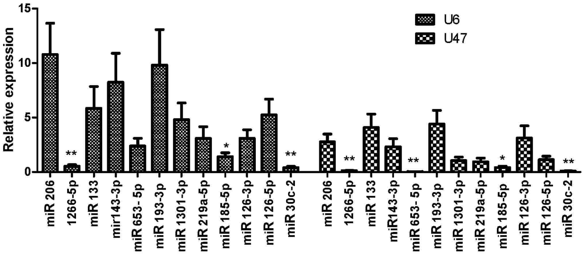
The expression of candidate miRNAs in PCa cancer and
BPH tissues was evaluated by RT-qPCR and the results were
normalized to U6 and U47 miRNA expression and mentioned earlier.
Among the 12 miRNAs that were evaluated for expression in clinical
samples, miR-1266-5p, miR-185-5p and miR-30c-2-3p were
downregulated in PCa samples compared with BPH samples (Fig. 1).
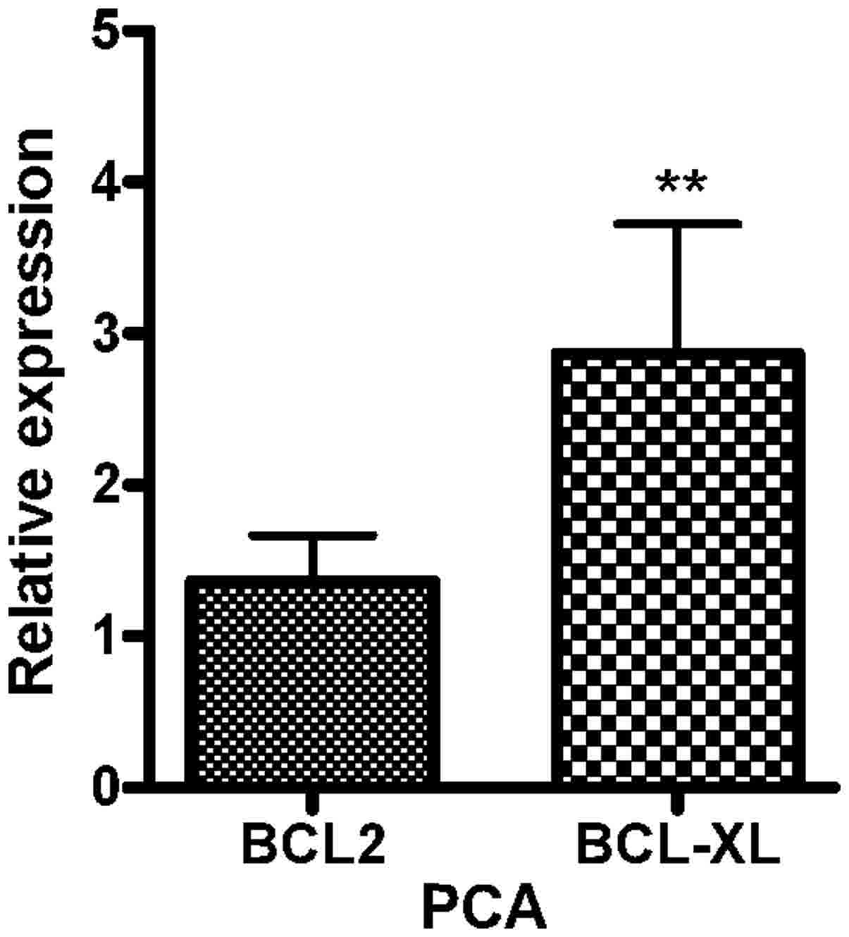
Furthermore, expression analysis of the BCL2
and BCL2L1 genes revealed that the two genes were
upregulated in PCa samples compared with expression in BPH samples
(Fig. 2).
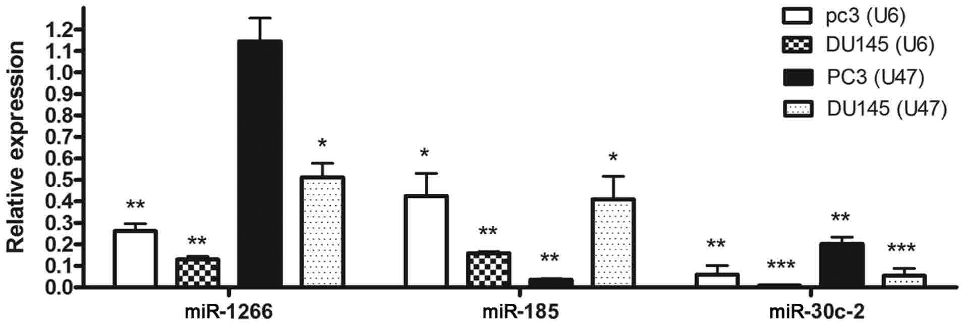
Expression analysis in PC3 and DU145
cell lines compared with HUVECs
The expression of miR-185-5p and miR-30c-2-3p was
downregulated in DU145 and PC3 cells compared with expression in
HUVECs when U6 or U47 housekeeping genes were used as the
normalizers. With respect to PC3 cells, miR-1266-5p expression was
also significantly downregulated when U47 was employed as the
normalizer (Fig. 3).
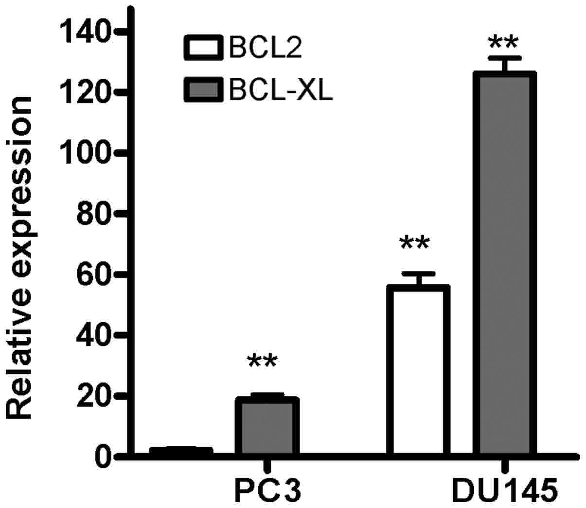
Expression of the BCL2 gene was upregulated
in the DU145 cells compared with expression in the HUVECs; however,
expression of this gene in the PC3 cells was not significantly
altered. On the other hand, expression of the BCL2L1 gene
was increased in the DU145 and PC3 cells compared with expression
in the HUVECs (Fig. 4).
In conclusion, the expression of 3 miRNAs
(miR-1266-5p, miR-185-5p and miR-30c-2-3p) was downregulated in
clinical PCa tissue samples and PCa cell lines.
Discussion
The principal aim of cancer treatment is the
complete removal of cancer cells without causing changes to the
rest of the body. Over the last few decades, microRNAs have
demonstrated potential in cancer treatment. These molecules are
able to regulate the expression of proteins involved in different
important cellular pathways, including the cell cycle and
apoptosis. Downregulation of certain miRNAs has been observed in
numerous types of cancer (23,24), and
molecular therapy studies have revealed the usefulness of miRNA
overexpression in cancer treatment in vitro and in
vivo (25).
The present study aimed to determine whether the
downregulation of miRNAs targeting two anti-apoptotic genes
(BCL2 and BCL2L1) may be associated with the
molecular pathogenesis of PCa. In order to examine this, a few
miRNA candidates were selected using bioinformatics tools, and
their expression was analyzed in clinical tissue samples obtained
from patients with PCa and compared with their expression in BPH
tissue samples. Among 12 miRNA candidates, 3 exhibited
downregulation that was counter-regulated with the expression of
two target genes.
The results of the present study are supported by
those of previous studies, which evaluated the expression of
selected miRNAs in various cancer tissues. In 2014, Chen et
al (23) demonstrated that
miR-1266 was downregulated in 58 patients with gastric cancer;
however, it was indicated that miR-1266 binds to the 3′-UTR of the
human telomerase reverse transcriptase gene and that its expression
in gastric cancer tissues was reduced in comparison with the normal
group (23).
In another study, the expression of miR-185 in
breast cancer tissues was reduced in comparison to the control
group. The authors demonstrated that overexpression of miR-185 may
inhibit the proliferation of breast cancer cells (26). The study demonstrated that miR-185
binds to the 3′-UTR region of the vascular endothelial growth
factor A (VEGF-A) gene and there was also a significant inverse
association between the expression of miR-185 and that of VEGF-A
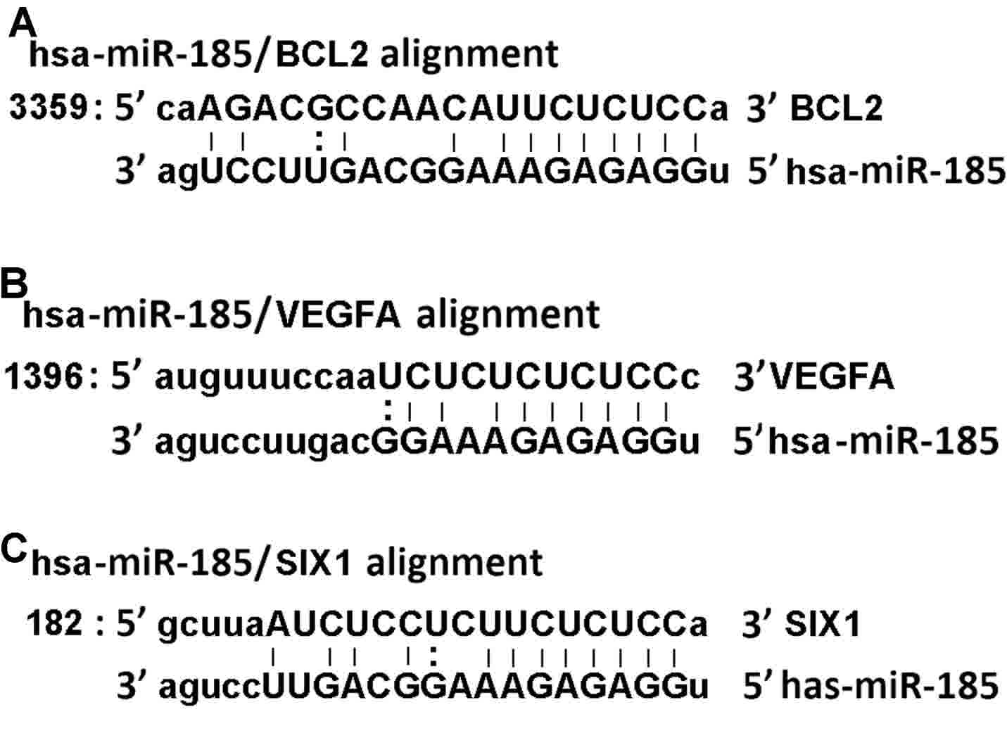
(26). Of note, the alignment of
miR-185 with the 3′-UTR sequences of VEGF-A and BCL2 mRNAs
exhibited similarity in the seed region sequence (www.microrna.org; Fig.
5). It is likely that miR-185 is also able to target the
BCL2 gene. In this case, overexpression of miR-185 in cancer
cells may inhibit angiogenesis while simultaneously promoting
apoptosis.
Furthermore, the reduced miR-185 expression has been
observed in another study performed in 2010 (27). The investigators observed
downregulation of miR-185 in ovarian cancer, pediatric renal tumors
and multiple breast cancer cell lines. Additionally, it was
demonstrated that overexpression of miR-185 was able to reduce the
expression of the SIX1 oncogene, which induces apoptosis in
apoptosis-resistant cancer cells (27). Furthermore, has-miR-185 exhibits an
identical binding sequence to the seed region sequences of SIX1 and
BCL2 mRNAs (www.microrna.org; Fig.
5).
In another study, Zhang et al (28) revealed a significant downregulation of
miR-30c in colon cancer tissue samples and in the colon cancer
HCT-116 cell line (28).
Additionally, overexpression of miR-30c resulted in the inhibition
of cancer cell proliferation, migration and invasion (28). Although they validated targeting of
the ADAM19 gene by miR-30c and proposed that ‘miR-30c inhibited
cancer cells via targeting ADAM19’, this observation may be, at
least in part, due to simultaneous targeting of the BCL2L1
gene by miR-30c, as the seed sequence binding sites of these genes
are similar (Fig. 5). The results of
the present study demonstrated that downregulation of miR-30c was
paralleled with overexpression of the BCL2L1 gene. However,
this assumption requires further confirmation in future
studies.
In 2012, researchers revealed that miR-491
overexpression may induce apoptosis in pancreatic cancer SW1990
cells by targeting the 3′-UTR of the BCL2L1 gene. The seed
match sequence region of the BCL2L1 gene was confirmed by
mutagenesis tests (29). Targeting of
the BCL2L1 anti-apoptotic gene by miR-491 and the subsequent
reduction of cancer cell phenotype (i.e., apoptosis), supports the
hypothesis of the present study, that downregulation of miR-30c may
cause overexpression of its target, the BCL2L1 gene,
eventually halting cell apoptosis.
In 2012, Zhao et al (30) confirmed by luciferase reporter assay
that miR-125b was able to bind to the 3′-UTR of the BCL2
gene, which had previously been predicted in bioinformatics
analysis. The same study also demonstrated that miR-125b suppressed
hepatocellular carcinoma cell proliferation and promoted apoptosis
by inhibiting the gene expression of BCL2 (30). Another study, which was performed in
2014, revealed that miR-16 was downregulated in human brain glioma
tissues and that overexpression of miR-16 suppressed the expression
of BCL2 and promoted cell apoptosis (31). Additionally, overexpression of miR-16
in a human glioma nude mouse model confirmed tumor growth
suppression (31). A 2011 study on
the human non-small cell lung cancer (NSCLC) A549 cell line
indicated that miR-7 expression in the NSCLC cell line was reduced,
while overexpression of miR-7 resulted in the suppression of A549
cell proliferation, as well as the induction of apoptosis (32). Furthermore, in the same study, miR-7
binding to the 3′-UTR of the BCL2 gene was been confirmed by
bioinformatics prediction and luciferase reporter assay (32).
In general, downregulation of miR-1266, miR-185 and
miR-30c has been previously reported in several different types of
cancer cells. However, interventional studies have demonstrated
that overexpression of various miRs targeting the BCL2 and
BCL2L1 genes may improve cell apoptosis. Furthermore, the
results of the present study demonstrated concordance between
bioinformatics prediction and/or literature review with expression
analysis of miR-1266, miR-185 and miR30c, as well as the
BCL2 and BCL2L1 genes, in PCa tissues and PCa cell
lines. It is likely that overexpression of these 3 microRNAs may
suppress the proliferation of tumor cells via upregulation of the
apoptosis pathway. Notably, further investigations are necessary in
order to confirm whether or not selected microRNAs are bound to the
3′-UTR of the BCL2 and BCL2-XL genes and reduce their
expression in PCa cell lines; and whether or not overexpression of
microRNAs reduces the expression of target genes, and if the latter
is co-incident with changes in cancerous phenotypes, including cell
apoptosis promotion, cell proliferation reduction or cell cycle
attenuation.
Acknowledgements
S. Ostadrahimi would like to thank the Pasteur
Institute of Iran for the grant supporting her PhD studentship. The
corresponding authors would also like to thank Dr. Hassan and Dr.
Abedi for their support.
Funding
The present study was supported by a grant from The
Pasteur Institute of Iran (grant no. 93/0110/7967).
Availability of data and materials
The datasets used and/or analyzed during the current
study are available from the corresponding author on reasonable
request.
Authors' contributions
Experimental design was performed by PFE, RM and SO.
The experiments were carried out by SO, SF and MP. Data were
analyzed by SO and PFE. Samples were gathered by MoA, HS and MaA.
Discussions, comments and clarification of the subject matter were
provided by MAV, MH, MK and LTT. MAV and MH contributed reagents,
materials and analytical tools. SO and PFE wrote the
manuscript.
Ethics approval and consent to
participate
The present study was approved by the Pasteur
Institute of Iran Ethical Review Board. Written informed consent
was obtained from all participants prior to the clinical samples
being obtained.
Consent for publication
Written informed consent was obtained from all
participants prior to publication.
Competing interests
The authors declare that they have no competing
interests.
References
|
1
|
Tabayoyong W and Abouassaly R: Prostate
cancer screening and the associated controversy. Surg Clin North
Am. 95:1023–1039. 2015. View Article : Google Scholar : PubMed/NCBI
|
|
2
|
Giovannucci E: Medical history and
etiology of prostate cancer. Epidemiol Rev. 23:159–162. 2001.
View Article : Google Scholar : PubMed/NCBI
|
|
3
|
Siegel RL, Miller KD and Jemal A: Cancer
statistics, 2015. CA Cancer J Clin. 65:5–29. 2015. View Article : Google Scholar : PubMed/NCBI
|
|
4
|
Verma A, St Onge J, Dhillon K and
Chorneyko A: PSA density improves prediction of prostate cancer.
Can J Urol. 21:7312–7321. 2014.PubMed/NCBI
|
|
5
|
Harris WP, Mostaghel EA, Nelson PS and
Montgomery B: Androgen deprivation therapy: Progress in
understanding mechanisms of resistance and optimizing androgen
depletion. Nat Clin Pract Urol. 6:76–85. 2009. View Article : Google Scholar : PubMed/NCBI
|
|
6
|
Xu B, Niu X, Zhang X, Tao J, Wu D, Wang Z,
Li P, Zhang W, Wu H, Feng N, et al: miR-143 decreases prostate
cancer cells proliferation and migration and enhances their
sensitivity to docetaxel through suppression of KRAS. Mol Cell
Biochem. 350:207–213. 2011. View Article : Google Scholar : PubMed/NCBI
|
|
7
|
Doench JG and Sharp PA: Specificity of
microRNA target selection in translational repression. Genes Dev.
18:504–511. 2004. View Article : Google Scholar : PubMed/NCBI
|
|
8
|
Sayed D and Abdellatif M: MicroRNAs in
development and disease. Physiol Rev. 91:827–887. 2011. View Article : Google Scholar : PubMed/NCBI
|
|
9
|
Xu T, Zhu Y, Xiong Y, Ge YY, Yun JP and
Zhuang SM: MicroRNA-195 suppresses tumorigenicity and regulates
G1/S transition of human hepatocellular carcinoma cells.
Hepatology. 50:113–121. 2009. View Article : Google Scholar : PubMed/NCBI
|
|
10
|
Fujita Y, Kojima T, Kawakami K, Mizutani
K, Kato T, Deguchi T and Ito M: miR-130a activates apoptotic
signaling through activation of caspase-8 in taxane-resistant
prostate cancer cells. Prostate. 75:1568–1578. 2015. View Article : Google Scholar : PubMed/NCBI
|
|
11
|
Liu XD, Zhang LY, Zhu TC, Zhang RF, Wang
SL and Bao Y: Overexpression of miR-34c inhibits high
glucose-induced apoptosis in podocytes by targeting Notch signaling
pathways. Int J Clin Exp Pathol. 8:4525–4534. 2015.PubMed/NCBI
|
|
12
|
Wang X, Qiu W, Zhang G, Xu S, Gao Q and
Yang Z: MicroRNA-204 targets JAK2 in breast cancer and induces cell
apoptosis through the STAT3/BCl-2/survivin pathway. Int J Clin Exp
Pathol. 8:5017–5025. 2015.PubMed/NCBI
|
|
13
|
Hanahan D and Weinberg RA: Hallmarks of
cancer: The next generation. Cell. 144:646–674. 2011. View Article : Google Scholar : PubMed/NCBI
|
|
14
|
Zhang J, Zhang T, Ti X, Shi J, Wu C, Ren X
and Yin H: Curcumin promotes apoptosis in A549/DDP
multidrug-resistant human lung adenocarcinoma cells through an
miRNA signaling pathway. Biochem Biophys Res Commun. 399:1–6. 2010.
View Article : Google Scholar : PubMed/NCBI
|
|
15
|
Hossini AM and Eberle J: Apoptosis
induction by Bcl-2 proteins independent of the BH3 domain. Biochem
Pharmacol. 76:1612–1619. 2008. View Article : Google Scholar : PubMed/NCBI
|
|
16
|
Li CY, Chu JY, Yu JK, Huang XQ, Liu XJ,
Shi L, Che YC and Xie JY: Regulation of alternative splicing of
Bcl-x by IL-6, GM-CSF and TPA. Cell Res. 14:473–479. 2004.
View Article : Google Scholar : PubMed/NCBI
|
|
17
|
Li J and Yuan J: Caspases in apoptosis and
beyond. Oncogene. 27:6194–6206. 2008. View Article : Google Scholar : PubMed/NCBI
|
|
18
|
Xu C, Lu Y, Pan Z, Chu W, Luo X, Lin H,
Xiao J, Shan H, Wang Z and Yang B: The muscle-specific microRNAs
miR-1 and miR-133 produce opposing effects on apoptosis by
targeting HSP60, HSP70 and caspase-9 in cardiomyocytes. J Cell Sci.
120:3045–3052. 2007. View Article : Google Scholar : PubMed/NCBI
|
|
19
|
Revil T, Toutant J, Shkreta L, Garneau D,
Cloutier P and Chabot B: Protein kinase C-dependent control of
Bcl-x alternative splicing. Mol Cell Biol. 27:8431–8441. 2007.
View Article : Google Scholar : PubMed/NCBI
|
|
20
|
Boise LH, González-García M, Postema CE,
Ding L, Lindsten T, Turka LA, Mao X, Nuñez G and Thompson CB:
bcl-x, a bcl-2-related gene that functions as a dominant regulator
of apoptotic cell death. Cell. 74:597–608. 1993. View Article : Google Scholar : PubMed/NCBI
|
|
21
|
Jovanovic M and Hengartner MO: miRNAs and
apoptosis: RNAs to die for. Oncogene. 25:6176–6187. 2006.
View Article : Google Scholar : PubMed/NCBI
|
|
22
|
Organization WH: National cancer control
programmes: Policies and managerial guidelines. World Health
Organization; 2002
|
|
23
|
Chen L, Lü MH, Zhang D, Hao NB, Fan YH, Wu
YY, Wang SM, Xie R, Fang DC, Zhang H, et al: miR-1207-5p and
miR-1266 suppress gastric cancer growth and invasion by targeting
telomerase reverse transcriptase. Cell Death Dis. 5:e10342014.
View Article : Google Scholar : PubMed/NCBI
|
|
24
|
Horsham JL, Kalinowski FC, Epis MR, Ganda
C, Brown RA and Leedman PJ: Clinical potential of microRNA-7 in
cancer. J Clin Med. 4:1668–1687. 2015. View Article : Google Scholar : PubMed/NCBI
|
|
25
|
Li Z and Rana TM: Therapeutic targeting of
microRNAs: Current status and future challenges. Nat Rev Drug
Discov. 13:622–638. 2014. View Article : Google Scholar : PubMed/NCBI
|
|
26
|
Wang R, Tian S, Wang HB, Chu DP, Cao JL,
Xia HF and Ma X: MiR-185 is involved in human breast carcinogenesis
by targeting Vegfa. FEBS Lett. 588:4438–4447. 2014. View Article : Google Scholar : PubMed/NCBI
|
|
27
|
Imam JS, Buddavarapu K, Lee-Chang JS,
Ganapathy S, Camosy C, Chen Y and Rao MK: MicroRNA-185 suppresses
tumor growth and progression by targeting the Six1 oncogene in
human cancers. Oncogene. 29:4971–4979. 2010. View Article : Google Scholar : PubMed/NCBI
|
|
28
|
Zhang Q, Yu L, Qin D, Huang R, Jiang X,
Zou C, Tang Q, Chen Y, Wang G, Wang X and Gao X: Role of
microRNA-30c targeting ADAM19 in colorectal cancer. PLoS One.
10:e01206982015. View Article : Google Scholar : PubMed/NCBI
|
|
29
|
Guo R, Wang Y, Shi WY, Liu B, Hou SQ and
Liu L: MicroRNA miR-491-5p targeting both TP53 and Bcl-XL induces
cell apoptosis in SW1990 pancreatic cancer cells through
mitochondria mediated pathway. Molecules. 17:14733–14747. 2012.
View Article : Google Scholar : PubMed/NCBI
|
|
30
|
Zhao A, Zeng Q, Xie X, Zhou J, Yue W, Li Y
and Pei X: MicroRNA-125b induces cancer cell apoptosis through
suppression of Bcl-2 expression. J Genet Genomics. 39:29–35. 2012.
View Article : Google Scholar : PubMed/NCBI
|
|
31
|
Yang TQ, Lu XJ, Wu TF, Ding DD, Zhao ZH,
Chen GL, Xie XS, Li B, Wei YX, Guo LC, et al: MicroRNA-16 inhibits
glioma cell growth and invasion through suppression of BCL2 and the
nuclear factor-κB1/MMP9 signaling pathway. Cancer Sci. 105:265–271.
2014. View Article : Google Scholar : PubMed/NCBI
|
|
32
|
Xiong S, Zheng Y, Jiang P, Liu R, Liu X
and Chu Y: MicroRNA-7 inhibits the growth of human non-small cell
lung cancer A549 cells through targeting BCL-2. Int J Biol Sci.
7:805–814. 2011. View Article : Google Scholar : PubMed/NCBI
|