Introduction
Acute lymphoblastic leukemia (ALL) is the most
common childhood cancer and the most frequent cancer in Hispanic
children (1). In Mexico, it has been
observed that childhood ALL has a relatively higher frequency
amongst patients with leukemia, accounting for ~85% of childhood
leukemia, whereas acute myeloid leukemia (AML) represents ~15%
(2). Childhood ALL in Mexico has a
mortality rate of 63.7 per million children, one of the highest
rates reported globally (2).
Previously, methotrexate (MTX) has been used to
target the folate metabolic pathway and is an important component
for the treatment of cancer (3)
including breast cancer (4), head and
neck cancer (5), lung cancer
(6) and osteosarcoma (7). Anti-folate therapies work against
folate-dependent enzymes by inhibiting de novo pyrimidine
and purine biosynthesis, resulting in cell death (8). At present, MTX is used in the
chemotherapeutic treatment of patients with ALL. MTX functions as a
binding inhibitor of dihydrofolate reductase (DHFR), an enzyme that
catalyzes the reduction of DHF to tetrahydrofolate (an essential
process for the biosynthesis of purines and thymidylate
precursors), and therefore inhibits de novo DNA synthesis
(9,10). The molecular mechanisms which underlie
MTX resistance are attributed to decreased accumulation of MTX due
to impaired transport and decreased retention of MTX, and an
increase in DHFR expression (10–12).
Previous studies have demonstrated that increased
levels of DHFR mRNA result in resistance to MTX therapy (13–15).
However, there are a limited number of studies on the function of
DHFR expression in patients with childhood acute leukemia. The
purpose of the present study was to determine whether the
expression of DHFR is associated with survival of patients with ALL
and analyze whether the expression of DHFR may be used as a
prognostic marker in acute leukemia.
Materials and methods
Patients
The present study was a hospital-based retrospective
study that analyzed 96 children (60 males, 36 females; mean age,
7.78±4.97 years), with ALL validated by bone marrow aspirates and
based on the French-American-British morphological criteria
cytochemical staining properties and immunophenotyping of blast
cells (16,17), who were admitted to the Pediatric
Oncology Service of the State Cancer Institute (Arturo Beltran
Ortega, Acapulco, Guerrero, Mexico), between September 2005 and
July 2015. The diagnosis of ALL was further subclassified as
T-lineage (CD3+, CD7+plus CD2+ or
CD5+ or both) or B-lineage (CD22+,
CD19+, CD20+, CD79A+,
HLA-DR+ and CD10+) according to the study by
Gómez-Gómez et al (18). In
addition, 100 healthy individuals were included in the present
study as controls (53 males, 47 females; mean age, 10.21±5.53
years), all of whom had no familial history of cancer. All
individuals (patients and controls) included in the study were aged
between 1 and 17 years. The study was approved by the Ethics
Committee of the State Cancer Institute (Arturo Beltran Ortega,
Acapulco, Mexico).
Overall survival (OS) time was determined according
to the study by Gómez-Gómez et al (18). Briefly, OS time was determined as the
time between the day of registration into the study and the day of
mortality (from any cause) or the day of last known contact.
Patients with ALL were classified into one of the following two
groups: Low-risk, aged between 1 and 10 years with <50,000
leucocytes/mm3 at diagnosis; high risk, aged <1 and
>10 years with >50,000 leucocytes/mm3 at
diagnosis.
Specimen collection and total RNA
extraction
Bone marrow (patients) and blood (controls) samples
from the 196 individuals were obtained and processed, according to
the study by Gómez-Gómez et al (18). Different samples were used between
patients and controls as bone marrow from healthy individuals was
deemed ethically unacceptable. Total RNA was isolated from the bone
marrow and/or blood samples using the TRIzol® method
(Invitrogen; Thermo Fisher Scientific, Inc., Waltham, MA, USA).
Quantification of mRNA using reverse
transcription-quantitative polymerase chain reaction (RT-qPCR)
Total RNA (1 µg) was converted to cDNA using the
Superscript III First-Strand Synthesis System (Invitrogen; Thermo
Fisher Scientific, Inc.), according to the manufacturer's protocol.
In brief, a 20 ml cDNA reaction mix contained extracted total RNA
(1 µg), 500 ng oligo (dT) (12–18) and
dNTP mix (0.5 mM each). The mixture was heated at 65°C for 5 min
and incubated on ice for 3 min. 1× first-strand RT buffer, 5 mM DTT
and 5 U of RNase Inhibitor (Invitrogen; Thermo Fisher Scientific,
Inc.) were added into each tube. The tubes were incubated at 42°C
for 2 min, 200 U of SuperScript III (Invitrogen; Thermo Fisher
Scientific, Inc.) was added and tubes were incubated at 42°C for 50
min, and finally 70°C for 15 min.
All PCR assays were carried out in triplicate in a
25 µl reaction volume, including the following: 5 µl cDNA template
(500 ng), 12.5 µl SYBR-Green PCR Master Mix (SYBR Green PCR
Reagents kit; Applied Biosystems; Thermo Fisher Scientific, Inc.),
0.5 µM of each oligonucleotide and ultrapure water. The following
oligonucleotides were used: DHFR forward,
5′-TTCCTGAGAAGAATCGACCTTTAAA-3′ and reverse,
5′-AAGGCATCATCTAGACTTCTGGAAA-3′; hypoxanthine-guanine
phosphoribosyltransferase (HPRT) forward, 5′-AAGCTTGCTGGTGAAAAGG-3′
and reverse, 5′-AAACATGATTCAAATCCCTGA-3′. The thermocycling
conditions were as follows: 95°C for 5 min followed by 40 cycles of
95°C for 30 sec, 60°C for 30 sec and 72°C for 30 sec. All RT-qPCR
reactions were performed in 96-well plates using the Applied
Biosystems 7500 system (Applied Biosystems; Thermo Fisher
Scientific, Inc.). The expression levels of mRNA were determined
according to the 2−ΔΔCq method (19), using HPRT mRNA expression as a
reference. The DHFR level in the ALL samples was defined as the
relative value, compared with that of samples from the healthy
individuals.
Detection of translocations
The detection of BCR-ABL, ETV6-RUNX1, AML1-ETO and
CBFΒ-MYH11 translocations was realized by PCR and according to the
protocol previously reported by Organista-Nava et al
(20).
Statistical analysis
SPSS version 20.0 (IBM Corp., Armonk, NY, USA) and
GraphPad Prism version 5.0 (GraphPad Software, Inc., La Jolla, CA,
USA) software were used to analyze the data obtained. The results
are presented as the mean ± standard deviation or median (25–75%
quartiles). To compare medians and frequencies between groups,
χ2 tests were used. The Mann-Whitney U test was used to
compare differences in the DHFR mRNA levels between ALL patients
and healthy individuals. OS time was determined using the
Kaplan-Meier estimator method. Univariate logistic regression
analysis was performed to define the risk of relapse, and
multivariable logistic regression analysis was used to identify
independent risk factors for ALL relapse. P<0.05 was considered
to indicate a statistically significant difference.
Results
Characteristics of the patients and
controls
The healthy individuals (controls) exhibited a mean
age of 10.21±5.53 years and a normal leukocyte count (between 4 and
10×103 leukocytes/mm3; median 8,000
leukocytes/mm3). The patients at diagnosis exhibited a
mean age of 7.78±4.97 years, with a mean leukocyte count at
diagnosis of 18,000 leukocytes/mm3. Characteristics of
the controls and patients are outlined in Table I. Of the patients included in the
present study, 44.79% were classified as low-risk (aged between 1
and 10 years with <50,000 leucocytes/mm3 at
diagnosis) and the remaining 55.21% were classified as high-risk
(aged <1 and >10 years with >50,000
leucocytes/mm3 at diagnosis). The results of the present
study demonstrated that B-lineage ALL was observed in 90.63%
patients. Breakpoint cluster region (BCR)/Abelson murine leukemia
viral oncogene homolog 1 (ABL) fusion gene was identified in 7.29%
(7/96) patients with ALL. The ETS variant 6 (ETV6)-runt-related
transcription factor 1 (RUNX1) rearrangement was revealed in only
0.01% (1/96) patients with ALL, and 60.42% (58/96) did not exhibit
either the BCR-ABL or ETV6-RUNX1 gene rearrangement. 30 of the 96
patients were not considered for rearrangement analysis as analysis
was not possible due to insufficient sample (Table I).
 | Table I.Characteristic and clinical data for
patients with ALL compared with those of healthy individuals. |
Table I.
Characteristic and clinical data for
patients with ALL compared with those of healthy individuals.
| Variable | ALL, n=96 | Healthy individuals,
n=100 | P-value |
|---|
| Mean age ± standard
deviation, years | 7.78±4.97 | 10.21±5.53 |
<0.001a |
|
Leukocytes/mm3 | 18,000
(4,700–42,875)a | 8,000
(7,000–9,000)a |
<0.001a |
| Sex |
|
|
0.195 |
|
Female | 36 (37.50) | 47 (47.00) |
|
| Male | 60 (62.50) | 53 (53.00) |
|
| Status of
participant |
|
|
|
|
Alive | 34 (35.42) | 100 (100.00) | – |
|
Deceased | 62 (64.58) | – |
|
| Risk group |
|
|
|
| Low | 44 (45.83) | – | – |
| High | 52 (54.17) | – |
|
|
Immunophenotype |
|
|
|
|
B-lineage | 87 (90.63) | – | – |
|
T-lineage | 9 (9.37) | – |
|
| Chromosomal
translocation |
|
|
|
|
ETV6-RUNX1 [t(12;21)] | 1 (1.04) | – |
|
| BCR-ABL
[t(9;22)] | 7 (7.29) | – |
|
|
None | 58 (60.42) | – |
|
| Not
determined | 30 (31.25) | – |
|
| DHFR level, median
(25–75 percentiles) | 9.38
(3.39–27.48) | 1.07
(0.84–1.24) |
<0.001a |
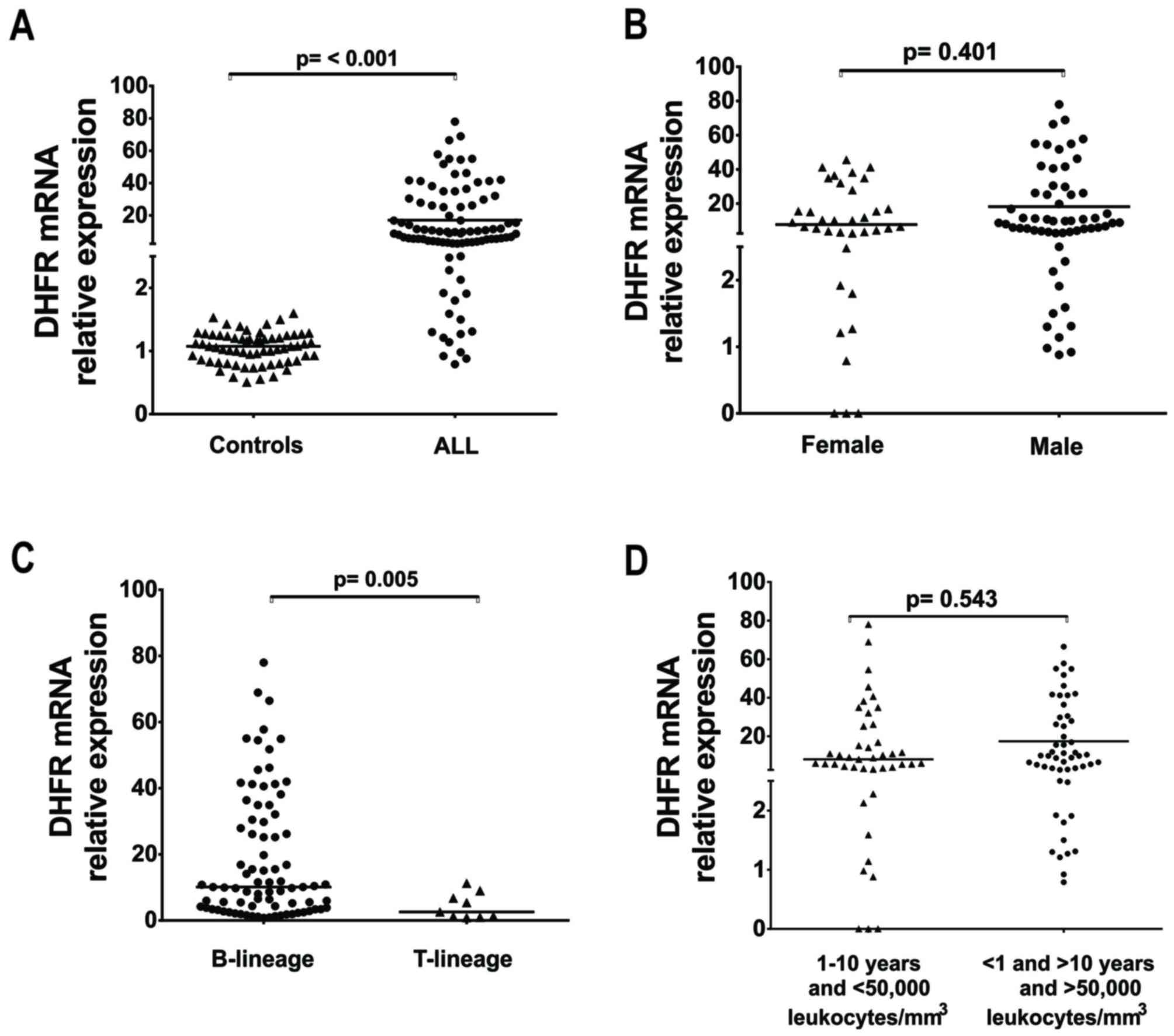
DHFR is significantly expressed
patients with ALL
As presented in Table
I and Fig. 1, the expression of
DHFR was significantly increased in ALL, compared with that in the
controls (Fig. 1A; P<0.001). The
median expression of DHFR in the control group was 1.07-fold (25–75
percentile, 0.84–1.24), whereas the median DHFR expression in the
ALL group was 9.38 (3.39–27.48). No statistically significant
difference was identified between the male and female patients
(P=0.401; Fig. 1B). it was observed
that DHFR was significantly increased in patients with B-lineage
ALL, compared in with patients with T-lineage ALL (P=0.005); the
mean was 18.63-fold (B-lineage) vs. 4.36-fold (T-lineage) (fold vs.
control mean DHFR expression; Fig.
1C). In addition, no statistically significant difference
between the low- and high-risk groups was observed (P=0.543;
Fig. 1D).
Patients with ALL who exhibit
increased DHFR expression have a higher risk of relapse during
treatment
The associations between DHFR expression and the
risk of relapse are presented in Table
II. The ALL patients were divided into either the high or low
DHFR expression groups, using as the median expression level of
DHFR (9.38) as the threshold value. A total of 43/96 patients with
ALL were classified in the low expression group and the remaining
53/96 were considered to exhibit high levels of DHFR expression.
Patients in the high expression group exhibited a 2.81-fold [95%
confidence interval (CI), 1.09–7.24; P=0.033] increased risk of
relapse during treatment, compared with the low expression group
(Table II).
| Table II.Association between DHFR expression
level, clinical features and the risk of relapse for patients with
ALL. |
Table II.
Association between DHFR expression
level, clinical features and the risk of relapse for patients with
ALL.
|
|
|
|
| Univariate
analysis | Multivariate
analysis |
|---|
|
|
|
|
|
|
|
|---|
| Categories | Without relapse, n
(%) | With relapse, n
(%) | P-value | OR | 95% CI |
P-valuea | OR | 95% CI | P-value |
|---|
| Sex |
|
| 0.84 | 1.38 | 0.56–3.38 | 0.49 | 1.43 | 0.55–3.71 | 0.46 |
|
Female | 12 | 24 |
|
|
|
|
|
|
|
|
Male | 16 | 44 |
|
|
|
|
|
|
|
| Risk group |
|
| 0.04a | 2.91 | 1.16–7.26 | 0.02a | 3.20 | 1.23–8.29 | 0.02a |
|
Low | 18 | 26 |
|
|
|
|
|
|
|
|
High | 10 | 42 |
|
|
|
|
|
|
|
| DHFR level |
|
| 0.04a | 2.50 | 1.01–6.16 | 0.047a | 2.81 | 1.09–7.24 | 0.03a |
|
Downregulated | 17 | 26 |
|
|
|
|
|
|
|
|
Upregulated | 11 | 42 |
|
|
|
|
|
|
|
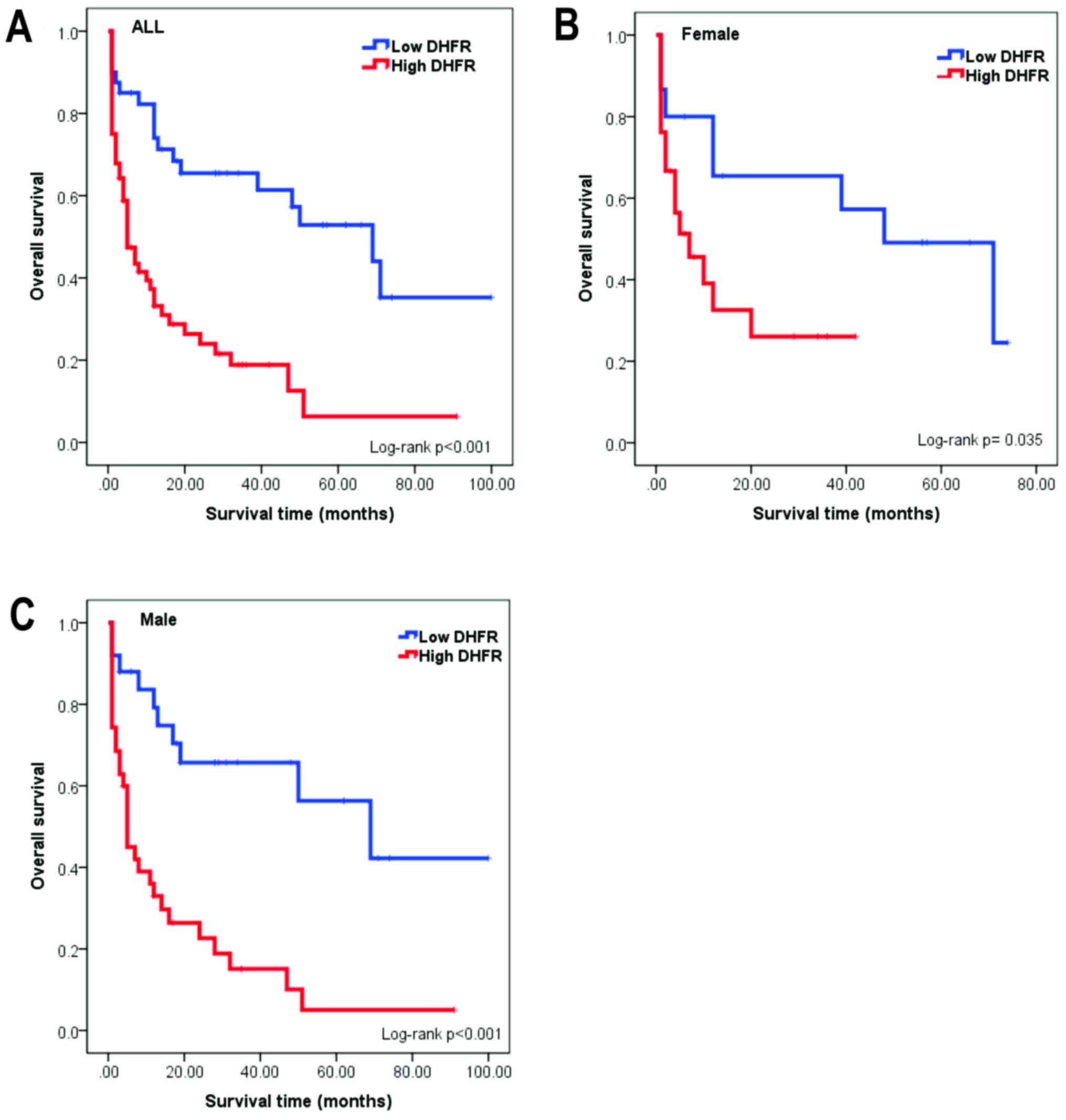
Expression of DHFR and survival in ALL
patients
The Kaplan-Meier estimator survival curves were
performed to calculate the differences between the survival of ALL
patients with high and low expression of DHFR. As presented in
Fig. 2A, ALL patients in the high
DHFR expression group exhibited poorer survival, compared with the
low expression group. Similarly, in Fig.
2B and C, male and female patients with high levels of DHFR
expression exhibited poorer survival, compared with patients with
low DHFR expression (log-rank test, P=0.035 in females; P<0.0001
in males).
Patients with B-lineage ALL and high DHFR expression
exhibited decreased survival, compared with patients with B-lineage
ALL and low DHFR expression (Fig.
3A). Although a decrease in OS was observed in patients with
T-lineage ALL and high DHFR expression, compared with patients with
T-lineage ALL and low DHFR expression, no significant association
was identified (log-rank test, P=0.115; Fig. 3B). In addition, patients with ALL
classified as low- or high-risk, and with high DHFR expression,
exhibited decreased survival, compared with patients with low DHFR
expression (log-rank test; P<0.05; Fig. 3C and D).
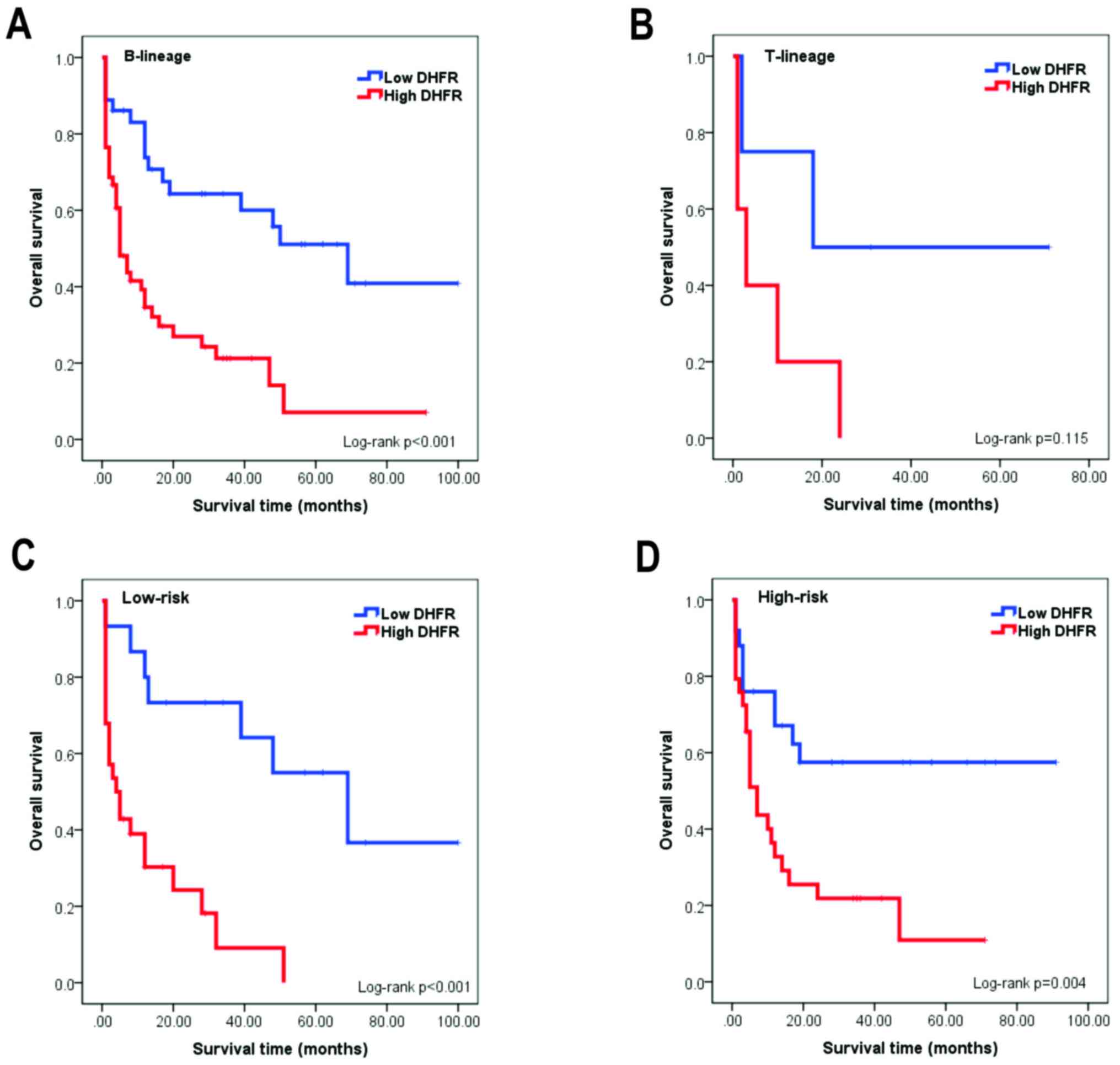 | Figure 3.Association between cell lineage (B or
T), risk-group, DHFR expression level and prognosis in patients
with childhood ALL. (A) The OS was significantly decreased for
patients with ALL with B-cell lineage and high DHFR expression,
compared with patients with B-cell lineage ALL and low DHFR
expression (P<0.001). (B) The OS was decreased for patients with
ALL of the T-cell lineage and high DHFR expression, compared with
patients with T-cell lineage ALL and low DHFR expression; however,
this difference was not identified to be statistically significant
(P>0.05). (C) In the low-risk group (aged between 1 and 10 years
with <50,000 leucocytes/mm3), the OS was
significantly increased for patients with ALL and low DHFR
expression, compared with that in patients with high DHFR
expression (P<0.001). (D) In the high-risk group (aged <1 and
>10 years with >50,000 leucocytes/mm3), the OS was
significantly decreased for patients with high DHFR expression,
compared with those with low DHFR expression (P=0.004). DHFR,
dihydrofolate reductase; ALL, acute lymphoblastic leukemia; OS,
overall survival. |
Discussion
MTX is an important component in the
chemotherapeutic treatment of several neoplasm types, including
childhood ALL (15). The primary
causes of MTX-resistance include alterations to its receptor
(reduced folate carrier) and increased DHFR expression (9,21). MTX
principally exhibits effects by inhibiting DHFR, which has an
important function in folate metabolism (11). DHFR is involved in DNA biosynthesis
and cell replication (9,22). If there is an increase in the
expression of DHFR, DNA becomes more unstable, which may lead to
abnormal cell proliferation (23).
Therefore, it is important to identify novel biomarkers for
patients with a high-risk of treatment failure (relapse), so the
appropriate chemotherapeutic scheme may be selected and the
survival time of these patients improved.
The overexpression of DHFR in certain cancer cell
lines was identified to be a mechanism underlying resistance to MTX
chemotherapy (9,21). However, limited information is
available regarding the function of DHFR expression in patients
with ALL. In the present study, the expression of DHFR was analyzed
in samples from patients with ALL, which determined an association
between clinical characteristics and patient survival. In addition,
the results of the present study revealed that DHFR mRNA was
expressed at a significantly increased level in patients with ALL,
compared with in the controls (P<0.001), which was similar to
the results of prior studies where high levels of DHFR were
observed in acute leukemia (13,21).
To determine the clinical significance of DHFR
expression in acute leukemia, a logistic regression analysis was
performed to determine associations between the clinical
characteristics of patients with ALL and the risk of relapse. The
results of the present study demonstrated a significant association
between the level of DHFR expression and the risk of relapse of
leukemia [odds ratio (OR), 2.50; 95% CI, 1.01–6.16; P=0.047).
Concordant with the results of Matheson et al (21), the results of the present study
revealed that increased expression of DHFR in patients with ALL
increased the risk of relapse. This suggests that DHFR expression
levels may be important factor in ALL.
Similar to the results of previous studies (18,20,24), an
association between age, sex, leukocyte count at diagnosis and
prognosis was identified in the present study. The results of the
present study revealed that patients in the high-risk group (aged
<1 and >10 years with >50,000 leucocytes/mm3 at
diagnosis) exhibited a poor prognosis, compared with patients in
the low-risk group (OR, 2.91; 95% CI, 1.16–7.26; P=0.02); these
values have been established by the National Cancer Institute as
the values to identify children with poor prognosis (25). Furthermore, DHFR expression was a poor
independent prognostic factor (Table
II).
The association between DHFR expression levels and
the survival of patients with ALL was investigated in the present
study. The results demonstrated that patients with high levels of
DHFR expression exhibited decreased survival compared with patients
with low DHFR expression levels (log-rank, P<0.05). Similar to
the reports of Matherly et al (13), Matheson et al (21) demonstrated an association between the
high expression of the DHFR and reduced survival in patients with
ALL; this indicates that high levels of DHFR expression are an
important prognostic factor in childhood ALL.
The results of the present study have supported the
use of genetic factors to complement known biological or
disease-based prognostic indicators in ALL. The demonstration of an
increase in the expression of the DHFR in ALL is indicative of poor
prognosis for patients with ALL.
Acknowledgements
The authors thank Dr Victor Hugo Garzón Barrientos
(State Cancer Institute, Arturo Beltran Ortega, Acapulco, Mexico)
for their contribution of biological material and facilitating
access to clinical data.
References
|
1
|
Crazzolara R and Bendall L: Emerging
treatments in acute lymphoblastic leukemia. Curr Cancer Drug
Targets. 9:19–31. 2009. View Article : Google Scholar : PubMed/NCBI
|
|
2
|
Pérez-Saldivar ML, Fajardo-Gutiérrez A,
Bernáldez-Ríos R, Martínez-Avalos A, Medina-Sanson A,
Espinosa-Hernández L, Jde Flores-Chapa D, Amador-Sánchez R,
Peñaloza-González JG, Alvarez-Rodríguez FJ, et al: Childhood acute
leukemias are frequent in Mexico City: Descriptive epidemiology.
BMC Cancer. 11:3552011. View Article : Google Scholar : PubMed/NCBI
|
|
3
|
Gonen N and Assaraf YG: Antifolates in
cancer therapy: Structure, activity and mechanisms of drug
resistance. Drug Resist Updat. 15:183–210. 2012. View Article : Google Scholar : PubMed/NCBI
|
|
4
|
Bonadonna G, Valagussa P, Moliterni A,
Zambetti M and Brambilla C: Adjuvant cyclophosphamide,
methotrexate, and fluorouracil in node-positive breast Cancer: The
results of 20 years of follow-up. N Engl J Med. 332:901–906. 1995.
View Article : Google Scholar : PubMed/NCBI
|
|
5
|
Guardiola E, Peyrade F, Chaigneau L,
Cupissol D, Tchiknavorian X, Bompas E, Madroszyk A, Ronchin P,
Schneider M, Bleuze JP, et al: Results of a randomised phase II
study comparing docetaxel with methotrexate in patients with
recurrent head and neck cancer. Eur J Cancer. 40:2071–2076. 2004.
View Article : Google Scholar : PubMed/NCBI
|
|
6
|
Scagliotti GV, Parikh P, von Pawel J,
Biesma B, Vansteenkiste J, Manegold C, Serwatowski P, Gatzemeier U,
Digumarti R, Zukin M, et al: Phase III study comparing cisplatin
plus gemcitabine with cisplatin plus pemetrexed in
chemotherapy-naive patients with advanced-stage non-small-cell lung
cancer. J Clin Oncol. 26:3543–3551. 2008. View Article : Google Scholar : PubMed/NCBI
|
|
7
|
Fuchs N, Bielack SS, Epler D, Bieling P,
Delling G, Körholz D, Graf N, Heise U, Jürgens H, Kotz R, et al:
Long-term results of the co-operative German-Austrian-Swiss
osteosarcoma study group's protocol COSS-86 of intensive multidrug
chemotherapy and surgery for osteosarcoma of the limbs. Ann Oncol.
9:893–899. 1998. View Article : Google Scholar : PubMed/NCBI
|
|
8
|
Visentin M, Zhao R and Goldman ID: The
Antifolates. Hematol Oncol Clin North Am. 26(629–648): ix2012.
|
|
9
|
Gorlick R, Goker E, Trippett T, Waltham M,
Banerjee D and Bertino JR: Intrinsic and acquired resistance to
methotrexate in acute leukemia. N Engl J Med. 335:1041–1048. 1996.
View Article : Google Scholar : PubMed/NCBI
|
|
10
|
de Jonge R, Tissing WJ, Hooijberg JH, et
al: Polymorphisms in folate-related genes and risk of pediatric
acute lymphoblastic leukemia. Blood. 113:2284–2289. 2009.
View Article : Google Scholar : PubMed/NCBI
|
|
11
|
Assaraf YG: Molecular basis of antifolate
resistance. Cancer Metastasis Rev. 26:153–181. 2007. View Article : Google Scholar : PubMed/NCBI
|
|
12
|
Bertino JR, Göker E, Gorlick R, Li WW and
Banerjee D: Resistance mechanisms to methotrexate in tumors. Stem
Cells. 14:5–9. 1996. View Article : Google Scholar : PubMed/NCBI
|
|
13
|
Matherly LH, Taub JW, Wong SC, Simpson PM,
Ekizian R, Buck S, Williamson M, Amylon M, Pullen J, Camitta B and
Ravindranath Y: Increased frequency of expression of elevated
dihydrofolate reductase in T-cell versus B-precursor acute
lymphoblastic leukemia in children. Blood. 90:578–589.
1997.PubMed/NCBI
|
|
14
|
Chauhan PS, Bhushan B, Singh LC, Mishra
AK, Saluja S, Mittal V, Gupta DK and Kapur S: Expression of genes
related to multiple drug resistance and apoptosis in acute
leukemia: Response to induction chemotherapy. Exp Mol Pathol.
92:44–49. 2012. View Article : Google Scholar : PubMed/NCBI
|
|
15
|
Dulucq S, St-Onge G, Gagné V, Ansari M,
Sinnett D, Labuda D, Moghrabi A and Krajinovic M: DNA variants in
the dihydrofolate reductase gene and outcome in childhood ALL.
Blood. 111:3692–3700. 2008. View Article : Google Scholar : PubMed/NCBI
|
|
16
|
Bennett JM, Catovsky D, Daniel MT,
Flandrin G, Galton DA, Gralnick HR and Sultan C: Proposals for the
Classification of the Acute Leukaemias French-American-British
(FAB) Co-operative Group. Br J Haematol. 33:451–458. 1976.
View Article : Google Scholar : PubMed/NCBI
|
|
17
|
Vardiman JW, Thiele J, Arber DA, Brunning
RD, Borowitz MJ, Porwit A, Harris NL, Le Beau MM,
Hellström-Lindberg E, Tefferi A and Bloomfield CD: The 2008
revision of the World health organization (WHO) classification of
myeloid neoplasms and acute leukemia: Rationale and important
changes. Blood. 114:937–951. 2009. View Article : Google Scholar : PubMed/NCBI
|
|
18
|
Gómez-Gómez Y, Organista-Nava J,
Saavedra-Herrera MV, Rivera-Ramírez AB, Terán-Porcayo MA, Del
Carmen Alarcón-Romero L, Illades-Aguiar B and Leyva-Vázquez MA:
Survival and risk of relapse of acute lymphoblastic leukemia in a
Mexican population is affected by dihydrofolate reductase gene
polymorphisms. Exp Ther Med. 3:665–672. 2012. View Article : Google Scholar : PubMed/NCBI
|
|
19
|
Nolan T, Hands RE and Bustin SA:
Quantification of mRNA using real-time RT-PCR. Nat Protoc.
1:1559–1582. 2006. View Article : Google Scholar : PubMed/NCBI
|
|
20
|
Organista-Nava J, Gómez-Gómez Y,
Illades-Aguiar B, Del Carmen Alarcón-Romero L, Saavedra-Herrera MV,
Rivera-Ramírez AB, Garzón-Barrientos VH and Leyva-Vázquez MA: High
miR-24 expression is associated with risk of relapse and poor
survival in acute leukemia. Oncol Rep. 33:1639–1649. 2015.
View Article : Google Scholar : PubMed/NCBI
|
|
21
|
Matheson EC, Hogarth LA, Case MC, Irving
JA and Hall AG: DHFR and MSH3 co-amplification in childhood acute
lymphoblastic leukaemia, in vitro and in vivo. Carcinogenesis.
28:1341–1346. 2007. View Article : Google Scholar : PubMed/NCBI
|
|
22
|
Abali EE, Skacel NE, Celikkaya H and Hsieh
YC: Regulation of human dihydrofolate reductase activity and
expression. Vitam Horm. 79:267–292. 2008. View Article : Google Scholar : PubMed/NCBI
|
|
23
|
Galbiatti AL, Castro R, Caldas HC,
Padovani JA Jr, Pavarino ÉC and Goloni-Bertollo EM: Alterations in
the expression pattern of MTHFR, DHFR, TYMS, and SLC19A1 genes
after treatment of laryngeal cancer cells with high and low doses
of methotrexate. Tumor Biol. 34:3765–3771. 2013. View Article : Google Scholar
|
|
24
|
Ng SM, Lin HP, Ariffin WA, Zainab AK, Lam
SK and Chan LL: Age, sex, haemoglobin level, and white cell count
at diagnosis are important prognostic factors in children with
acute lymphoblastic leukemia treated with BFM-type protocol. J Trop
Pediatr. 46:338–343. 2000. View Article : Google Scholar : PubMed/NCBI
|
|
25
|
Smith M, Arthur D, Camitta B, Carroll AJ,
Crist W, Gaynon P, Gelber R, Heerema N, Korn EL, Link M, et al:
Uniform approach to risk classification and treatment assignment
for children with acute lymphoblastic leukemia. J Clin Oncol.
14:18–24. 1996. View Article : Google Scholar : PubMed/NCBI
|