Introduction
Hysterocarcinoma is the most common malignant tumor
of the female reproductive system and it is the main cause of death
for female tumor patients in developing countries (1). The occurrence, proliferation and
invasion of the tumor are regulated by many factors and complex
signaling pathways (2).
Hysterocarcinoma is a common gynecological tumor worldwide. There
are approximately 500,000 new cases of hystero carcinoma worldwide
every year (3,4). Globally, there are approximately 275,000
patients who succumb to cervical cancer every year (5). Therefore, positive intervention measures
for hystero carcinoma are of great significance (6–8). At
present, treatments for hysterocarcinoma focus mainly on surgical
operation, which is assisted by radio- and chemotherapy, but the
prognosis for patients is still not high enough.
Adrenergic receptor α1 (ADRA1A) is located on
chromosome 8p (9). Previous findings
suggested that adrenergic signals on cell pathways can promote the
development of cancer (10–12). However, sufficient evidence on the
possible effects of ADRA1A in hysterocarcinoma is still
lacking.
This study determined the expression levels of serum
ADRA1A in hysterocarcinoma patients in order to analyze their
potential clinical significance.
Materials and methods
Clinical material
In total, 455 patients diagnosed with
hysterocarcinoma in the Gynecology and Oncology Department of Hubei
Cancer Hospital (Wuhan, China) were selected for the study from
May, 2007 to May, 2012. The patients were confirmed with
hysterocarcinoma by pathological section biopsies. All the patients
were being treated for the first time and had complete clinical and
follow-up visit data. Patients who received adjuvant therapy, and
those who suffered local recurrences and complications with other
tumors were excluded. The patient were aged from 35 to 65 years
(51.6±2.5 on average); individuals in the control group were
healthy as confirmed by standard health examinations. There were
380 cases in the control group, ranging in age from 37 to 65 years
(52.8±2.3 on average). General data, such as age or socioeconomic
status of the individuals in the two groups showed no statistically
significant differences (P>0.05) ensuring the comparability of
the levels of ADRA1A among the groups.
The Ethics Committee of Hubei Cancer Hospital
approved this research. All enrolled individuals or their legal
guardians signed informed consent forms.
Sample collection
Fasting venous blood (5 ml) from each subject were
collected in the morning and placed in coagulation-promoting vacuum
tubes. After centrifugation for 20 min at 8,000 × g, the separated
sera were saved at 20°C. All samples were tested within 24 h.
Testing methods
Serum ADRA1A level was tested using ELISA kits
(Shanghai Guangrui Biological Technology, Shanghai, China). All the
operations were performed following aseptic operation principles
and were conducted according to the manual standards.
Statistical methods
SPSS 20.0 software (IBM Corp., Armonk, NY, USA) was
used for data processing. Measurement data were expressed by mean ±
standard deviation. Comparisons were made using the t-test. The
Kaplan-Meier survival analysis was used to determine the median
survival time of patients. P<0.05 was considered to indicate a
statistically significant difference. For correlative analyses, the
Spearman's rank correlation was adopted. The numeration correlation
coefficient was rs.
Results
Expression of ADRA1A in peripheral
serum
Compared with the normal control group, the
expression levels of ADRA1A in the peripheral serum of
hysterocarcinoma patients were obviously increased (P<0.05)
(Table I).
 | Table I.Average expression levels of ADRA1A in
peripheral serum of individuals in the two groups. |
Table I.
Average expression levels of ADRA1A in
peripheral serum of individuals in the two groups.
| Groups | No. of cases | ADRA1A (pmol/l) |
|---|
| Hysterocarcinoma | 455 | 4.164±1.682 |
| Healthy control | 380 | 1.243±0.986 |
Correlation between the ADRA1A
expression level and the FIGO staging for hysterocarcinoma
The levels of ADRA1A in serum of hysterocarcinoma
patients increased gradually with higher FIGO stages (from stages I
to IV). However, the average level difference between stages II and
I and that between stages IV and III had no statistical difference
(P>0.05). Notably, the expression level of ADRA1A was positively
correlated with the hysterocarcinoma FIGO staging groups I–II or
III–IV (r=0.312, P=0.014) (Table
II).
| Table II.Average ADRA1A expression levels in
each subgroup of hysterocarcinoma staging in patients. |
Table II.
Average ADRA1A expression levels in
each subgroup of hysterocarcinoma staging in patients.
| Hysterocarcinoma
staging | No. of cases | Average expression
levels of ADRA1A in peripheral serum |
|---|
| I | 126 | 2.689±1.568 |
| II | 150 | 3.455±1.764 |
| III | 128 | 4.802±1.634 |
| IV | 51 | 5.623±1.792 |
Correlation between the serum ADRA1A
level (high or low) in the hysterocarcinoma group and
clinicopathological characteristics
The 455 hysterocarcinoma patients were divided into
two groups, according to their serum ADRA1A expression level. A
total of 248 cases had expression levels that were lower than the
median expression level (4.16 pmol/l) and were allocated to a low
expression group. The remaining 207 cases whose expression level of
ADRA1A in serum was higher than the median expression level, were
allocated to a high expression group. Thus, within the
hysterocarcinoma group, comparison of the relative expression
levels of ADRA1A between patients with different FIGO staging
classification and pelvic lymph node metastasis revealed
differences of statistical significance (P<0.05). However, a
comparison of the relative expression levels between patients with
different age, tissue type, lesion size, invasive depth, and
differentiation degrees revealed no differences of statistical
significance (P>0.05). Finally, a comparison of the expression
levels of ADRA1A between patients with different FIGO staging and
those with and without lymph node metastasis revealed that patients
with lower staging and no metastasis had lower levels than patients
with higher staging and presence of metastasis (P<0.05)
(Table III).
| Table III.Correlation between the average serum
ADRA1A levels in the hysterocarcinoma group and clinicopathological
characteristics. |
Table III.
Correlation between the average serum
ADRA1A levels in the hysterocarcinoma group and clinicopathological
characteristics.
| Characteristics | No. of cases (n) | Low expression level
of serum ADRA1A | High expression level
of serum ADRA1A | P-value |
|---|
| Age (years) |
|
|
| 0.158 |
|
<50 | 269 | 140 | 129 |
|
| ≥50 | 186 | 108 | 78 |
|
| Lesion size (cm) |
|
|
| 0.176 |
|
<4 | 231 | 98 | 133 |
|
| ≥4 | 224 | 150 | 74 |
|
| Stage (FIGO) |
|
|
| 0.019 |
| I–II | 276 | 178 | 98 |
|
|
III–IV | 179 | 29 | 150 |
|
| Differentiation
degree |
|
|
| 0.201 |
| High | 172 | 70 | 102 |
|
| Middle
and low | 283 | 178 | 105 |
|
| Myometrial invasion
depth |
|
|
| 0.191 |
|
<1/2 | 242 | 102 | 140 |
|
| ≥1/2 | 213 | 146 | 67 |
|
| Pelvic lymph node
metastasis |
|
|
| 0.031 |
| Yes | 179 | 39 | 140 |
|
| No | 276 | 168 | 108 |
|
| Tissue type |
|
|
| 0.215 |
| Squamous
cell carcinoma | 368 | 176 | 192 |
|
|
Non-squamous cell
carcinoma | 87 | 72 | 15 |
|
Correlation between the relative
expression level of serum ADRA1A and the prognosis of
hysterocarcinoma patients
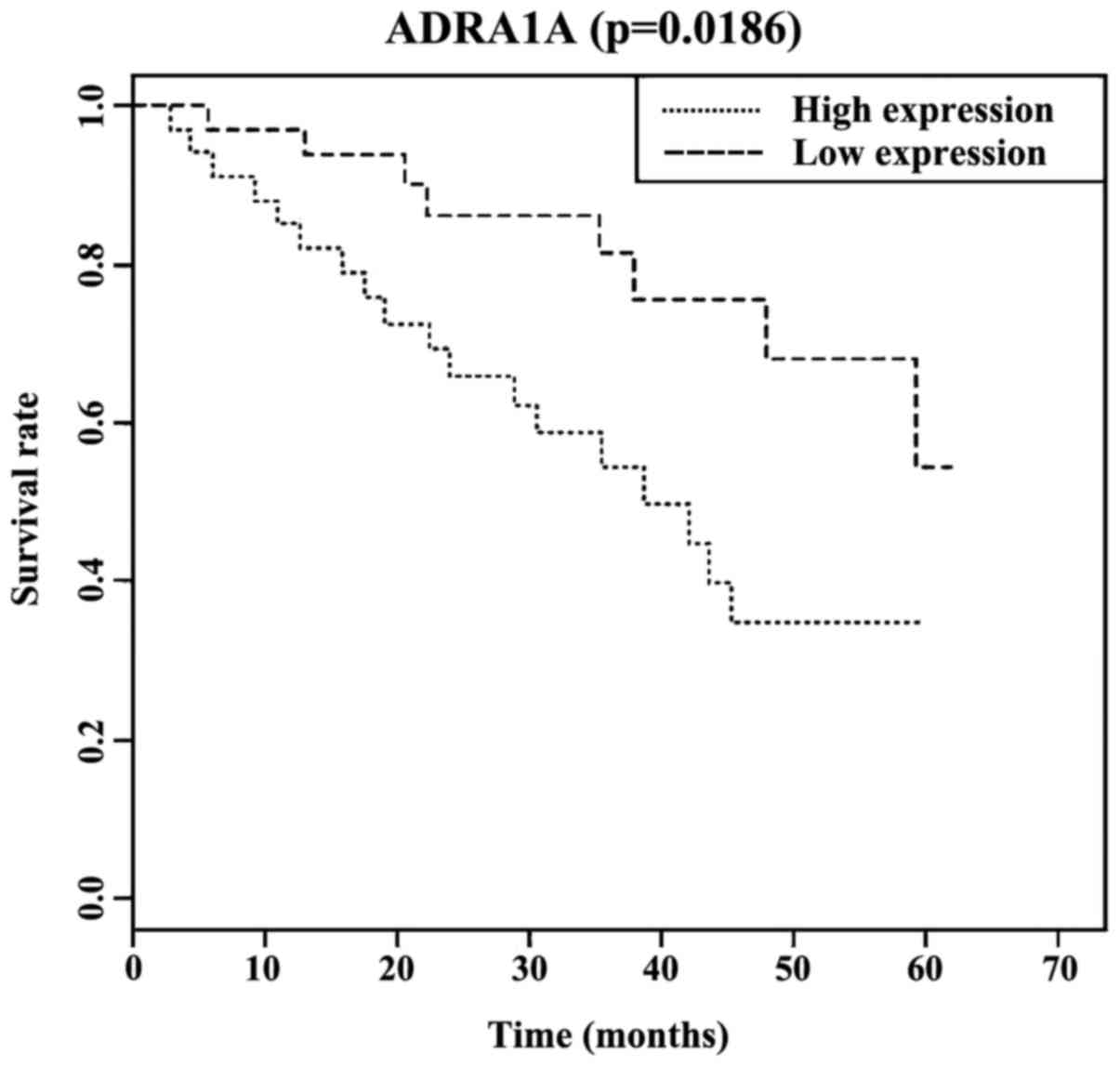
The Kaplan-Meier survival analysis indicated that
the median survival time (37.1 months) in patients with high
expression of serum ADRA1A was lower than that in patients with low
expression of serum ADRA1A (68 months), with differences of
statistical significance (P<0.05). Additionally, the three-year
and five-year survival rates of patients with low expression of
serum ADRA1A were, respectively, 74.00 and 62.00%; and the three-
and five-year survival rates of patients with a high expression of
serum ADRA1A were, respectively, 52.00 and 32.00% (Fig. 1).
Discussion
The ADRA1A gene encodes the α-epinephrine
receptors (13) for ligands such as
epinephrine, catecholamine and norepinephrine. These receptors play
important roles in the functioning of the sympathetic nervous
system (14). Previous findings have
revealed that the expression of ADRA1A receptors can be fine-tuned
to regulate the differentiation and apoptosis of epithelial cells
(15,16). Additionally, He and Huang suggested
that the high expression of ADRA1A exerts a carcinogenetic effect
(17). The pathogenesis mechanisms of
hysterocarcinoma are dependent on the development of metaplasia of
local epithelial cells. The results of the present study suggest
that the expression level of serum ADRA1A in patients with
hysterocarcinoma is higher than that in healthy individuals, which
is in agreement with a published study on the high expression of
ADRA1A in prostate cancer (18). We
believe the simple testing of ADRA1A in the peripheral blood of
hysterocarcinoma patients can be of use as a new reference for
hysterocarcinoma screening.
Clinically, the definition of hysterocarcinoma
staging usually follows the standard of the International
Federation of Gynecology and Obstetrics (FIGO). In this study, it
was found that the expression level of ADRA1A was positively
correlated with the FIGO staging of hysterocarcinoma (r=0.312,
P=0.014). Furthermore, the serum ADRA1A level was closely
correlated with the prognosis in patients. High expression levels
of serum ADRA1A indicate a poorer prognosis and a significant
reduction in the survival rate. A literature review revealed it is
possible that the inhibition of adrenergic receptors can inhibit
metastasis in liver cancer, which means that the expression level
of adrenergic receptors is increased in liver cancer metastasis
patients (19). The results of the
present study showed that the level of serum ADRA1A in patients
with hysterocarcinoma is higher and the total prognosis is poorer,
which may be related to the fact that the increased expression of
ADRA1A somehow promotes tumor metastasis. Moreover, the survival
analysis results of the present study showed the three-and
five-year survival rates of patients with low expression of serum
ADRA1A were respectively 52.00 and 32.00%, and this is basically in
agreement with reported results on the survival rate of
hysterocarcinoma patients (20,21). On
the other hand, the increased serum ADRA1A expression levels
predict a poor prognosis for patients. Accordingly, the levels of
ADRA1A in serum could be used to predict prognosis in patients.
From the fact that the serum ADRA1A level was
positively correlated with the FIGO staging status, it is possible
to indirectly associate the correlation between serum ADRA1A level
and survival prognosis. However, the conventional FIGO staging
adopted in clinical practice does not take into consideration the
presence or absence of lymph node metastasis. Accuracy of clinical
staging is imperative in treatment guidance and prognosis.
Nevertheless, imaging examination for the condition of lymph node
metastasis (22) facilitates
prognosis evaluation. Therefore, in the present study, we included
this variable and also found that patients with lymph node
metastasis demonstrated higher serum ADRA1A levels, which obviously
occurs in conjunction with a poorer prognosis. Similarly, the serum
ADRA1A level seemed to indicate a poor prognosis in cervical cancer
(23). We therefore suggest that the
determination of serum ADRA1A expression levels should aid doctors
in correctly evaluating the hysterocarcinoma staging, lymph node
metastasis and prognosis. Nevertheless, limitations of this
approach include the lack of data needed to correctly establish
standard high and low expression levels of ADRA1A or survival
analysis. Consequently, further studies should be carried out to
determine these factors.
To conclude, the results of the present study have
shown that the serum ADRA1A level is closely correlated with the
occurrence and development of hysterocarcinoma. The level can serve
as a potential index for the screening for hysterocarcinoma, assist
in its diagnosis and serves as a biological index for prognosis
evaluation. Testing of serum ADRA1A should therefore be popularized
and expanded to include a wider population.
Acknowledgements
Not applicable.
Funding
No funding was received.
Availability of data and materials
The datasets used and/or analyzed during the present
study are available from the corresponding author on reasonable
request.
Authors' contributions
LP wrote the manuscript and analyzed blood sample.
WP contributed to ELISA. PH and HFZ helped with statistical
analysis. All authors read and approved the final manuscript.
Ethics approval and consent to
participate
The Ethics Committee of the Hubei Cancer Hospital
(Wuhan, China) approved the present. All enrolled individuals or
their legal guardians signed informed consent forms.
Consent for publication
Not applicable.
Competing interests
The authors declare that they have no competing
interests.
References
|
1
|
Fei R, Chen J and Du P: The expression of
miR-574-5p in cervical cancer tissue and its downregulation
influence on cervical cancer SiHa cell. Basic Clin Med. 35:458–462.
2015.
|
|
2
|
Berridge MJ, Bootman MD and Roderick HL:
Calcium signalling: Dynamics, homeostasis and remodelling. Nat Rev
Mol Cell Biol. 4:517–529. 2003. View
Article : Google Scholar : PubMed/NCBI
|
|
3
|
Zong S, Wang X, Yang Y, Wu W, Li H, Ma Y,
Lin W, Sun T, Huang Y, Xie Z, et al: The use of cisplatin-loaded
mucoadhesive nanofibers for local chemotherapy of cervical cancers
in mice. Eur J Pharm Biopharm. 93:127–135. 2015. View Article : Google Scholar : PubMed/NCBI
|
|
4
|
Shazly SA, Murad MH, Dowdy SC, Gostout BS
and Famuyide AO: Robotic radical hysterectomy in early stage
cervical cancer: A systematicreview and meta-analysis. Gynecol
Oncol. 138:457–471. 2015.https://doi.org/10.1016/j.ygyno.2015.06.009
View Article : Google Scholar : PubMed/NCBI
|
|
5
|
Belhadj H, Rasanathan JJ, Denny L and
Broutet N: Sexual and reproductive health and HIV services:
Integrating HIV/AIDS and cervical cancer prevention and control.
Int J Gynaecol Obstet. 121 Suppl 1:S29–S34. 2013. View Article : Google Scholar : PubMed/NCBI
|
|
6
|
Mabeya H, Khozaim K, Liu T, Orango O,
Chumba D, Pisharodi L, Carter J and Cu-Uvin S: Comparison of
conventional cervical cytology versus visual inspection with acetic
acid among human immunodeficiency virus-infected women in Western
Kenya. J Low Genit Tract Dis. 16:92–97. 2012.7. View Article : Google Scholar : PubMed/NCBI
|
|
7
|
Mwanahamuntu MH, Sahasrabuddhe VV,
Pfaendler KS, Mudenda V, Hicks ML, Vermund SH, Stringer JS and
Parham GP: Implementation of ‘see-and-treat’ cervical cancer
prevention services linked to HIV care in Zambia. AIDS. 23:N1–N5.
2009. View Article : Google Scholar : PubMed/NCBI
|
|
8
|
Byrd TL, Peterson SK, Chavez R and Heckert
A: Cervical cancer screening beliefs among young Hispanic women.
Prev Med. 38:192–197. 2004. View Article : Google Scholar : PubMed/NCBI
|
|
9
|
Loo SK, Fisher SE, Francks C, Ogdie MN,
MacPhie IL, Yang M, McCracken JT, McGough JJ, Nelson SF, Monaco AP,
et al: Genome-wide scan of reading ability in affected sibling
pairs with attention-deficit/hyperactivity disorder: Unique and
shared genetic effects. Mol Psychiatry. 9:485–493. 2004. View Article : Google Scholar : PubMed/NCBI
|
|
10
|
Cole SW and Sood AK: Molecular pathways:
Beta-adrenergic signaling in cancer. Clin Cancer Res. 18:1201–1206.
2012. View Article : Google Scholar : PubMed/NCBI
|
|
11
|
Fitzgerald PJ: Beta blockers,
norepinephrine, and cancer: An epidemiological viewpoint. Clin
Epidemiol. 4:151–156. 2012. View Article : Google Scholar : PubMed/NCBI
|
|
12
|
Fitzgerald PJ: Is norepinephrine an
etiological factor in some types of cancer? Int J Cancer.
124:257–263. 2009. View Article : Google Scholar : PubMed/NCBI
|
|
13
|
Freitas SR, Pereira AC, Floriano MS, Mill
JG and Krieger JE: Association of alpha1a-adrenergic receptor
polymorphism and blood pressure phenotypes in the Brazilian
population. BMC Cardiovasc Disord. 8:402008. View Article : Google Scholar : PubMed/NCBI
|
|
14
|
Liu YR, Loh EW, Lan TH, Chen SF, Yu YH,
Chang YH, Huang CJ, Hu TM, Lin KM, Yao YT, et al: ADRA1A gene is
associated with BMI in chronic schizophrenia patients exposed to
antipsychotics. Pharmacogenomics J. 10:30–39. 2010. View Article : Google Scholar : PubMed/NCBI
|
|
15
|
Higgins JR and Gosling JA: Studies on the
structure and intrinsic innervation of the normal human prostate.
Prostate Suppl. 2(S2): 5–16. 1989. View Article : Google Scholar : PubMed/NCBI
|
|
16
|
Chapple CR, Burt RP, Andersson PO,
Greengrass P, Wyllie M and Marshall I: Alpha 1-adrenoceptor
subtypes in the human prostate. Br J Urol. 74:585–589. 1994.
View Article : Google Scholar : PubMed/NCBI
|
|
17
|
He L and Huang C: MiR-19b and miR-16
cooperatively signaling target the regulator ADRA1A in Hypertensive
heart disease. Biomed Pharmacother. 91:1178–1183. 2017. View Article : Google Scholar : PubMed/NCBI
|
|
18
|
Katsogiannou M, El Boustany C, Gackiere F,
Delcourt P, Athias A, Mariot P, Dewailly E, Jouy N, Lamaze C,
Bidaux G, et al: Caveolae contribute to the apoptosis resistance
induced by the α(1A)-adrenoceptor in androgen-independent prostate
cancer cells. PLoS One. 4:e70682009. View Article : Google Scholar : PubMed/NCBI
|
|
19
|
Li J, Yang XM, Wang YH, Feng MX, Liu XJ,
Zhang YL, Huang S, Wu Z, Xue F, Qin WX, et al: Monoamine oxidase A
suppresses hepatocellular carcinoma metastasis by inhibiting the
adrenergic system and its transactivation of EGFR signaling. J
Hepatol. 60:1225–1234. 2014. View Article : Google Scholar : PubMed/NCBI
|
|
20
|
Baalbergen A, Ewing-Graham PC, Hop WC,
Struijk P and Helmerhorst TJ: Prognostic factors in adenocarcinoma
of the uterine cervix. Gynecol Oncol. 92:262–267. 2004. View Article : Google Scholar : PubMed/NCBI
|
|
21
|
Taheri-Kadkhoda Z, Pettersson N,
Björk-Eriksson T and Johansson KA: Superiority of
intensity-modulated radiotherapy over three-dimensional conformal
radiotherapy combined with brachytherapy in nasopharyngeal
carcinoma: a planning study. Br J Radiol. 81:397–405. 2008.
View Article : Google Scholar : PubMed/NCBI
|
|
22
|
Irie T, Kigawa J, Minagawa Y, Itamochi H,
Sato S, Akeshima R and Terakawa N: Prognosis and
clinicopathological characteristics of Ib-IIb adenocarcinoma of the
uterine cervix in patients who have had radical hysterectomy. Eur J
Surg Oncol. 26:464–467. 2000. View Article : Google Scholar : PubMed/NCBI
|
|
23
|
Douine M, Roue T, Fior A, Adenis A, Thomas
N and Nacher M: Survival of patients with invasive cervical cancer
in French Guiana, 2003–2008. Int J Gynaecol Obstet. 125:166–167.
2014. View Article : Google Scholar : PubMed/NCBI
|