Introduction
Ovarian cancer is a common malignant tumor in women
and its morbidity and mortality are high (1). At present, the main therapeutic regime
is surgical therapy first, followed by chemotherapy using platinum
drugs. Previous findings showed that chemotherapy can prolong the
lifetime of patients (2). Multi-drug
resistance occurs in approximately 25% of ovarian cancer patients,
which leads to the decreased curative effect of chemotherapy, thus
reducing the survival rate and quality of life of patients
(3).
High mobility group box 1 (HMGB1) is a non-histone
nuclear protein that exists in nucleus extensively. At the same
time, a small amount of HMGB1 is present in the cytoplasm and
cytomembrane (4). HMGB1 is known to
play a key role in the occurrence, progression, metastasis and drug
resistance of tumors (5). Breast
cancer susceptibility gene 1 (BRCA1) is a kind of repair
gene for DNA damage, and can lead to the drug resistance of tumor
cells by repairing the cell damage caused by chemotherapy drugs
(6,7).
Clinical studies found that the sensitivity to cisplatin
chemotherapy and the prognosis of patients with a specifically high
expression of BRCA1 gene are worse than those of patients
with a specifically low expression of BRCA1 gene (8). P62, a multi-functional protein, is
abnormally expressed in most tumors and play a key role in
proliferation, differentiation, anti-apoptosis and induced
autophagy (9).
In order to analyze the expression of HMGB1, BRCA1
and P62 in ovarian carcinoma tissues and the association with
chemotherapy sensitivity of ovarian cancer patients, reverse
transcription-quantitative polymerase chain reaction (RT-qPCR) and
immunohistochemistry were used to investigate the expression of
HMGB1, BRCA1 and P62 in tumor and para-carcinoma normal tissues and
analyze the association with chemotherapy sensitivity of cisplatin
in order to provide a research basis for clinical rational drug
use.
Materials and methods
Materials
In the present study, tumor tissues and the
corresponding para-carcinoma normal tissues of 60 ovarian cancer
patients admitted into the Department of Surgery in Dongying
Hospital (Dongying, China) from June, 2012 to June, 2015, were
selected. The patients were definitively diagnosed with ovarian
cancer by clinical pathology and did not receive chemotherapy and
surgical therapy. Patients were aged 24–78 years, and the median
age was 52 years. This study was approved by the Clinical Ethics
Committee of Dongying People's Hospital. At the same time, all the
patients enrolled signed the informed consent form.
RNA extraction, reverse transcription, and RT-qPCR
kit (all from Invitrogen; Thermo Fisher Scientific, Inc., Waltham,
MA, USA), HMGB1, BRCA1, P62, GAPDH antibodies (1:800; cat. nos.
10829-1-AP, 22362-1-AP, 18420-1-AP, 10494-1-AP) and HRP-labeled
secondary antibodies (1:1000; cat. no. SA00001-2) (all from
Proteintech Biotechnology Co., Ltd.; Wuhan Sanying Biotechnology,
Wuhan, China); immunohistochemical staining kit SP-9001 (Beijing
Zhongshan Goldenbridge Biotechnology Co., Ltd.; OriGene
Technologies, Inc., Beijing, China), primer synthesis (Takara
Biotechnology Co., Ltd., Dalian, China) were used in the present
study.
Detection of the mRNA expression of
HMGB1, BRCA1 and P62 in tissue specimens of patients via
RT-qPCR
Approximately 100 mg tumor and para-carcinoma normal
tissues of patients frozen in liquid nitrogen were taken and the
total RNA was extracted according to the instructions of the RNA
extraction kit. An ultraviolet and visible spectrophotometer
(Hitachi Ltd., Tokyo, Japan) was used to detect the absorbance
value of the total RNA extracted at 260 and 280 nm, and the total
RNA samples with the A260/A280 value within 1.8–2.0 were selected
for the follow-up experiment.
The reverse transcription reaction was carried out
according to the instructions of the reverse transcription kit, and
the cDNA obtained served as the template. The mRNA expression of
HMGB1, BRCA1 and P62 was detected according to the instructions of
the PCR kit with GAPDH as the control gene. The primer
sequences are shown in Table I. The
reaction conditions were: 95°C for 10 min, 95°C for 15 sec, 60°C
for 30 sec, 72°C for 30 sec, a total of 42 cycles for
amplification; 72°C for 5 min. The Cq value was read,
and the 2−ΔCq method was used to calculate the relative
expression level. The formula used was: ΔCq (target
gene) = Cq (target gene) - Cq (control
gene).
 | Table I.RT-qPCR primer sequence. |
Table I.
RT-qPCR primer sequence.
| Genes | Primer sequence |
|---|
| HMGB1 | F
5′-TGGACTGCTCAGGAAAC-3′ |
|
| R
5′-AGGGGCAAACCGTAAT-3′ |
| BRCA1 | F
5′-CCCATTTTCCTCCCGCA-3′ |
|
| R
5′-GGACCTTGGTGGTTTCTTCCA-3′ |
| P62 | F
5′-CCAGCACCAAGAGCACGGACAGCG-3′ |
|
| R
5′-TGGGGAGAAGAAGGGGACCACGAA-3′ |
| GAPDH | F
5′-ATGGCACCGTCAAGGCTGAG-3′ |
|
| R
5′-GCAGTGATGGCATGGACTGT-3′ |
Detection of the protein expression of
HMGB1, BRCA1 and P62 in tissue specimens of patients via
immunohistochemistry
After the tissues were obtained during surgery, they
were fixed using 4% formalin, and then embedded in paraffin,
followed by conventional sections. The operation was carried out
according to the instructions of the SP-9001 immuno-histochemical
kit. The paraffin sections were dewaxed and hydrated for
pretreatment, and incubated at 3% H2O2 at
room temperature for 10 min. Then they were heated and repaired by
citric acid buffer and sealed using 10% goat serum. The primary
antibody (dilution 1:100) was added for incubation at 4°C
overnight. After the sections were washed with phosphate-buffered
saline (PBS), the biotin-labeled secondary antibody was added for
incubation at room temperature for 20 min, followed by washing
using PBS, color development via diaminobenzidine, hematoxylin
staining, dehydration, transparency via xylene, sealing via neutral
resin, and microscopic image (TE2000-U; Nikon Instruments Europe
BV, Amsterdam, The Netherlands).
Five fields (×400) were selected randomly in each
section. The staining results were scored according to the staining
intensity and the percentage of positive cells: The number of
positive cells were scored as: <5%: 0 point; 5–25%: 1 point;
26–50%: 2 points; >50%: 3 points. The staining intensity was
scored as: No staining: 0 point; light yellow: 1 point; brown
yellow: 2 points; dark brown: 3 points. Both scores above were
added: ≤3 points indicated a negative expression, while >3
points indicated a positive expression, and the statistical results
were analyzed.
Detection of patient's drug resistance
to cisplatin via in vitro resin droplet experiment
Tumor tissues of the patients were obtained during
surgery, immediately digested using the digestive enzyme, filtered
and centrifuged to collect cells. RPMI-1640 culture medium
containing 10% fetal calf serum was added to prepare the cell
suspension. Cell-collagen mixture was prepared in an ice bath, then
inoculated onto a 24-well plate and cultured for 24 h. The cells
were divided into the administration group (cisplatin) and blank
group (no drug) and cultured for 24 h. Then neutral red solution
was added and cells were fixed with 10% formaldehyde solution. The
in vitro drug sensitivity was evaluated by calculating the
number of cells in the administration group/the number of cells in
blank group, with <50% indicating drug sensitivity, and ≥50%
indicating cell drug resistance (10).
Association of the expression of
HMGB1, BRCA1 and P62 with drug resistance of patients
According to the expression status of HMGB1, BRCA1
and P62 in ovarian carcinoma tissue, 60 ovarian cancer patients
were divided into the positive and negative expression groups. A
Chi-square test was used to analyze the association of the
expression of HMGB1, BRCA1 and P62 in ovarian carcinoma tissue with
the drug resistance of patients to cisplatin.
Statistical analysis
SPSS 17.0 software (SPSS, Inc., Chicago, IL, USA)
was used for data processing. Measurement data were expressed as
mean ± standard deviation. A t-test was used for intergroup
comparison, the Chi-square test was used for intergroup comparisons
of enumeration data. P<0.05 was considered to indicate a
statistically significant difference.
Results
Detection of the mRNA expression of
HMGB1, BRCA1 and P62 in tissue specimens via RT-qPCR
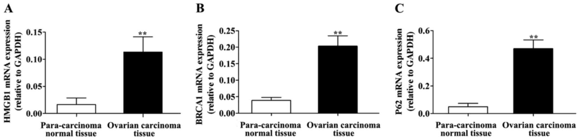
The RT-qPCR detection results are shown in Fig. 1. Compared with those in para-carcinoma
normal tissues, the mRNA expression of HMGB1, BRCA1 and P62 was
significantly higher in ovarian carcinoma tissues, and the
differences were statistically significant (P<0.01).
Detection of the protein expression of
HMGB1, BRCA1 and P62 in tissue specimens by
immunohistochemistry
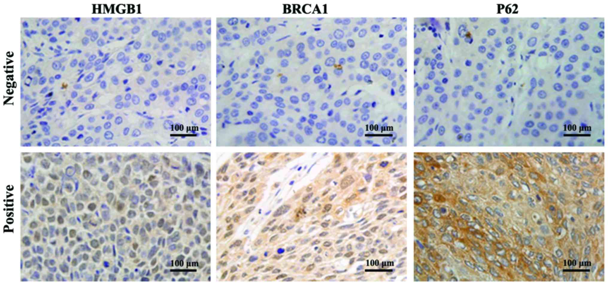
Immunohistochemistry detection results are shown in
Fig. 2. The positive
immunohistochemical staining of HMGB1, BRCA1 and P62 were all
yellow brown, with HMGB1 protein mainly being identified in the
cytoplasm, BRCA1 mainly in the cell nucleus, and partially in the
cytoplasm; and P62 mainly being identified in the cytoplasm.
The scores of statistical staining are shown in
Table II. The positive expression
rates of HMGB1 in ovarian carcinoma and para-carcinoma normal
tissues were 61.67% (37/60) and 13.33% (8/60), and the difference
was statistically significant (P<0.01); the positive expression
rates of BRCA1 in ovarian carcinoma and para-carcinoma normal
tissues were 78.33% (47/60) and 8.33% (5/60), and the difference
was statistically significant (P<0.01). The positive expression
rates of P62 in ovarian carcinoma and para-carcinoma normal tissues
were 71.67% (43/60) and 11.67% (7/60), and the difference was
statistically significant (P<0.01).
| Table II.Protein expression of HMGB1, BRCA1 and
P62 in para-carcinoma normal tissues and ovarian carcinoma
tissues. |
Table II.
Protein expression of HMGB1, BRCA1 and
P62 in para-carcinoma normal tissues and ovarian carcinoma
tissues.
|
|
| HMGB1 | BRCA1 | P62 |
|---|
|
|
|
|
|
|
|---|
| Groups | No. | Positive | Positive rate | P-value | Positive | Positive rate | P-value | Positive | Positive rate | P-value |
|---|
| Ovarian carcinoma
tissue | 60 | 37 | 61.67% | <0.01 | 47 | 78.33% | <0.01 | 43 | 71.67% | <0.01 |
| Para-carcinoma normal
tissue | 60 | 8 | 13.33% | <0.01 | 5 | 8.33% | <0.01 | 7 | 11.67% | <0.01 |
Drug resistance to cisplatin of 60
ovarian cancer patients
The results of in vitro resin droplet
experiment showed that 38 out of 60 ovarian cancer patients had
drug resistance to cisplatin, while 22 cases were sensitive to
cisplatin (Table III).
| Table III.Drug resistance to cisplatin of 60
ovarian cancer patients. |
Table III.
Drug resistance to cisplatin of 60
ovarian cancer patients.
| Groups | Case (n) | Ratio (%) |
|---|
| Drug-resistance | 38 | 63.33 |
| Sensitive | 22 | 36.67 |
Association of protein expression of
HMGB1, BRCA1 and P62 with drug resistance to cisplatin in ovarian
cancer
The results showed that the positive rates of the
protein expression of HMGB1, BRCA1 and P62 in 38 patients in the
drug resistance group were 84.21, 94.74 and 92.11%, respectively,
while those in the sensitive group were 22.73, 50.00 and 36.36%,
respectively. The results of the Chi-square test showed that the
positive rates of the protein expression of HMGB1, BRCA1 and P62 in
the drug resistance group were significantly higher than those in
the sensitive group (P<0.01) (Table
IV).
| Table IV.Association of protein expression of
HMGB1, BRCA1 and P62 with drug-resistance to cisplatin in ovarian
cancer. |
Table IV.
Association of protein expression of
HMGB1, BRCA1 and P62 with drug-resistance to cisplatin in ovarian
cancer.
|
|
| HMGB1 | BRCA1 | P62 |
|---|
|
|
|
|
|
|
|---|
| Groups | No. | Positive | Positive rate | P-value | Positive | Positive rate | P-value | Positive | Positive rate | P-value |
|---|
| Drug resistance | 38 | 32 | 84.21% | <0.01 | 36 | 94.74% | <0.01 | 35 | 92.11% | <0.01 |
| Sensitive | 22 | 5 | 22.73% | <0.01 | 11 | 50.00% | <0.01 | 8 | 36.36% | <0.01 |
Discussion
Clinical study statistics found that the 5-year
survival rate of patients with advanced ovarian cancer is lower
than 20%, and an important reason leading to poor prognosis is the
drug resistance of patients to chemotherapy drugs (11). Therefore, identification of the
specific indexes to predict the sensitivity of ovarian cancer
patients to chemotherapy drugs are issues to be solved, which can
provide powerful assistance for clinical doctors in formulating
therapeutic regimens and selecting chemotherapy drugs (12).
Cisplatin, a common chemotherapy drug in clinical
tumor therapy, is the first-line chemotherapy drug in ovarian
cancer treatment, and drug resistance to cisplatin is the main
reason affecting the curative effect of chemotherapy. The drug
resistance to cisplatin of ovarian cancer patients can be divided
into inherent or acquired drug resistance, and the drug resistance
mechanism of tumor is very complex. Currently, the molecular
mechanism of patient's drug resistance to cisplatin remains unclear
(13).
HMBG1 can inhibit cell apoptosis, and when HMBG1 is
overexpressed in cells, tumor cell apoptosis is markedly decreased.
When siRNA is used to silence HMGB1 expression, the sensitivity of
tumor cells to chemotherapy drugs can be elevated (14). BRCA1 gene is located on human
chromosome 17q21, and constitutes the susceptibility gene for
breast and ovarian cancer (15).
BRCA1 has the effect of inhibiting cell growth and plays a key role
in gene transcription, DNA damage repair and apoptosis, especially
in maintaining the genome stability (16). P62 is a kind of autophagy-induced
protein, and cells can relieve the endoplasmic reticulum stress by
autophagy, thus decreasing the tumor cell sensitivity to cisplatin.
Subsequently, when P62 is highly expressed, intracellular autophagy
activity is activated, eventually reducing the drug resistance of
ovarian carcinoma cells to cisplatin (17,18).
In order to further explore the expression of HMGB1,
BRCA1 and P62 in the tumor tissues of ovarian cancer patients and
its effect on the drug resistance of ovarian cancer patients to
cisplatin, RT-qPCR was performed to detect the mRNA expression of
HMGB1, BRCA1 and P62 in the tumor tissues of ovarian cancer
patients. The results showed that the mRNA expression levels of
HMGB1, BRCA1 and P62 were significantly higher in ovarian carcinoma
tissues compared with those in para-carcinoma normal tissues. In
addition, immunohistochemical results showed that the positive
protein expression rates of HMGB1, BRCA1 and P62 in ovarian
carcinoma tissues were 61.67% (37/60), 76.33% (47/60) and 71.67%
(43/60), respectively, which were significantly higher than those
in para-carcinoma normal tissues. Moreover, the in vitro
resin droplet experiment revealed that 38 out of 60 ovarian cancer
patients had drug resistance to cisplatin and 22 cases were
sensitive to cisplatin. The positive rates of the protein
expression of HMGB1, BRCA1 and P62 in the drug resistance group
were 84.21, 94.74 and 92.11%, respectively, while those in the
sensitive group were 22.73, 50.00 and 36.36%, respectively. Thus,
the positive protein expression rates of HMGB1, BRCA1 and P62 in
drug resistance were significantly higher than those in the
sensitive group.
Wang et al reported that the higher the
intracellular expression level of BRCA is, the lower the cell
sensitivity to cisplatin will be (19). Liu et al confirmed that after
HMGB1 is added into the cell culture fluid, it can induce cells to
develop drug resistance to chemotherapy drugs (14). Yu et al found that P62 protein
is overexpressed in tissues of ovarian cancer patients who have
drug resistance to cisplatin, and it is involved in the formation
mechanism of drug resistance of cells to cisplatin (20). This experiment further confirmed the
overexpression of HMGB1, BRCA1 and P62 in tumor tissues of ovarian
cancer patients, and it was found that the expression of HMGB1,
BRCA1 and P62 was associated with the drug resistance of ovarian
cancer patients to cisplatin.
In conclusion, the overexpression of HMGB1, BRCA1
and P62 exists in tumor tissues of ovarian cancer patients.
Furthermore, its positive expression is associated with
chemotherapy sensitivity of ovarian cancer patients. Thus, HMGB1,
BRCA1 and P62 may be molecular markers for the prediction of
chemotherapy sensitivity of ovarian cancer patients.
Competing interests
The authors declare that they have no competing
interests.
References
|
1
|
Wang Y, Liu P, Qiu L, Sun Y, Zhu M, Gu L,
Di W and Duan Y: Toxicity and therapy of cisplatin-loaded EGF
modified mPEG-PLGA-PLL nanoparticles for SKOV3 cancer in mice.
Biomaterials. 34:4068–4077. 2013. View Article : Google Scholar : PubMed/NCBI
|
|
2
|
Wu YJ, Neuwelt AJ, Muldoon LL and Neuwelt
EA: Acetaminophen enhances cisplatin- and paclitaxel-mediated
cytotoxicity to SKOV3 human ovarian carcinoma. Anticancer Res.
33:2391–2400. 2013.PubMed/NCBI
|
|
3
|
Ong PS, Chan SY and Ho PC: Microarray
analysis revealed dysregulation of multiple genes associated with
chemoresistance to As2O3 and increased tumor
aggressiveness in a newly established arsenic-resistant ovarian
cancer cell line, OVCAR-3/AsR. Eur J Pharm Sci. 45:367–378. 2012.
View Article : Google Scholar : PubMed/NCBI
|
|
4
|
Tang D, Kang R, Zeh HJ III and Lotze MT:
High-mobility group box 1 and cancer. Biochim Biophys Acta.
1799:131–140. 2010. View Article : Google Scholar : PubMed/NCBI
|
|
5
|
Wu D, Ding Y, Wang S, Zhang Q and Liu L:
Increased expression of high mobility group box 1 (HMGB1) is
associated with progression and poor prognosis in human
nasopharyngeal carcinoma. J Pathol. 216:167–175. 2008. View Article : Google Scholar : PubMed/NCBI
|
|
6
|
Beeghly A, Katsaros D, Chen H, Fracchioli
S, Zhang Y, Massobrio M, Risch H, Jones B and Yu H: Glutathione
S-transferase polymorphisms and ovarian cancer treatment and
survival. Gynecol Oncol. 100:330–337. 2006. View Article : Google Scholar : PubMed/NCBI
|
|
7
|
Stengel C, Newman SP, Leese MP, Potter BV,
Reed MJ and Purohit A: Class III beta-tubulin expression and in
vitro resistance to microtubule targeting agents. Br J Cancer.
102:316–324. 2010. View Article : Google Scholar : PubMed/NCBI
|
|
8
|
Quinn JE, James CR, Stewart GE, Mulligan
JM, White P, Chang GK, Mullan PB, Johnston PG, Wilson RH and Harkin
DP: BRCA1 mRNA expression levels predict for overall survival in
ovarian cancer after chemotherapy. Clin Cancer Res. 13:7413–7420.
2007. View Article : Google Scholar : PubMed/NCBI
|
|
9
|
Moscat J, Diaz-Meco MT and Wooten MW:
Signal integration and diversification through the p62 scaffold
protein. Trends Biochem Sci. 32:95–100. 2007. View Article : Google Scholar : PubMed/NCBI
|
|
10
|
Kobayashi H, Tanisaka K, Doi O, Kodama K,
Higashiyama M, Nakagawa H, Miyake M, Taki T, Hara S, et al: An in
vitro chemosensitivity test for solid human tumors using collagen
gel droplet embedded cultures. Int J Oncol. 11:449–455.
1997.PubMed/NCBI
|
|
11
|
Siegel RL, Miller KD and Jemal A: Cancer
statistics, 2015. CA Cancer J Clin. 65:5–29. 2015. View Article : Google Scholar : PubMed/NCBI
|
|
12
|
Sakamoto M, Kondo A, Kawasaki K, Goto T,
Sakamoto H, Miyake K, Koyamatsu Y, Akiya T, Iwabuchi H, Muroya T,
et al: Analysis of gene expression profiles associated with
cisplatin resistance in human ovarian cancer cell lines and tissues
using cDNA microarray. Hum Cell. 14:305–315. 2001.PubMed/NCBI
|
|
13
|
Takano M, Kudo K, Goto T, Yamamoto K, Kita
T and Kikuchi Y: Analyses by comparative genomic hybridization of
genes relating with cisplatin-resistance in ovarian cancer. Hum
Cell. 14:267–271. 2001.PubMed/NCBI
|
|
14
|
Liu L, Yang M, Kang R, Wang Z, Zhao Y, Yu
Y, Xie M, Yin X, Livesey KM, Lotze MT, et al: HMGB1-induced
autophagy promotes chemotherapy resistance in leukemia cells.
Leukemia. 25:23–31. 2011. View Article : Google Scholar : PubMed/NCBI
|
|
15
|
Miki Y, Swensen J, Shattuck-Eidens D,
Futreal PA, Harshman K, Tavtigian S, Liu Q, Cochran C, Bennett LM,
Ding W, et al: A strong candidate for the breast and ovarian cancer
susceptibility gene BRCA1. Science. 266:66–71. 1994. View Article : Google Scholar : PubMed/NCBI
|
|
16
|
Garvin AM, Attenhofer-Haner M and Scott
RJ: BRCA1 and BRCA2 mutation analysis in 86 early onset
breast/ovarian cancer patients. J Med Genet. 34:990–995. 1997.
View Article : Google Scholar : PubMed/NCBI
|
|
17
|
Greggio E, Lewis PA, van der Brug MP,
Ahmad R, Kaganovich A, Ding J, Beilina A, Baker AK and Cookson MR:
Mutations in LRRK2/dardarin associated with Parkinson disease are
more toxic than equivalent mutations in the homologous kinase
LRRK1. J Neurochem. 102:93–102. 2007. View Article : Google Scholar : PubMed/NCBI
|
|
18
|
Haugarvoll K, Toft M, Ross OA, White LR,
Aasly JO and Farrer MJ: Variants in the LRRK1 gene and
susceptibility to Parkinson's disease in Norway. Neurosci Lett.
416:299–301. 2007. View Article : Google Scholar : PubMed/NCBI
|
|
19
|
Wang L, Wei J, Qian X, Yin H, Zhao Y, Yu
L, Wang T and Liu B: ERCC1 and BRCA1 mRNA expression levels in
metastatic malignant effusions is associated with chemosensitivity
to cisplatin and/or docetaxel. BMC Cancer. 8:972008. View Article : Google Scholar : PubMed/NCBI
|
|
20
|
Yu H, Su J, Xu Y, Kang J, Li H, Zhang L,
Yi H, Xiang X, Liu F and Sun L: p62/SQSTM1 involved in cisplatin
resistance in human ovarian cancer cells by clearing ubiquitinated
proteins. J Eur Cancer. 47:1585–1594. 2011. View Article : Google Scholar
|