Introduction
Ovarian cancer is one of the most common types of
cancer in women, and the American cancer society estimated there
would be approximately 22,000 new cases of ovarian cancer and
14,000 mortalities caused by the disease in 2017 in the United
States, since ovarian cancer causes more deaths than any other
cancer of the female reproductive system (1). The most common ovarian cancer types are
known as epithelial ovarian cancer, which have the highest
mortality rate amongst gynecological malignant tumors in females
(2). More than 70% of patients with
ovarian cancer have been found in advanced stage (International
Federation of Gynecology and Obstetrics, FIGO stage III or IV)
(3,4).
Cis-dichlorodiamine platinum (cisplatin) is the main treatment for
advanced ovarian cancer. However, the majority of patients with
cisplatin resistance cannot benefit from chemotherapy (5). A variety of factors are involved in the
emergence of cisplatin resistance, including increased drug efflux,
abnormal drug targeting, enhanced DNA repair and altered apoptotic
pathway (6–11). The basic mechanism for the emergence
of chemotherapeutic drug resistance remains poorly understood. At
present, there is a lack of effective drugs to reduce resistance to
chemotherapy. Circadian genes are important genes, which regulate
biological activity and includes period circadian clock (PER)1,
PER2, PER3, timeless circadian clock (TIM), clock
circadian regulator (CLOCK), brain and muscle Arnt-like
protein (BMAL), cryptochrome circadian clock 1 (CRY)1
and CRY2 (11). Of these
genes, CLOCK gene was the first gene identified, and has
been revealed to be strongly associated with sensitivity of various
tumor types to chemotherapy, including gastric cancer,
cholangiocarcinoma and colorectal cancer (12–15). There
are a limited number of studies that have investigated the
association between the CLOCK gene and resistance to
chemotherapy in ovarian cancer.
The present study observed CLOCK gene
expression in cisplatin-sensitive cell line A2780 and
cisplatin-resistant cell line CP70.
The effects of cisplatin on the proliferation and
apoptosis of cisplatin-resistant cell line CP70 following the
knockdown of CLOCK in cisplatin-resistant CP70 cells were
investigated, and the effects of CLOCK on chemotherapy
resistance in ovarian cancer were discussed.
Materials and methods
Cells and main reagents
Cisplatin-sensitive A2780 and cisplatin-resistant
CP70 cell lines (Shanghai Bogoo Biotechnology Co., Ltd., Shanghai,
China) were incubated in Dulbecco's modified Eagle's medium (Gibco;
Thermo Fisher Scientific, Inc., Waltham, MA, USA) containing 10%
fetal bovine serum (Gibco; Thermo Fisher Scientific, Inc.). Rat
anti-human CLOCK antibody and horseradish peroxidase-labeled goat
anti-rat immunoglobulin G (IgG) were purchased from Santa Cruz
Biotechnology, Inc., Dallas, TX, USA. Quantitative polymerase chain
reaction (qPCR) primers were synthesized by Invitrogen (Thermo
Fisher Scientific, Inc.). CytoBuster protein extraction reagent was
purchased from Novagen (Merck KGaA, Darmstadt, Germany), and
cisplatin was obtained from Sigma-Aldrich (Merck KGaA).
Additionally, small-interfering (si)CLOCK small RNA fragments
(Ambion; Thermo Fisher Scientific, Inc.), cell transfection reagent
(Lonza Group, Ltd., Basel, Switzerland) and protease inhibitor
phenylmethanesulfony fluoride (Thermo Fisher Scientific, Inc.) were
also used. Phenylmethanesulfony fluoride was added to PBS prior to
use.
Reverse transcription (RT)-qPCR
Total RNA was extracted using Trizol reagent (Thermo
Fisher Scientific). cDNAs were synthesized using a transcriptor
first strand cDNA synthesis kit (Roche Applied Science, Penzberg,
Germany), according to the manufacturer's protocol. mRNA levels
were measured using using a FastStart Universal SYBR Green Master
kit (Roche Applied Science) in a Light Cycler 96 (Roche Applied
Science, Penzberg, Germany). Primer sequences were as follows:
CLOCK forward, 5′-ACACCCAGAAGGAAGAGCAA-3′ reverse,
5′-GCGAGAACGCTTTGCTTTAG-3′; GAPDH forward,
5′-ATGTCGTGGAGTCTACTGGC-3′, reverse 5′-AGGATGCATTGCTGACAATC-3′.
Reaction conditions are as follows: Pre-denaturation at 94°C for 5
min, 40 cycles of 94°C for 30 sec, 60°C for 40 sec and 72°C for 40
sec, followed by 72°C for 10 min. Target gene fragments were
amplified with a DNA thermal cycler. GAPDH was used as an internal
reference. Relative mRNA levels were calculated using the
2−ΔΔCq method (16). Data
were calculated from three independent experiments.
Cell treatment
Cisplatin-sensitive A2780 and cisplatin-resistant
cell line CP70 cells (1×10⁵/ml) (Shanghai Gefan
Biotechnology Co., Ltd., Shanghai, China) were treated with 0, 32
and 64 µM dipeptidyl peptidase-4 (Exalpha Biologicals, Inc.,
Shirley, MA, USA) for 48 h at 37°C.
Western blot assay
Total protein was extracted from cisplatin-sensitive
A2780 and cisplatin-resistant CP70 cells in the logarithmic phase.
Cisplatin-treated cells were digested with 0.25% trypsin, and total
protein was extracted using CytoBuster protein extraction reagent.
An equal volume of the protein (30 µg) was loaded onto 8% SDS-PAGE
separating gel and 5% stacking gel and transferred to
nitrocellulose membranes by semi-dry method. The membranes were
blocked with Tris-buffered saline and Tween-20 containing 5% bovine
serum albumin (Gibco; Thermo Fisher Scientific, Inc.) for 2 h at
room temperature, and incubated with rat anti-human CLOCK antibody
(1:1,500; cat no. 3896-100; BioVision, Inc., Milpitas, CA, USA) at
4°C overnight. On the following day, the membranes were washed
three times with 0.1% Tris-buffered saline and Tween-20 for 5 min
each, incubated with horseradish peroxidase-labeled IgG secondary
antibody at room temperature for 1 h and washed three times with
0.1% Tris-buffered saline and Tween-20. The membranes were
incubated in SuperSignal West Pico substrate (Thermo Fisher
Scientific, Inc.) for visualization, and were incubated with
β-actin at 55°C for 20 min, densitometry of the western blotting
was analyzed with Image Pro Plus 6.0 (cat no. 20910; Media
Cybernetics, Inc., Rockville, MD, USA). β-actin (1:1,500; cat no.
MAB8929; R&D Systems, Inc., Minneapolis, MN, USA), served as
the internal reference. The experiments were conducted at least in
triplicate.
Cell transfection
The cell transfection reagent as mentioned
previously was used. The cells were transfected with 100 nM of
siCLOCK small RNA fragments for 48 h according to the
manufacturer's protocol. At 24 h after transfection, an MTT assay
was performed.
MTT assay
Following the knockdown of CLOCK protein expression,
cisplatin-resistant CP70 cells at a density of 1×10⁵/ml
and control cells (cells without gene knockout) at a density of
1×10⁵/ml were incubated in a 96-well plate at 37°C. A
total of 20 µl MTT was added in each well for 4 h at 37°C.
Following removal of the supernatant, 150 µl dimethyl sulfoxide was
added, followed by a low oscillation speed for 10 min. Optical
density was measured at 490 nm.
Statistical analysis
All data were analyzed with SPSS software (version
16.0; SPSS, Inc., Chicago, IL, USA). t-test was used to compare the
differences in data between two groups. One-way analysis of
variance was utilized to compare the differences among multiple
groups. P<0.05 was considered to indicate a statistically
significant difference.
Results
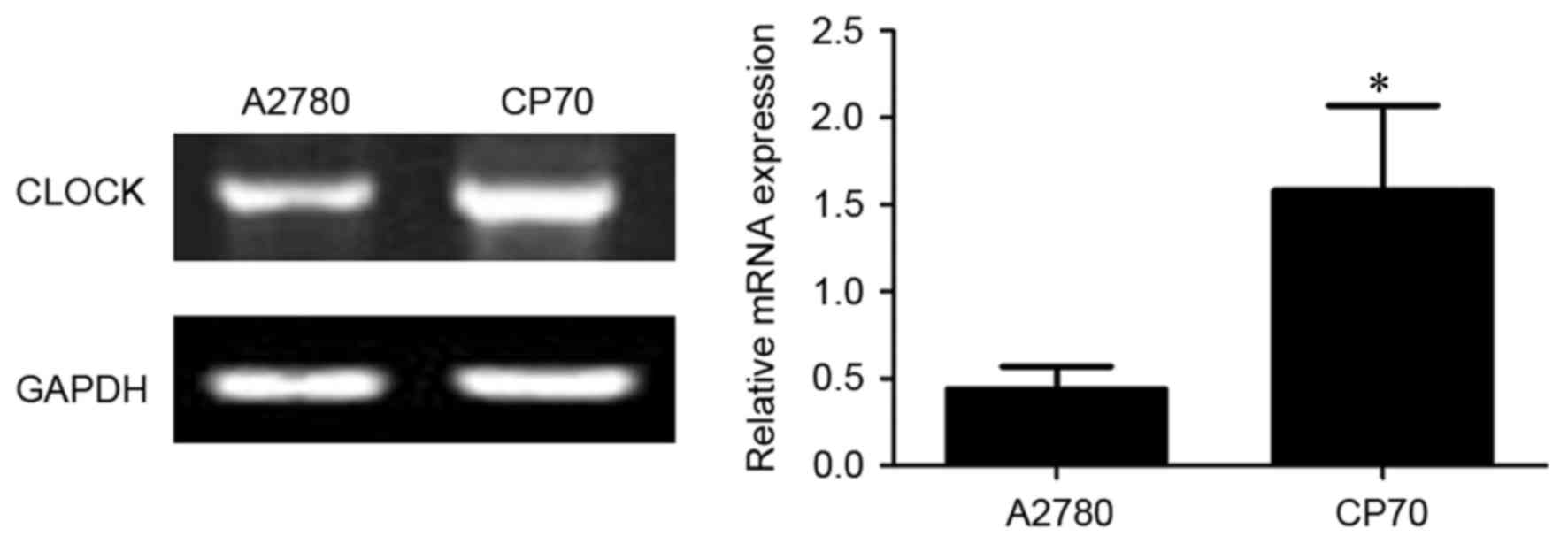
CLOCK mRNA expression in two types of
ovarian cancer cells
As shown in Fig. 1,
the expression of CLOCK mRNA was significantly higher in
cisplatin-resistant CP70 cells (1.58±0.49) compared with
cisplatin-sensitive A2780 cells (0.44±0.13) (P<0.01).
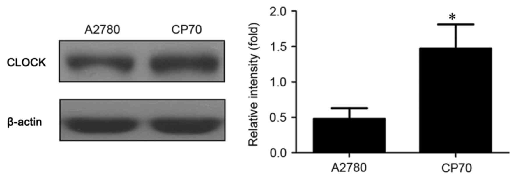
CLOCK protein expression in two types
of ovarian cancer cells
As exhibited in Fig.
2, the expression of CLOCK protein was significantly higher in
cisplatin-resistant CP70 cells (1.47±0.34) compared with
cisplatin-sensitive A2780 cells (0.48±0.15) (P<0.01).
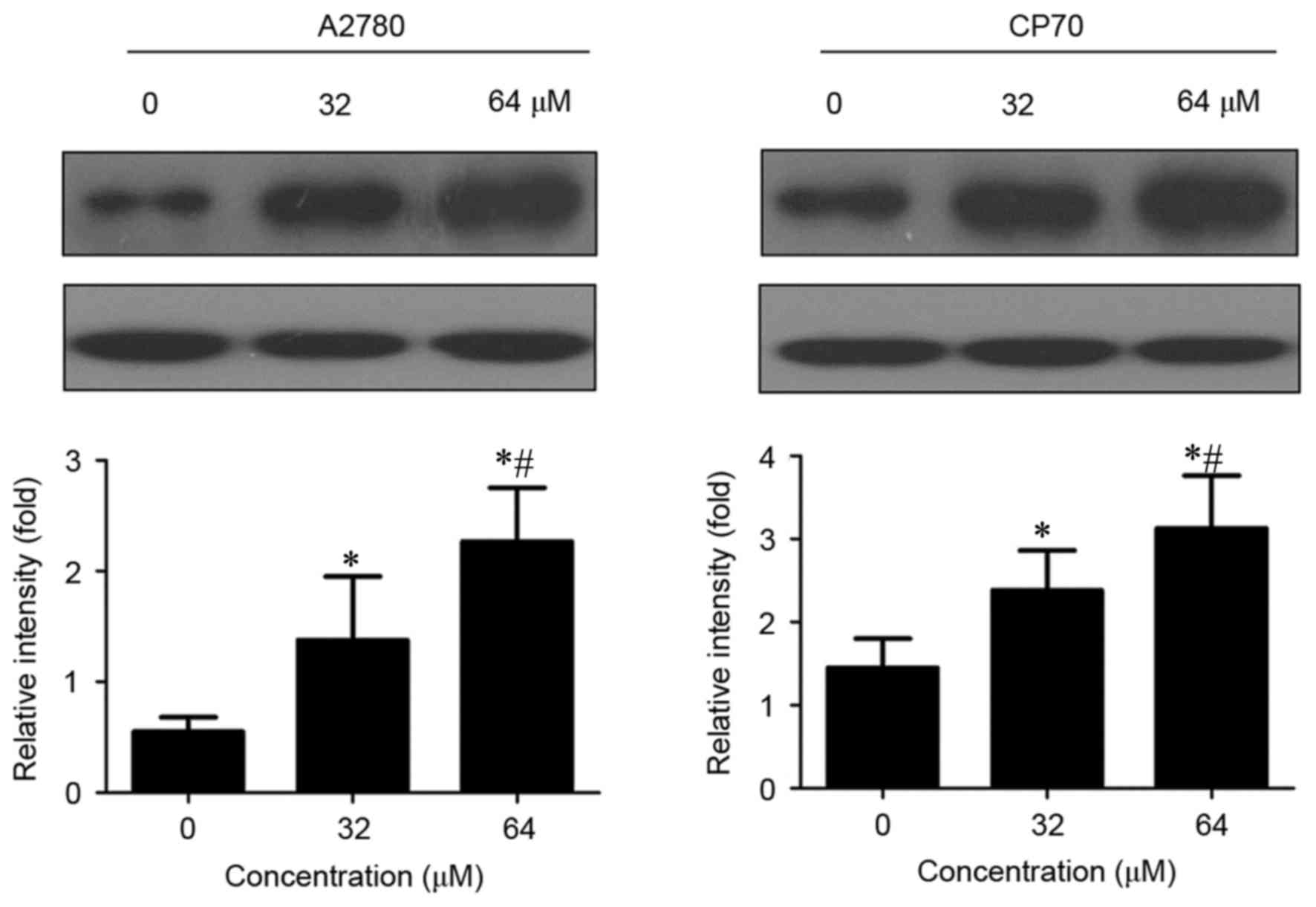
Effects of cisplatin on CLOCK protein
expression in two types of ovarian cancer cells
CLOCK protein expression was significantly increased
in cisplatin-treated A2780 and CP70 cells compared with untreated
cells (P<0.01; Fig. 3).
Furthermore, CLOCK protein expression gradually increased with an
increased concentration of cisplatin (P<0.01).
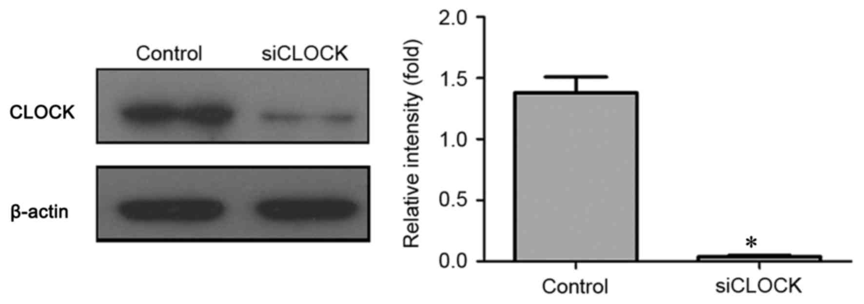
Confirming the effects of CLOCK
knockdown on protein expression
As illustrated in Fig.
4, CLOCK siRNA transfection was able to significantly
knockdown CLOCK protein expression in cisplatin-resistant CP70
cells compared with control cells (P<0.01).
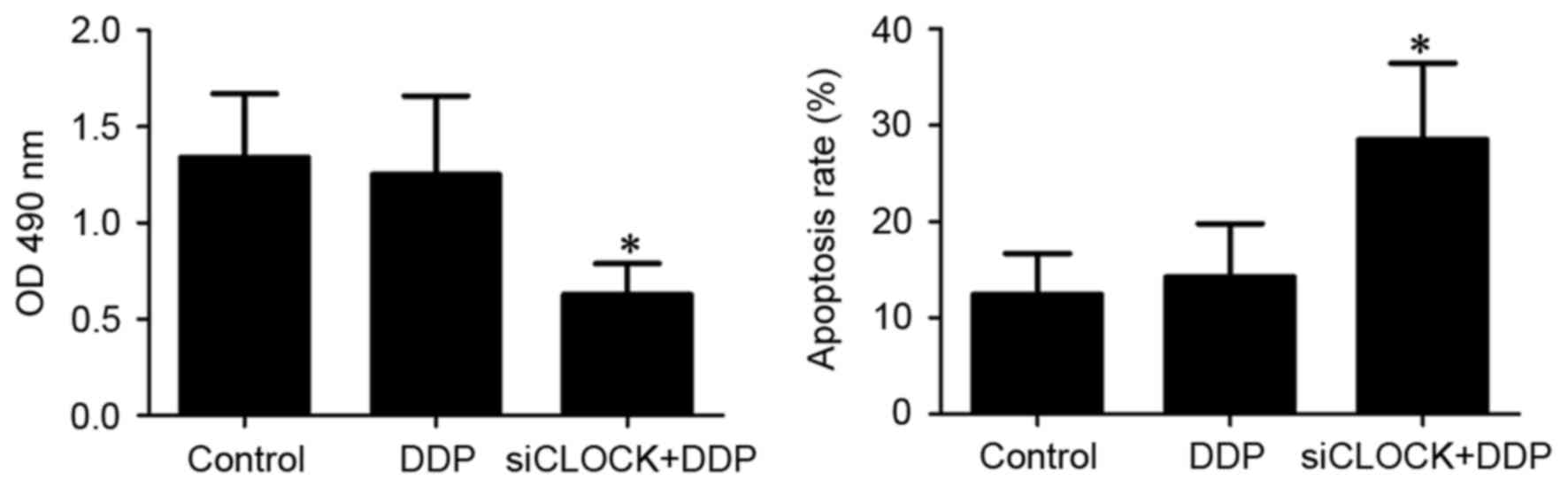
Proliferation and apoptosis of
cisplatin-resistant CP70 cells following CLOCK knockdown
As shown in Fig. 5,
treatment with cisplatin did not significantly affect the
proliferation and apoptosis of cisplatin-treated CP70 cells
(P>0.05). By contrast following CLOCK knockdown, treatment with
cisplatin was able to significantly inhibit the proliferation of
CP70 cells and induce its apoptosis (P<0.01).
Discussion
Ovarian cancer is the fourth most common malignant
tumor in women and is the leading cause of mortality from
gynecologic malignancies. Due to its high mortality rate, ovarian
cancer has become a global public health problem (17–20). The
overall 5-year survival rate in the United States is 45%, the
1-year survival rate is 72% and the 10-year survival rate is 35%
(21). For cases where a diagnosis is
made early in the disease, when the cancer is still confined to the
primary site, the 5-year survival rate is 92.7% (22). Approximately 70% of women with the
advanced disease respond to initial treatment, a majority of whom
attain complete remission, but half of these women experience
recurrence 1–4 years following treatment (23). Therefore, if ovarian cancer can be
diagnosed at an early stage, patients can get better treatment.
Nevertheless, >70% of the patients with ovarian cancer have been
diagnosed at advanced stages (FIGO stage III or IV) (4). The main method for advanced ovarian
cancer is cisplatin treatment. However, the emergence of cisplatin
resistance in the majority of patients with ovarian cancer reduces
the effects of the chemotherapeutics (5).
A variety of different factors are involved in the
emergence of cisplatin resistance, including increased drug efflux,
abnormal drug targeting, enhanced DNA repair and altered apoptotic
pathway (6–11). Molecular mechanisms of drug resistance
in cisplatin-based chemotherapy remain unclear. There is a lack of
effective drugs to reduce the resistance to chemotherapy.
Circadian rhythm is an endogenous adaptation
mechanism in the process of long-term biological evolution and a
basic characteristic of life activity. Circadian genes are
important genes, which regulate biological activity and include
PER1, PER2, PER3, TIM, CLOCK, BMAL, CRY1 and CRY2
(11). The CLOCK gene was the
first gene discovered and was identified as a circadian clock gene.
CLOCK gene is located on the long arm of chromosome 4 (4p12)
and contains >20 exons (24). The
CLOCK gene is not only expressed in normal tissue and cells.
Abnormal expression of CLOCK gene can also be detected in a
number of types of tumors (25–27). It
was also demonstrated that the knockdown of CLOCK was able to
increase the apoptosis of glioma cells (28). CLOCK gene has been revealed to
be strongly associated with sensitivity to chemotherapy in various
tumors, including gastric cancer, cholangiocarcinoma and colorectal
cancer (13–15).
However, there are a limited number of studies that
have investigated the association between CLOCK gene and
chemotherapy resistance in ovarian cancer. Therefore, the present
study observed CLOCK gene expression in cisplatin-sensitive
A2780 and cisplatin-resistant CP70 cells and investigated the
effects of cisplatin treatment on the proliferation and apoptosis
of cisplatin-resistant CP70 cells following CLOCK knockdown.
The effects of CLOCK gene on chemotherapy resistance in
ovarian cancer were also discussed.
The present study first compared CLOCK mRNA
and protein expression in two ovarian cancer cell lines (A2780 and
CP70). Results demonstrated that CLOCK mRNA and protein
expression was significantly lower in cisplatin-sensitive A2780
cells compared with cisplatin-resistant CP70 cells (P<0.01),
indicating that CLOCK gene expression was strongly
associated with cisplatin resistance in ovarian cancer cells. To
further verify the association between CLOCK gene and
cisplatin resistance in ovarian cancer cells, cisplatin-sensitive
A2780 and cisplatin-resistant CP70 cells were treated with
different concentrations of cisplatin. Results showed that CLOCK
protein expression increased with an increased concentration of
cisplatin in the two cell lines following cisplatin treatment in a
dose-dependent manner (P<0.01), which further suggested that
CLOCK gene was associated with cisplatin resistance in
ovarian cancer cells. To confirm the precise effect of CLOCK
gene on cisplatin resistance in ovarian cancer cells, the present
study knocked down the expression of CLOCK protein in
cisplatin-resistant CP70 cells by RNA interference. The results
showed that cisplatin treatment was able to significantly suppress
the proliferation of CP70 cells, and induce its apoptosis following
the knockdown of CLOCK protein (P<0.01). In summary, the
expression of circadian gene CLOCK was strongly associated
with cisplatin resistance in ovarian cancer cells. The increase in
the expression of circadian gene CLOCK may reduce the
sensitivity to cisplatin treatment in ovarian cancer cells.
Acknowledgements
Not applicable.
Funding
The project was supported by the China Postdoctoral
Science Foundation grant (grant no. 2013M542498).
Availability of data and materials
All relevant data are included in the present
study.
Authors' contributions
HX conceived and designed the experiments, and wrote
the manuscript. ZW, GM and HC conducted the experiments, collected,
analyzed and interpreted the data. All authors reviewed the
manuscript.
Ethics approval and consent to
participate
Not applicable.
Consent for publication
Not applicable.
Competing interests
The authors declare that they have no competing
interests.
References
|
1
|
Smith RA, Andrews KS, Brooks D, Fedewa SA,
Manassaram-Baptiste D, Saslow D, Brawley OW and Wender RC: Cancer
screening in the United States, 2017: A review of current American
Cancer Society guidelines and current issues in cancer screening.
CA Cancer J Clin. 67:100–121. 2017. View Article : Google Scholar : PubMed/NCBI
|
|
2
|
Siegel R, Naishadham D and Jemal A: Cancer
statistics, 2013. CA Cancer J Clin. 63:11–30. 2013. View Article : Google Scholar : PubMed/NCBI
|
|
3
|
Knutson KL, Karyampudi L, Lamichhane P and
Preston C: Targeted immune therapy of ovarian cancer. Cancer
Metastasis Rev. 34:53–74. 2015. View Article : Google Scholar : PubMed/NCBI
|
|
4
|
Killedar A, Stutz MD, Sobinoff AP,
Tomlinson CG, Bryan TM, Beesley J, Chenevix-Trench G, Reddel RR and
Pickett HA: A common cancer risk-associated allele in the hTERT
locus encodes a dominant negative inhibitor of telomerase. PLoS
Genet. 11:e10052862015. View Article : Google Scholar : PubMed/NCBI
|
|
5
|
Yan C, Yang F, Zhou C, Chen X, Han X, Liu
X, Ma H and Zheng W: MCT1 promotes the cisplatin-resistance by
antagonizing Fas in epithelial ovarian cancer. Int J Clin Exp
Pathol. 8:2710–2718. 2015.PubMed/NCBI
|
|
6
|
Vallo S, Michaelis M, Rothweiler F,
Bartsch G, Gust KM, Limbart DM, Rödel F, Wezel F, Haferkamp A and
Cinatl J Jr: Drug-resistant urothelial cancer cell lines display
diverse sensitivity profiles to potential second-line therapeutics.
Transl Oncol. 8:210–216. 2015. View Article : Google Scholar : PubMed/NCBI
|
|
7
|
Takano M, Kakizoe S, Kawami M, Nagai J,
Patanasethnont D, Sripanidkulchai B and Yumoto R: Modulation of
P-glycoprotein function and multidrug resistance in cancer cells by
Thai plant extracts. Pharmazie. 69:823–828. 2014.PubMed/NCBI
|
|
8
|
Territo PR, Maluccio M, Riley AA, McCarthy
BP, Fletcher J, Tann M, Saxena R and Skill NJ: Evaluation of
11C-acetate and 18F-FDG PET/CT in mouse multidrug resistance gene-2
deficient mouse model of hepatocellular carcinoma. BMC Med Imaging.
15:152015. View Article : Google Scholar : PubMed/NCBI
|
|
9
|
Jing X, Zhang H, Hu J, Su P, Zhang W, Jia
M, Cheng H, Li W and Zhou G: β-arrestin 2 is associated with
multidrug resistance in breast cancer cells through regulating MDR1
gene expression. Int J Clin Exp Pathol. 8:1354–1363.
2015.PubMed/NCBI
|
|
10
|
Sun Y, Liu JH, Jin L, Sui YX, Han LL and
Huang Y: Effect of autophagy-related beclin1 on sensitivity of
cisplatin-resistant ovarian cancer cells to chemotherapeutic
agents. Asian Pac J Cancer Prev. 16:2785–2791. 2015. View Article : Google Scholar : PubMed/NCBI
|
|
11
|
Sun Y, Liu JH, Jin L, Sui YX, Lai L and
Yang Y: Inhibition of Beclin 1 expression enhances
cisplatin-induced apoptosis through a mitochondrial-dependent
pathway in human ovarian cancer SKOV3/DDP cells. Oncol Res.
21:261–269. 2014. View Article : Google Scholar : PubMed/NCBI
|
|
12
|
Song JM, Hu X, Fu X, Liu K and Li GM:
Effects of circadian genes hClock and hBmal1 on migration and
invasion of SGC-7901 cells. Xiandai Zhongliu Yixue. 21:2678–2681.
2013.
|
|
13
|
Hu ML, Yeh KT, Lin PM, Hsu CM, Hsiao HH,
Liu YC, Lin HY, Lin SF and Yang MY: Deregulated expression of
circadian clock genes in gastric cancer. BMC Gastroenterol.
14:672014. View Article : Google Scholar : PubMed/NCBI
|
|
14
|
Filipski E, Subramanian P, Carrière J,
Guettier C, Barbason H and Lévi F: Circadian disruption accelerates
liver carcinogenesis in mice. Mutat Res. 680:95–105. 2009.
View Article : Google Scholar : PubMed/NCBI
|
|
15
|
Fang L, Yang Z, Zhou J, Tung JY, Hsiao CD,
Wang L, Deng Y, Wang P, Wang J and Lee MH: Circadian clock gene
cry2 degradation is involved in chemoresistance of colorectal
cancer. Mol Cancer Ther. 14:1476–1487. 2015. View Article : Google Scholar : PubMed/NCBI
|
|
16
|
Livak KJ and Schmittgen TD: Analysis of
relative gene expression data using real-time quantitative PCR and
the 2(-Delta Delta C(T)) method. Methods. 25:402–408. 2001.
View Article : Google Scholar : PubMed/NCBI
|
|
17
|
Yan-Hong H, Jing L, Hong L, Shan-Shan H,
Yan L and Ju L: Association between alcohol consumption and the
risk of ovarian cancer: A meta-analysis of prospective
observational studies. BMC Public Health. 15:2232015. View Article : Google Scholar : PubMed/NCBI
|
|
18
|
Cui X, Li L, Yan G, Meng K, Lin Z, Nan Y,
Jin G and Li C: High expression of NQO1 is associated with poor
prognosis in serous ovarian carcinoma. BMC Cancer. 15:2442015.
View Article : Google Scholar : PubMed/NCBI
|
|
19
|
Gao L, Ye X, Ma RQ, Cheng HY, Han HJ, Cui
H, Wei LH and Chang XH: Low programmed cell death 5 expression is a
prognostic factor in ovarian cancer. Chin Med J (Engl).
128:1084–1090. 2015. View Article : Google Scholar : PubMed/NCBI
|
|
20
|
Lambrechts S, Lambrechts D, Despierre E,
Van Nieuwenhuysen E, Smeets D, Debruyne PR, Renard V, Vroman P,
Luyten D, Neven P, et al: Genetic variability in drug transport,
metabolism or DNA repair affecting toxicity of chemotherapy in
ovarian cancer. BMC Pharmacol Toxicol. 16:22015. View Article : Google Scholar : PubMed/NCBI
|
|
21
|
Rooth C: Ovarian cancer: Risk factors,
treatment and management. Br J Nurs. 22:S23–S30. 2013. View Article : Google Scholar : PubMed/NCBI
|
|
22
|
Zhang Q, Burdette JE and Wang JP:
Integrative network analysis of TCGA data for ovarian cancer. BMC
Syst Biol. 8:13382014. View Article : Google Scholar : PubMed/NCBI
|
|
23
|
Longoria TC and Eskander RN: Immune
checkpoint inhibition: Therapeutic implications in epithelial
ovarian cancer. Recent Pat Anticancer Drug Discov. 10:133–144.
2015. View Article : Google Scholar : PubMed/NCBI
|
|
24
|
Jung H, Choe Y, Kim H, Park N, Son GH,
Khang I and Kim K: Involvement of CLOCK: BMAL1 heterodimer in
serum-responsive mPer1 induction. Neuroreport. 14:15–19. 2003.
View Article : Google Scholar : PubMed/NCBI
|
|
25
|
Bjarnason GA, Jordan RC and Sothern RB:
Circadian variation in the expression of cell-cycle proteins in
human oral epithelium. Am J Pathol. 154:613–622. 1999. View Article : Google Scholar : PubMed/NCBI
|
|
26
|
Uth K and Sleigh R: Deregulation of the
circadian clock constitutes a significant factor in tumorigenesis:
A clockwork cancer. Part II. In vivo studies. Biotechnol Biotechnol
Equip. 28:379–386. 2014. View Article : Google Scholar : PubMed/NCBI
|
|
27
|
Tavano F, Pazienza V, Fontana A, Burbaci
FP, Panebianco C, Saracino C, Lombardi L, De Bonis A, di Mola FF,
di Sebastiano P, et al: SIRT1 and circadian gene expression in
pancreatic ductal adenocarcinoma: Effect of starvation. Chronobiol
Int. 32:497–512. 2015. View Article : Google Scholar : PubMed/NCBI
|
|
28
|
Wang F, Li C, Yongluo and Chen L: The
circadian gene clock plays an important role in cell apoptosis and
the DNA damage response in vitro. Technol Cancer Res Treat.
15:480–486. 2016. View Article : Google Scholar : PubMed/NCBI
|