Introduction
Oral squamous cell carcinoma (OSCC) is one of the
most common subtypes of all malignant head and neck tumors
worldwide (1). OSCC is a highly
invasive lesion that frequently metastasizes to cervical lymph
nodes (2). Metastasis to regional
lymph nodes and distant sites occurs in ~40% of patients with oral
cancer and is associated with a poor prognosis (3,4). At
present, only 25–40% of patients with lymph node metastasis (LNM)
achieve 5-year survival, compared with ~90% of patients without
metastasis (5).
Metastasis is the result of complicated events, in
which the dysfunction of epithelial cell-cell adhesion is known to
be critical for tumor invasion and metastasis (6). Cadherins, as a major class of cell-cell
adhesion molecules, regulate the adhesion and migration of cells in
a calcium-dependent manner (7).
Reduction of E-cadherin expression predicts the loss of epithelial
cell adhesion (8), which is a
prognostic factor for head and neck squamous cell carcinoma,
according to a systematic review with meta-analysis (9). Cadherin 6 (CDH6) is a class II cadherin
and its upregulation reportedly promotes the motility and invasion
of several cancer cells (10–12). Cadherin 11 (CDH11), a mesenchymal
cadherin, is upregulated during prostate cancer (PCa) progression
and bone metastasis (13). CDH11
expression increased the migration and invasion of PCa cells and
enabled PCa cells to intercalate into an osteoblast monolayer
(14). However, the roles of these
two proteins in mediating cell migration in OSCC remain unclear.
CD44 is a transmembrane glycoprotein and a cell adhesion molecule
that serves a crucial role in the differentiation, invasion and
metastasis of various types of tumor cell (15,16). CD44
is also involved in the epithelial-mesenchymal transition (EMT),
which is associated with metastasis.
Recent studies have suggested that the increase in
CD44 expression in OSCC is associated with increased metastasis and
decreased survival (17,18). However, the association between CDH6,
CDH11 and CD44 expression in OSCC has not previously been
investigated. Therefore, the aim of the present study was to
determine whether these adhesion factors may serve as potential
prediction factors for OSCC metastasis and prognosis, in order to
better understand the underlying pathogenesis of this malignancy
and potentially to improve the prognosis and treatment outcome of
patients with OSCC.
Materials and methods
Patients and tumor procurement
A total of 101 patients with OSCC who were treated
at the Department of Oral and Maxillofacial Surgery, Peking Union
Medical College Hospital (Beijing, China) between March 2006 and
December 2010 were retrospectively enrolled in the present study.
Tumors were pathologically staged according to the American Joint
Cancer Committee staging system (19)
Within 30 min after surgical extirpation, tissues were frozen in
liquid nitrogen, and the study was approved by the Ethics Committee
of Peking Union Medical College. The clinicopathological
information of the patients from whom the tissues were obtained is
presented in Table I.
 | Table I.Clinicopathological characteristics
in patients with oral squamous cell carcinoma (n=101). |
Table I.
Clinicopathological characteristics
in patients with oral squamous cell carcinoma (n=101).
| Variables | No. patients
(%) |
|---|
| Age, years |
|
|
>60 | 53 (52.5) |
|
<60 | 48 (47.5) |
| Sex |
|
|
Male | 65 (85.1) |
|
Female | 36 (14.9) |
| Location |
|
|
Tongue | 45 (44.6) |
|
Gingiva | 23 (22.8) |
| Floor
of mouth | 17 (16.8) |
|
Buccae | 14 (13.9) |
| Hard
palate | 2 (1.9) |
| Lymph node
metastasis |
|
|
Yes | 35 (34.7) |
| No | 66 (65.3) |
| Tumor stage |
|
| I | 25 (24.8) |
| II | 34 (33.6) |
|
III | 19 (18.8) |
| IV | 23 (22.8) |
|
Differentiation |
|
|
Well | 38 (37.6) |
|
Moderate | 50 (49.5) |
|
Poor | 13 (12.9) |
| Smoking |
|
|
Yes | 70 (69.3) |
| No | 31 (30.7) |
| Alcohol |
|
|
Yes | 65 (64.4) |
| No | 36 (35.6) |
Reverse transcription-quantitative
polymerase chain reaction (RT-qPCR)
Tumor tissue samples with or without LNM
(n=10/group) were homogenized, and total RNA was extracted using
TRIzol reagent (Invitrogen; Thermo Fisher Scientific, Inc.,
Waltham, MA, USA) and was further purified using the RNeasy RNA
Extraction Mini kit (Qiagen GmbH, Hilden, Germany). RNA samples
were converted into first strand cDNA using an RT2 First
Strand kit, according to the manufacturer's protocols
(SABiosciences; Qiagen GmbH). Quantitative gene expression was
analyzed by qPCR, performed on an ABI Prism 7900 HT (Applied
Biosystems; Thermo Fisher Scientific, Inc.). The optimized
parameters for PCR were: 95°C for 2 min, 94°C for 10 sec, 61.5°C
for 30 sec and 72°C for 40 sec for 40 cycles). The assay used an
RT2 Profiler PCR array system (RT2 Profiler™
PCR Array Human Tumor Metastasis (PAHS-028Z); SABiosciences; Qiagen
GmbH) that contained a panel of 84 genes and SYBRGreen qPCR
detection method and was performed according to the manufacturer's
protocols. Quality controls were all within the recommended range.
Data were analyzed by the 2−∆ΔCq method (20).
Immunohistochemical (IHC)
staining
IHC staining was performed by the
streptavidin-biotin complex (SABC) method using 4% formalin-fixed
(fixed for 48 h at 25°C), paraffin-embedded 4-µm tissue sections.
Tissue sections were heat-immobilized at 60°C for 30 min and
deparaffinized in xylene and rehydrated through a descending
ethanol series (100, 95, 90, 80 and 70%) at room temperature for 5
min at each concentration. Following the blocking of endogenous
peroxidase activity with 0.3% hydrogen peroxide for 10 min, the
sections were blocked for 1 h at room temperature with 5% bovine
serum albumin, prior to being incubated with primary rabbit
anti-CDH6 antibody (dilution, 1:100; cat. no. ab64917; Abcam,
Cambridge, MA, USA), mouse anti-CDH11 antibody (dilution, 1:50;
cat. no. ab151446; Abcam) or rabbit anti-CD44 antibody (dilution,
1:100; cat. no. ab51037; Abcam) for 24 h at 4°C. The slides were
rinsed with phosphate-buffered saline (PBS) three times and were
incubated with a goat anti-rabbit horseradish peroxidase-conjugated
secondary antibody (dilution, 1:500; cat. no. ZB-2010; OriGene
Technologies, Inc., Beijing, China) or a goat anti-mouse secondary
antibody (cat. no. ZB-2305; OriGene Technologies, Inc.) for 30 min
at room temperature. Following incubation with SABC for 20 min at
room temperature, the sections were exposed to
3,3′-diaminobenzidine for 1 min, counterstained with hematoxylin at
room temperature for 15 sec, mounted in neutral gum and images were
captured using a Nikon Eclipse E600 microscope (Nikon Corporation,
Tokyo, Japan). PBS was used instead of the primary antibody as a
negative control.
Evaluation of immunohistochemistry was quantified by
two pathologists, who were blinded to all patient diagnoses. The
staining intensity was estimated in a 4-step scale as previously
reported (0, no staining; 1, weak intensity; 2, moderate intensity;
and 3, strong intensity) (21). The
fraction of stained cells was scored according to the following
criteria: Score 0, <10% positive cancer cells; score 1, 11–50%
positive cancer cells; score 2, 51–80% positive cancer cells; and
score 3, >80% positive cancer cells. The final staining score
was assigned based on the multiplication of the staining intensity
and the percentage of positive cells, and was graded as follows: 0,
0; 1, 1–3; 2, 5–6; and 3, 7–9. Scores of 0 or 1 were classed as low
expression, and scores 2 or 3 were classed as high expression for
further analysis.
Statistical analysis
All experiments were repeated at least three times.
Data were analyzed using SPSS software (version 20.0; SPSS, Inc.,
Chicago, IL, USA) and GraphPad prism software (GraphPad Software,
Inc., La Jolla, CA, USA). Significance values were assigned using
Student's t-tests and the c2 test was used to compare
the results between the two groups and the associations with
clinicopathological features, including age, sex, histopathological
grade, LNM and tumor stage. P<0.05 was considered to indicate a
statistically significant difference.
Results
Patient characteristics
The patient cohort included 65 males and 36 females,
with a median age of 62 years (range, 34–85 years). Primary OSCC
tumors were most frequently identified on the tongue (45/101;
44.6%), followed by the gingiva (23/101; 22.8%), the floor of the
mouth (17/101; 16.8%), the buccal (14/101; 13.9%) and the hard
palate (2/101; 1.9%). LNM occurred in 35/101 patients (34.7%).
Further patient characteristics are presented in Table I.
Expression of tumor metastasis
cytokines involved in cell adhesion and inflammation is increased
in OSCC tumors with LNM
The expression of 84 tumor metastasis cytokines was
analyzed using RT-qPCR analysis. CDH6, CDH11 and CD44 mRNA
expression was significantly increased in the OSCC patients with
LNM compared with the OSCC patients without LNM (2.93-, 2.01- and
1.92-fold; P<0.05), which was assessed by Student's t-test.
Including CDH6, CDH11 and CD44, 21 genes were upregulated and 5
others were downregulated in OSCC tumors with LNM compared with
OSCC tumors without LNM (Table II).
Of the genes that were upregulated, 7 were associated with
cell-cell adhesion (adenomatous polyposis coli, CD44, CDH6, CDH11,
catenin α1, FAT tumor suppressor homolog 1 and integrin subunit
α7). The expression of 3 matrix metallopeptidases (MMPs) was
increased (MMP7, MMP9 and MMP13), which was reported to be
associated with tumor LNM (22–24). The
majority of the upregulated chemokines belong to the chemokine
ligand subfamily and are involved in macrophage recruitment
[monocyte chemoattractant protein (MCP)-1, macrophage inflammatory
protein-1α, regulated on activation, normal T cell expressed and
secreted and MCP-3] (25), while the
other three are chemokine receptors (C-X-C chemokine receptor type
4, fibroblast growth factor receptor 4 and thyroid-stimulating
hormone receptor). Three genes involved in apoptosis
(metastasis-associated 1, tumor necrosis factor superfamily member
10 and tumor protein p53) were also upregulated. In contrast to the
increased expression of MMPs, two extracellular matrix proteins
(heparanase precursor and tissue inhibitor of metalloproteinase-3),
which are MMP inhibitors, were downregulated. The expression of
gonadotrophin-releasing hormone 1, a growth factor included in the
array, was decreased (Table II).
| Table II.Expression profiling of human tumor
metastasis-related gene in oral squamous cell carcinoma tissues
with or without metastasis, as determined by reverse
transcription-quantitative polymerase chain reaction. Fold-change
was calculated using the 2-∆ΔCq method. n=10/group. |
Table II.
Expression profiling of human tumor
metastasis-related gene in oral squamous cell carcinoma tissues
with or without metastasis, as determined by reverse
transcription-quantitative polymerase chain reaction. Fold-change
was calculated using the 2-∆ΔCq method. n=10/group.
| Gene
abbreviations | Full gene name | Fold-change | P-value |
|---|
| Upregulated |
|
|
|
|
APC | Adenomatous
polyposis coli | 1.25 | 0.035a |
|
CD44 | CD44 molecule | 1.92 | 0.011a |
|
CDH6 | Cadherin 6 | 2.93 | 0.007b |
|
CDH11 | Cadherin 11 | 2.01 | 0.009b |
|
CTNNA1 | Catenin α1 | 1.49 | 0.034a |
|
FAT1 | FAT tumor
suppressor homolog 1 | 1.76 | 0.032a |
|
ITGA7 | Integrin, α7 | 2.14 | 0.022a |
|
FGFR4 | Fibroblast growth
factor receptor 4 | 1.34 | 0.040a |
|
MDM2 | Mdm2 p53 binding
protein homolog | 1.92 | 0.009b |
|
MMP7 | Matrix
metallopeptidase 7 (matrilysin) | 2.36 | 0.029a |
|
MMP9 | Matrix
metallopeptidase 9 | 5.11 | 0.001b |
|
MMP13 | Matrix
metallopeptidase 13 | 2.51 | 0.008b |
|
CCL2 | Chemokine (C-C
motif) 2(MCP-1) | 1.98 | 0.013a |
|
CCL3 | Chemokine (C-C
motif) 3(MIP-1A) | 2.16 | 0.021a |
|
CCL5 | Chemokine (C-C
motif) 5(RANTES) | 2.71 | 0.004a |
|
CCL7 | Chemokine (C-C
motif) 7(MCP-3) | 1.74 | 0.023a |
|
CXCR4 | Chemokine (C-X-C
motif) receptor 4 | 1.72 | 0.019a |
|
MTA1 |
Metastasis-associated 1 | 1.32 | 0.061 |
|
TNFSF10 | Tumor necrosis
factor superfamily, member 10 | 1.13 | 0.081 |
|
TP53 | Tumor protein
p53 | 1.43 | 0.021a |
|
TSHR | Thyroid stimulating
hormone receptor | 1.65 | 0.018a |
| Downregulated |
|
|
|
|
CDH1 | E-cadherin
(epithelial) | −1.13 | 0.083a |
|
BRMS1 | Breast cancer
metastasis suppressor 1 | −1.42 | 0.021a |
|
TIMP3 | TIMP
metallopeptidase inhibitor 3 | −0.92 | 0.102 |
|
GNRH1 |
Gonadotropin-releasing hormone 1 | −0.61 | 0.050a |
|
HPSE | Heparanase | −0.66 | 0.034a |
CDH6, CDH11 and CD44 expression,
detected by IHC staining, exhibited a significant positive
association with cervical LNM in tumor tissues
The expression of CDH6, CDH11 and CD44 in specimens
from patients with OSCC was determined using IHC staining. As
demonstrated by Fig. 1, there was
almost no staining in normal mucosa for CDH6 (Fig. 1A), CDH11 (Fig. 1B) or CD44 (Fig. 1C). However, these adhesion factors
were localized to the nucleus and the cytoplasm of tumor cells, and
high expression of CDH6 (44/66, 66%; Fig.
1D), CDH11 (33/66, 50%; Fig. 1E)
and CD44 (37/66, 56%; Fig. 1F) was
observed in 66 OSCC cases without LNM (Table III). Additionally, the expression of
CDH6 (31/35, 89%; Fig. 1G), CDH11
(25/35, 71%; Fig. 1H) and CD44
(31/35, 89%; Fig. 1I) was
significantly increased in tumor cells with LNM, compared with
those without LNM (n=35; P<0.001; Table III). These results indicated that
high expression of CDH6, CDH11 and CD44 was significantly
associated with LNM in OSCC (Table
III).
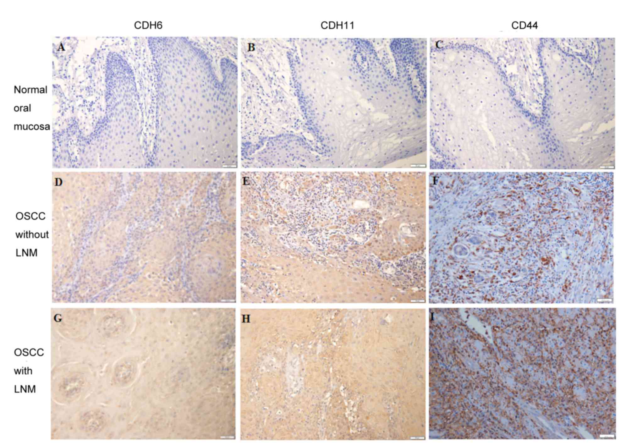 | Figure 1.Expression of CDH6, CDH11 and CD44 in
normal tissue and in OSCC with and without LNM. Photomicrographs
demonstrate representative examples of (A) CDH6, (B) CDH11 and (C)
CD44 expression in normal oral mucosa. Representative examples of
(D) CDH6, (E) CDH11 and (F) CD44 expression in OSCC without LNM.
Representative examples of (G) CDH6, (H) CDH11 and (I) CD44
expression in OSCC with LNM. Magnification, ×200; scale bar, 50 µm.
CDH, cadherin; CD44, cluster of differentiation 44; OSCC, oral
squamous cell carcinoma; LNM, lymph node metastasis. |
| Table III.Immunohistochemical analysis of CDH6,
CDH11 and CD44 expression in OSCC. Association between expression
of CDH6, CDH11 or CD44 and tumor metastasis was analyzed using SPSS
software. |
Table III.
Immunohistochemical analysis of CDH6,
CDH11 and CD44 expression in OSCC. Association between expression
of CDH6, CDH11 or CD44 and tumor metastasis was analyzed using SPSS
software.
|
| No. of patients
(%) |
|
|---|
|
|
|
|
|---|
| Variables | High | Low | P-value |
|---|
| CDH6 |
|
|
|
| OSCC
without LNM | 44 (66) | 22 (34) | 0.017a |
| OSCC
with LNM | 31 (89) | 4 (11) |
|
| CDH11 |
|
|
|
| OSCC
without LNM | 33 (50) | 33 (50) | 0.038a |
| OSCC
with LNM | 25 (71) | 10 (29) |
|
| CD44 |
|
|
|
| OSCC
without LNM | 37 (56) | 29 (44) | 0.001b |
| OSCC
with LNM | 31 (89) | 4 (11) |
|
Associations between CDH6, CDH11 and
CD44 expression and clinicopathological factors
The associations between CDH6, CDH11 and CD44
protein expression and clinicopathological data were assessed using
the c2 test and are summarized in Table IV. CDH6, CDH11 and CD44 protein
expression was significantly associated with LNM (P=0.017, P=0.038
and P=0.001, respectively). OSCC cases with LNM exhibited higher
rates of elevated CDH6, CDH11 and CD44 protein expression. In
addition, tumor stage was significantly associated with these
adhesion factors (P=0.002, P=0.016 and P=0.018, respectively).
However, no significant differences were identified between the
expression of these proteins and the degree of tumor
differentiation, age or sex (P>0.05).
| Table IV.Association between expression of
CDH6, CDH11 or CD44 and clinicopathological characteristics in
patients with oral squamous cell carcinoma (n=101). |
Table IV.
Association between expression of
CDH6, CDH11 or CD44 and clinicopathological characteristics in
patients with oral squamous cell carcinoma (n=101).
|
| CDH6 | CDH11 | CD44 |
|---|
|
|
|
|
|
|---|
| Variable | High | Low | P-value | High | Low | P-value | High | Low | P-value |
|---|
| Patient number | 75 | 26 |
| 58 | 43 |
| 68 | 33 |
|
| Age, years |
|
|
|
|
|
|
|
|
|
|
>60 | 40 | 13 | 0.769 | 32 | 21 | 0.528 | 37 | 16 | 0.576 |
|
<60 | 35 | 13 |
| 26 | 22 |
| 31 | 17 |
|
| Sex |
|
|
|
|
|
|
|
|
|
|
Male | 47 | 18 | 0.547 | 40 | 25 | 0.261 | 40 | 25 | 0.096 |
|
Female | 28 | 8 |
| 18 | 18 |
| 28 | 8 |
|
| Lymph node
metastasis |
|
|
|
|
|
|
|
|
|
|
Yes | 31 | 4 | 0.017a | 25 | 10 | 0.038a | 31 | 4 | 0.001b |
| No | 44 | 22 |
| 33 | 33 |
| 37 | 29 |
|
| Tumor (AJCC)
stage |
|
|
|
|
|
|
|
|
|
|
>II | 38 | 4 | 0.002b | 30 | 12 | 0.016a | 33 | 9 | 0.018a |
|
<II | 37 | 22 |
| 28 | 31 |
| 33 | 26 |
|
|
Differentiation |
|
|
|
|
|
|
|
|
|
|
Well | 26 | 12 | 0.067 | 23 | 15 | 0.859 | 29 | 9 | 0.853 |
|
Moderately | 35 | 15 |
| 26 | 24 |
| 30 | 20 |
|
|
Poorly | 10 | 3 |
| 9 | 4 |
| 9 | 4 |
|
| Smoking |
|
|
|
|
|
|
|
|
|
|
Yes | 52 | 18 | 0.992 | 42 | 28 | 0.278 | 47 | 23 | 0.953 |
| No | 23 | 8 |
| 15 | 16 |
| 21 | 10 |
|
| Alcohol |
|
|
|
|
|
|
|
|
|
| No | 45 | 15 | 0.836 | 33 | 27 | 0.551 | 38 | 22 | 0.301 |
|
Yes | 30 | 11 |
| 25 | 16 |
| 30 | 11 |
|
Associations among CDH6, CDH11 and
CD44 protein expression in OSCC
In the present study, the associations among the
protein expression of CDH6, CDH11 and CD44 were assessed using the
χ2 test. As demonstrated by Table V, 56/101 patients with OSCC exhibited
high expression and 14 exhibited low expression of CD44 and CDH6.
The association between CD44 and CDH6 expression was statistically
significant (r=0.266; P=0.008). Similarly, there was a
statistically significant association between the expression of
CD44 and CDH11 (r=0.254; P=0.011), with 45/101 patients with OSCC
exhibiting a high expression. These results indicated the
co-association between CDH6, CDH11 and CD44.
| Table V.Associations between CD44 protein
expression and the expression of CDH6 and CDH11 in patients with
oral squamous cell carcinoma (n=101). |
Table V.
Associations between CD44 protein
expression and the expression of CDH6 and CDH11 in patients with
oral squamous cell carcinoma (n=101).
|
| CD44 |
|
|
|---|
|
|
|
|
|
|---|
| Protein expression
(no.) | High (n=68) | Low (n=33) | rΦ | P-value |
|---|
| CDH6 |
|
| 0.266 | 0.007a |
| High
(75) | 56 | 19 |
|
|
| Low
(26) | 12 | 14 |
|
|
| CDH11 |
|
| 0.254 | 0.01a |
| High
(58) | 45 | 13 |
|
|
| Low
(43) | 23 | 20 |
|
|
Association between CDH6, CDH11 and
CD44 expression and survival in patients with OSCC
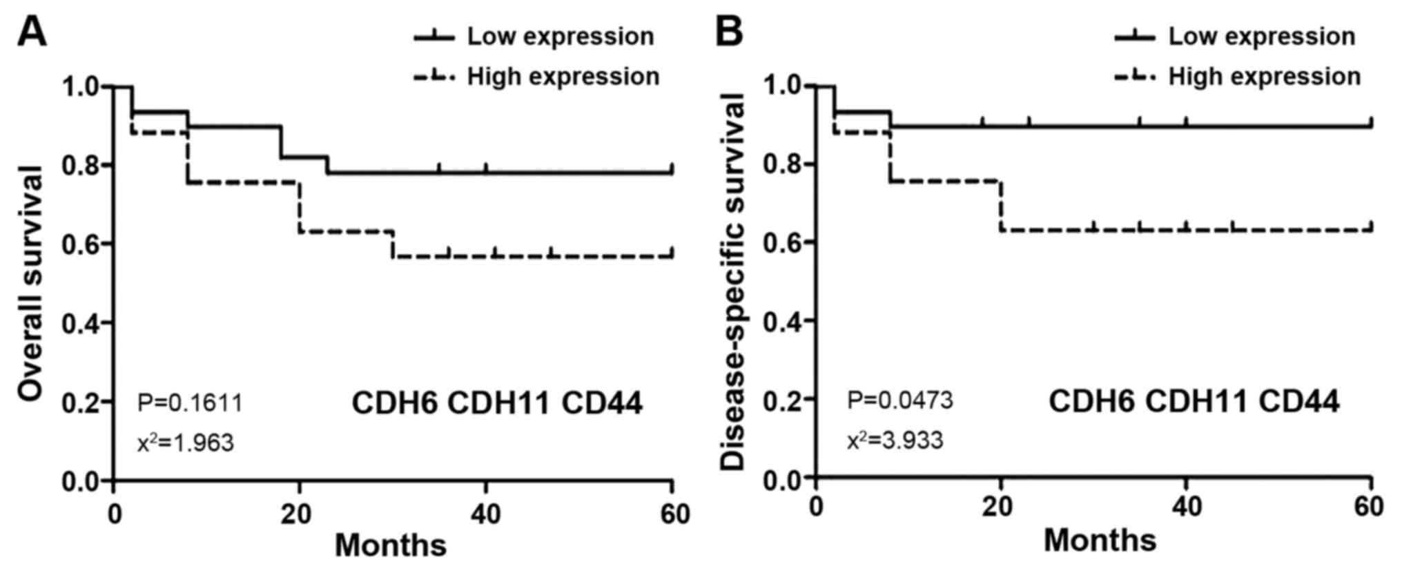
No significant associations between the 5-year
overall survival rate of patients with OSCC, and CDH6, CDH11 or
CD44 expression were identified (Fig.
2A-C). However, the 5-year disease-specific survival rate of
OSCC patients with high CDH6 and CD44 expression was significantly
decreased when compared with those exhibiting low expression [CDH6
(P=0.018; χ2=5.534) and CD44 (P=0.031;
χ2=4.622); Fig. 2D and F].
As CDH6, CDH11 and CD44 were revealed to be associated with LNM in
the present study, the association between patient survival and the
co-expression of these factors was also investigated. The results
revealed that the co-expression of CDH6, CDH11 and CD44 was not
significantly associated with overall survival (Fig. 3A). However, the 5-year
disease-specific survival rate of patients with high co-expression
of these proteins was decreased when compared with that of patients
exhibiting low co-expression (P=0.047; χ2=3.933;
Fig. 3B).
Discussion
The primary cause of mortality in OSCC is
metastasis, occurring primarily via the lymphatic system. The
impact of this depends on the size and site of the primary tumor,
the Tumor-Node-Metastasis (TNM) stage (19), the depth of invasion, perineural
invasion, patient compliance, biological tumor markers and tumor
grade (26). Intercellular adhesion
is mediated by a family of glycoproteins known as cadherins, which
serve an important role in the migration and dissemination of cells
during tumor progression and metastasis (27). It is well-known that loss of
E-cadherin and increased expression of N-cadherin is associated
with tumor cell invasion in oral and ovarian cancer (28,29). By
contrast, other mesenchymal-associated cadherins and adhesion
factors, including CDH6, CDH11 and CD44, which have been proven to
be able to interfere with epithelial cell-cell adhesion and to
promote cancer cell invasion and metastasis, are often
overexpressed in ovarian cancer (30–32). Until
now, however, the precise role of the three factors in OSCC has not
received a great deal of attention. The present study investigated
the role of three factors in the metastasis of OSCC using IHC
staining and RT-qPCR. It was revealed that CDH6, CDH11 and CD44
expression in tumor cells was associated with LNM in patients with
OSCC.
In the present study, the mRNA expression of 84
potential human tumor metastatic factors, including CDH6, CDH11 and
CD44, was observed in OSCC tissues with or without LNM. CDH6, CDH11
and CD44 mRNA expression was significantly increased in the OSCC
patients with LNM, and 18 other upregulated and 5 downregulated
cytokines were observed. CDH6, CDH11 and CD44 protein expression
was subsequently examined in 10 normal oral mucosae and 101 OSCC
tissues, including 35 cases with LNM, using IHC staining. These
results demonstrated that no cases of normal oral mucosa exhibited
high protein expression of these factors. However, high protein
expression of CDH6, CDH11 and CD44 was observed in 89, 71 and 89%
of 35 cases, respectively, of OSCC with LNM compared with 66, 50
and 56% of 66 OSCC cases, respectively, without LNM. The
association between CDH6, CDH11 or CD44 protein expression and
several clinicopathological indicators was assessed. The
overexpression of these factors was associated with aggressive
histopathological features, including LNM and advanced TNM stages,
whereby OSCC patients with an advanced TNM stage (III–IV) and LNM
exhibited higher expression of these proteins. However, there was
no association between protein expression and age, sex or tumor
differentiation. Furthermore, the present study observed that the
prognostic value may be largely enhanced by co-evaluating the
expression of these three proteins in patients with OSCC. These
results were in line with those of previous studies in other types
of cancer (29–31), indicating that CDH6, CDH11 and CD44
may be either directly or indirectly involved in the metastasis of
OSCC. Therefore, it is reasonable to combine the expression of
these three proteins as predictive parameters for improved
evaluation of the progression of OSCC.
CDH6 and CDH11 expression have been revealed to be
associated with aggressive tumor cell migration and a poorer
patient outcome (30–32). However, the underlying mechanisms of
CDH6 and CDH11 in the progression of several types of cancer are
not completely understood. CDH6 has been suggested to promote EMT
during tumor development and progression by mediating pro-EMT
signals (33). CDH6 affects the
activity of F-actin and Rho GTPase, which is important for cell
motility and EMT (34). EMT is a
complex change in the cell phenotype that is important for cell
migration and carcinoma metastasis. A previous study also
demonstrated that CDH6 is a novel transforming growth factor
(TGF)-β target gene during EMT in thyroid tumors and that its
expression is controlled by TGF-β pathway molecules, including the
Smad pathway or the phosphoinositide 3-kinase (PI3K)/protein kinase
B (Akt) pathway (35).
During EMT, cadherin expression changes (cadherin
switching), including E- to N-cadherin switching, is associated
with cancer metastatic occurrence (36). CDH6 to CDH11 switching is also
involved in the EMT (37). The
present study also indicated that CDH6 is associated with CDH11,
and that overexpression of CDH11 is associated with LNM. Although
high expression of CDH11 alone was not associated with a low
overall survival rate in the OSCC cohort of the present study, it
was a statistically significant prognostic factor in several types
of cancer, through its participation in EMT (38). However, the prognostic value was
enhanced by co-evaluating the expression of CDH6, CDH11 and CD44 in
the present study. Previous studies have reported that CDH11
interacts with angiomotin (Amot) to promote the migration of cancer
cells. Amot is known to be involved in cell polarity and migration
(39,40). The Amot p80 isoform was revealed to
regulate apical polarity by interacting with Rich1 (39), a small GTPase-activating protein, and
to affect proliferation (41). CDH11
also regulates α-catenin turnover at the adherens junctions,
facilitating the dynamic remodeling of cell-cell interactions,
which are important for inter-cellular invasion (42,43). CDH11
was associated with signal transduction molecules, including the
PI3K/Akt pathway (44).
In the present study, CD44 was revealed to be
significantly associated with LNM, suggesting that CD44 is also
involved in OSCC metastasis. These results are consistent with
those of previous studies, which revealed that upregulation of CD44
represents a crucial event in the development of metastasis and
that silencing CD44 expression suppressed its tumorigenic effects,
including proliferation, migration and invasion in ovarian cancer
and breast cancer cells (45,46). CD44 interacts with c-Src kinase to
regulate the mitogen-activated protein kinase (MAPK), PI3K and
signal transducer and activator of transcription 3 pathways
(47). The MAPK and
PI3K/Akt/mechanistic target of rapamycin pathways have been
demonstrated to promote cancer cell proliferation and to enhance
invasiveness and angiogenesis (18).
In addition, a previous study revealed that CD44 expression is
associated with EMT and LNM in patients with OSCC (48). The present study revealed that CD44
was significantly associated with cadherins, including CDH6 and
CDH11, and that its expression was also associated with the
PI3K/Akt pathway. Therefore, it may be beneficial to combine these
adhesion factors as metastasis-predictive parameters in order to
improve their prognostic value, as multiple signal pathways,
including the MAPK and PI3K/Akt pathways, may be involved in the
metastasis of OSCC.
In conclusion, the present study demonstrated that
the high expression of CDH6, CDH11 and CD44 in OSCC cells was
significantly associated with an increased propensity to develop
LNM and an advanced tumor stage. A positive association between
CD44, CDH6 and CDH11 protein expression in OSCC patients was also
observed in the present study. The combined use of these factors
may improve the accuracy of metastasis and prognosis prediction.
The results of the present study highlighted the important role of
these adhesion factors in the progression of OSCC, which may
provide a novel perspective in the prediction and prevention of
metastasis in OSCC.
Acknowledgements
The authors would like to thank Miss Xiaoliang Jiang
for providing editorial and technical assistance.
Funding
The present study was supported by grants from the
National Natural Science Foundation of China (grant no. 81250043)
and the Natural Science Foundation of Beijing Municipality (grant
no. 7142135).
Availability of data and materials
The datasets used and/or analyzed during the current
study are available from the corresponding author on reasonable
request.
Authors' contributions
CM drafted the article, designed the study and
analyzed the data. JZ, RL, LZ, YC and LY collected and analyzed the
data. TS, MW, ML and YL performed the experiments and collected the
data. TZ designed the study, secured the funding and performed
critical revision of the article.
Ethics statement and consent to
participate
The study was approved by the Ethics Committee of
Peking Union Medical College.
Consent for publication
All patients provided written informed consent for
the publication of associated data and accompanying images.
Competing interests
The authors declare that they have no competing
interests.
Glossary
Abbreviations
Abbreviations:
|
OSCC
|
oral squamous cell carcinoma
|
|
LNM
|
lymph node metastasis
|
|
CDH6
|
cadherin 6
|
|
CDH11
|
cadherin 11
|
|
PCa
|
prostate cancer
|
|
IHC
|
immunohistochemical
|
|
SABC
|
streptavidin-biotin complex
|
|
RT-qPCR
|
reverse transcription-quantitative
polymerase chain reaction
|
|
MMPs
|
matrix metalloproteinases
|
|
EMT
|
epithelial-mesenchymal transition
|
|
Akt
|
protein kinase B
|
|
PI3K
|
phosphatidylinositol 3-kinase
|
|
MAPK
|
mitogen-activated protein kinase
|
|
STAT
|
signal transducer and activator of
transcription
|
References
|
1
|
Slootweg PJ and Eveson JW: Tumours of the
oral cavity and oropharynx. In: World Health Organization
Classification of TumoursPathology & Genetics of Head and Neck
Tumours. IARC Press; Lyon: pp. 166–167. 2005
|
|
2
|
Okura M, Aikawa T, Sawai N, Lida S and
Kogo M: Decision analysis and ent threshold in a management for the
N0 neck of the oral cavity carcinoma. Oral Oncol. 45:908–911. 2009.
View Article : Google Scholar : PubMed/NCBI
|
|
3
|
Lea J, Bachar G, Sawka AM, Lakra DC,
Gilbert RW, Irish JC, Brown DH, Gullane PJ and Goldstein DP:
Metastases to level IIb in squamous cell carcinoma of the oral
cavity: A systematic review and meta-analysis. Head Neck.
32:184–190. 2010.PubMed/NCBI
|
|
4
|
Fan S, Tang QL, Lin YJ, Chen WL, Li JS,
Huang ZQ, Yang ZH, Wang YY, Zhang DM, Wang HJ, et al: A review of
clinical and histological parameters associated with contralateral
neck metastases in oral squamous cell carcinoma. Int J Oral Sci.
3:180–191. 2011. View Article : Google Scholar : PubMed/NCBI
|
|
5
|
Noguti J, De Moura CF, De Jesus GP, Da
Silva VH, Hossaka TA, Oshima CT and Ribeiro DA: Metastasis from
oral cancer: An overview. Cancer Genomics Proteomics. 9:329–336.
2012.PubMed/NCBI
|
|
6
|
Thiery JP, Acloque H, Huang RY and Nieto
MA: Epithelial-mesenchymal transitions in development and disease.
Cell. 139:871–890. 2009. View Article : Google Scholar : PubMed/NCBI
|
|
7
|
Nelson WJ and Nusse R: Convergence of Wnt,
beta-catenin, and cadherin pathways. Science. 303:1483–1487. 2004.
View Article : Google Scholar : PubMed/NCBI
|
|
8
|
Huber GF, Züllig L, Soltermann A, Roessle
M, Graf N, Haerle SK, Studer G, Jochum W, Moch H and Stoeckli SJ:
Down regulation of E-Cadherin (ECAD)-a predictor for occult
metastatic disease in sentinel node biopsy of early squamous cell
carcinomas of the oral cavity and oropharynx. BMC Cancer.
11:217:1–8. 2011. View Article : Google Scholar
|
|
9
|
Zhao Z, Ge J, Sun Y, Tian L, Lu J, Liu M
and Zhao Y: Is E-cadherin immunoexpression a prognostic factor for
head and neck squamous cell carcinoma (HNSCC)? A systematic review
and meta-analysis. Oral Oncol. 48:761–767. 2012. View Article : Google Scholar : PubMed/NCBI
|
|
10
|
Thedieck C, Kuczyk M, Klingel K, Steiert
I, Müller CA and Klein G: Expression of Ksp-cadherin during kidney
development and in renal cell carcinoma. Br J Cancer. 92:2010–2017.
2005. View Article : Google Scholar : PubMed/NCBI
|
|
11
|
Fluge Ø, Bruland O, Akslen LA, Lillehaug
JR and Varhaug JE: Gene expression in poorly differentiated
papillary thyroid carcinomas. Thyroid. 16:161–175. 2006. View Article : Google Scholar : PubMed/NCBI
|
|
12
|
Sancisi V, Gandolfi G, Ragazzi M, Nicoli
D, Tamagnini I, Piana S and Ciarrocchi A: Cadherin 6 is a new RUNX2
target in TGF-β signalling pathway. PLoS One. 12:e754892013.
View Article : Google Scholar
|
|
13
|
Chu K, Cheng C J, Ye X, Lee YC, Zurita AJ,
Chen DT, Yu-Lee LY, Zhang S, Yeh ET, Hu MC, et al: Cadherin-11
promotes the metastasis of prostate cancer cells to bone. Mol
Cancer Res. 6:1259–1267. 2008. View Article : Google Scholar : PubMed/NCBI
|
|
14
|
Huang CF, Lira C, Chu K, Bilen MA, Lee YC,
Ye X, Kim SM, Ortiz A, Wu FL, Logothetis CJ, et al: Cadherin-11
increases migration and invasion of prostate cancer cells and
enhances their interaction with osteoblasts. Cancer Res.
70:4580–4589. 2010. View Article : Google Scholar : PubMed/NCBI
|
|
15
|
Chou YE, Hsieh MJ, Hsin CH, Chiang WL, Lai
YC, Lee YH, Huang SC, Yang SF and Lin CW: CD44 gene polymorphisms
and environmental factors on oral cancer susceptibility in Taiwan.
PLoS One. 9:e936922014. View Article : Google Scholar : PubMed/NCBI
|
|
16
|
Shakib PA, Ensani F, Abdirad A, Valizadeh
B, Seyedmajidi M and Sum S: CD44 and CD74: The promising candidates
for molecular targeted therapy in oral squamous cell carcinoma.
Dent Res J (Isfahan). 12:181–186. 2015.PubMed/NCBI
|
|
17
|
Joshua B, Kaplan MJ, Doweck I, Pai R,
Weissman IL, Prince ME and Ailles LE: Frequency of cells expressing
CD44, a head and neck cancer stem cell marker: Correlation with
tumor aggressiveness. Head Neck. 34:42–49. 2012. View Article : Google Scholar : PubMed/NCBI
|
|
18
|
Judd NP, Winkler AE, Murillo-Sauca O,
Brotman JJ, Law JH, Lewis JS Jr, Dunn GP, Bui JD, Sunwoo JB and
Uppaluri R: ERK1/2 regulation of CD44 modulates oral cancer
aggressiveness. Cancer Res. 72:365–374. 2012. View Article : Google Scholar : PubMed/NCBI
|
|
19
|
Edge SB, Byrd DR, Compton CC, Fritz AG,
Greene F and Trotti A: AJCC cancer staging manual. 7th edition.
Springer-Verlag; New York: pp. 347–377. 2010
|
|
20
|
Livak KJ and Schmittgen TD: Analysis of
relative gene expression data using real-time quantitative PCR and
the 2(-Delta Delta C(T)) method. Methods. 25:402–408. 2001.
View Article : Google Scholar : PubMed/NCBI
|
|
21
|
Yoshihama R, Yamaguchi K, Imajyo I, Mine
M, Hiyake N, Akimoto N, Kobayashi Y, Chigita S, Kumamaru W,
Kiyoshima T, et al: Expression levels of SOX2, KLF4 and brachyury
transcription factors are associated with metastasis and poor
prognosis in oral squamous cell carcinoma. Oncol Lett.
11:1435–1446. 2016. View Article : Google Scholar : PubMed/NCBI
|
|
22
|
Yang H, Liang J, Zhou J, Mi J, Ma K, Fan
Y, Ning J, Wang C, Wei X and Li E: Knockdown of RHOC by shRNA
suppresses invasion and migration of cholangiocellular carcinoma
cells via inhibition of MMP2, MMP3, MMP9 and epithelial-mesenchymal
transition. Mol Med Rep. 13:5255–5261. 2016. View Article : Google Scholar : PubMed/NCBI
|
|
23
|
Bradbury R, Jiang WG and Cui YX: MDM2 and
PSMA Play inhibitory roles in metastatic breast cancer cells
through regulation of matrix metalloproteinases. Anticancer Res.
36:1143–1151. 2016.PubMed/NCBI
|
|
24
|
Mendonsa AM, VanSaun MN, Ustione A, Piston
DW, Fingleton BM and Gorden DL: Host and tumor derived MMP13
regulate extravasation and establishment of colorectal metastases
in the liver. Mol Cancer. 14:492015. View Article : Google Scholar : PubMed/NCBI
|
|
25
|
Bonecchi R, Galliera E, Borroni EM, Corsi
MM, Locati M and Mantovani A: Chemokines and chemokine receptors:
An overview. Front Biosci. 14:540–551. 2009. View Article : Google Scholar
|
|
26
|
Chinn SB and Myers JN: Oral cavity
carcinoma: current management, controversies, and future
directions. J Clin Oncol. 33:3269–3276. 2015. View Article : Google Scholar : PubMed/NCBI
|
|
27
|
Ashaie MA and Chowdhury EH: Cadherins: The
superfamily critically involved in breast cancer. Curr Pharm Des.
22:616–638. 2016. View Article : Google Scholar : PubMed/NCBI
|
|
28
|
Zhou J, Tao D, Xu Q, Gao Z and Tang D:
Expression of E-cadherin and vimentin in oral squamous cell
carcinoma. Int J Clin Exp Pathol. 8:3150–3154. 2015.PubMed/NCBI
|
|
29
|
Alaee M, Danesh G and Pasdar M:
Plakoglobin reduces the in vitro growth, migration and invasion of
ovarian cancer cells expressing N-cadherin and mutant p53. PLoS
One. 11:e01543232016. View Article : Google Scholar : PubMed/NCBI
|
|
30
|
Paul R, Necknig U, Busch R, Ewing CM,
Hartung R and Isaacs WB: Cadherin-6: A new prognostic marker for
renal cell carcinoma. J Uro. 171:97–101. 2004. View Article : Google Scholar
|
|
31
|
Satcher RL, Pan T, Bilen MA, Li X, Lee YC,
Ortiz A, Kowalczyk AP, Yu-Lee LY and Lin SH: Cadherin-11
endocytosis through binding to clathrin promotes
cadherin-11-mediated migration in prostate cancer cells. J Cell
Sci. 128:4629–4641. 2015. View Article : Google Scholar : PubMed/NCBI
|
|
32
|
Ortiz A, Lee YC, Yu G, Liu HC, Lin SC,
Bilen MA, Cho H, Yu-Lee LY and Lin SH: Angiomotin is a novel
component of cadherin-11/β-catenin/p120 complex and is critical for
cadherin-11-mediated cell migration. FASEB J. 3:1080–1091. 2015.
View Article : Google Scholar
|
|
33
|
Clay MR and Halloran MC: Cadherin 6
promotes neural crest cell detachment via F-actin regulation and
influences active Rho distribution during epithelial-to-mesenchymal
transition. Development. 141:2506–2515. 2014. View Article : Google Scholar : PubMed/NCBI
|
|
34
|
Bravo-Cordero JJ, Moshfegh Y, Condeelis J
and Hodgson L: Live cell imaging of RhoGTPase biosensors in tumor
cell. Methods Mol Biol. 1046:359–370. 2013. View Article : Google Scholar : PubMed/NCBI
|
|
35
|
Sancisi V, Gandolfi G, Ragazzi M, Nicoli
D, Tamagnini I, Piana S and Ciarrocchi A: Cadherin 6 is a new RUNX2
target in TGF-β signalling pathway. PLoS One. 8:e754892013.
View Article : Google Scholar : PubMed/NCBI
|
|
36
|
Wheelock MJ, Shintani Y, Maeda M, Fukumoto
Y and Johnson KR: Cadherin switching. J Cell Sci. 121:727–735.
2008. View Article : Google Scholar : PubMed/NCBI
|
|
37
|
Bringuier PP, Schalken JA, Hervieu V and
Giroldi LA: Involvement of orphan nuclear receptor COUP-TFII in
cadherin-6 and cadherin-11 regulation: Implications in development
and cancer. Mech Dev. 136:64–72. 2015. View Article : Google Scholar : PubMed/NCBI
|
|
38
|
Satcher RL, Pan T, Cheng CJ, Lee YC, Lin
SC, Yu G, Li X, Hoang AG, Tamboli P, Jonasch E, et al: Cadherin-11
in renal cell carcinoma bone metastasis. PLoS One. 9:e898802014.
View Article : Google Scholar : PubMed/NCBI
|
|
39
|
Wells CD, Fawcett JP, Traweger A, Yamanaka
Y, Goudreault M, Elder K, Kulkarni S, Gish G, Virag C, Lim C, et
al: A Rich1/Amot complex regulates the Cdc42 GTPase and
apical-polarity proteins in epithelial cells. Cell. 125:535–548.
2006. View Article : Google Scholar : PubMed/NCBI
|
|
40
|
Ernkvist M, Luna Persson N, Audebert S,
Lecine P, Sinha I, Liu M, Schlueter M, Horowitz A, Aase K, Weide T,
et al: The Amot/Patj/Syx signaling complex spatially controls RhoA
GTPase activity in migrating endothelial cells. Blood. 113:244–253.
2009. View Article : Google Scholar : PubMed/NCBI
|
|
41
|
Yi C, Troutman S, Fera D,
Stemmer-Rachamimov A, Avila JL, Christian N, Persson NL, Shimono A,
Speicher DW, Marmorstein R, et al: A tight junction-associated
Merlin-angiomotin complex mediates Merlin's regulation of mitogenic
signaling and tumor suppressive functions. Cancer Cell. 19:527–540.
2011. View Article : Google Scholar : PubMed/NCBI
|
|
42
|
Feltes CM, Kudo A, Blaschuk O and Byers
SW: An alternatively spliced cadherin-11 enhances human breast
cancer cell invasion. Cancer Res. 62:6688–6697. 2002.PubMed/NCBI
|
|
43
|
Kiener HP, Stipp CS, Allen PG, Higgins JM
and Brenner MB: The cadherin-11 cytoplasmic juxtamembrane domain
promotes alpha-catenin turnover at adherens junctions and
intercellular motility. Mol Biol Cell. 17:2366–2375. 2006.
View Article : Google Scholar : PubMed/NCBI
|
|
44
|
Wu M, Xu T, Zhou Y, Lu H and Gu Z:
Pressure and inflammatory stimulation induced increase of
cadherin-11 is mediated by PI3K/Akt pathway in synovial fibroblasts
from temporomandibular joint. Osteoarthritis Cartilage.
21:1605–1612. 2013. View Article : Google Scholar : PubMed/NCBI
|
|
45
|
Gao Y, Foster R, Yang X, Feng Y, Shen JK,
Mankin HJ, Hornicek FJ, Amiji MM and Duan Z: Up-regulation of CD44
in the development of metastasis, recurrence and drug resistance of
ovarian cancer. Oncotarget. 6:9313–9326. 2015. View Article : Google Scholar : PubMed/NCBI
|
|
46
|
Xu H, Tian Y, Yuan X, Liu Y, Wu H, Liu Q,
Wu GS and Wu K: Enrichment of CD44 in basal-type breast cancer
correlates with EMT, cancer stem cell gene profile, and prognosis.
Onco Targets Ther. 9:431–444. 2016.PubMed/NCBI
|
|
47
|
Hynes NE and Lane HA: ERBB receptors and
cancer: The complexity of targeted inhibitors. Nat Rev Cancer.
5:341–354. 2005. View Article : Google Scholar : PubMed/NCBI
|
|
48
|
Ghuwalewala S, Ghatak D, Das P, Dey S,
Sarkar S, Alam N, Panda CK and Roychoudhury S: CD44(high)CD24(low)
molecular signature determines the Cancer Stem Cell and EMT
phenotype in oral squamous cell carcinoma. Stem Cell Res.
16:405–417. 2016. View Article : Google Scholar : PubMed/NCBI
|