Introduction
Cervical cancer is one of the most common
malignancies in females worldwide. Mortality rates associated with
uterine cervical cancer have declined due to the widespread use of
cancer screening for the prevention and early detection of cervical
cancer (1). Moreover, since
concurrent chemoradiotherapy (CCRT) has been established as a
standard treatment, the prognosis of those patients has improved
(2,3).
However, about one third of patients experience recurrence within
five years (4), with a median
survival after recurrence of 15 months (5), and less than 5% of them survive for 5
years (6). Thus, the oncologic
outcome is far from satisfactory. Especially, the prognosis of
patients with recurrent disease within a previously irradiated
field is unfavorable (7). In
addition, earlier studies indicated that response rates to
chemotherapy in those patients were poorer than that of those with
out-of-field recurrence (8,9). Therefore, the oncologic outcome of
patients with post-radiotherapy relapsed/persistent cervical cancer
(PRRCC) is still poor. Recently, bevacizumab, a humanized
monoclonal antibody targeting vascular endothelial growth factor A
(VEGF-A), has been approved for this tumor, and moreover
immunotherapy is under investigation (10).
VEGF-A is a multifunctional and an important
molecule in endothelial signaling and angiogenesis. VEGF-A binds to
its receptor VEGFR-1, and the downstream signaling is thought to be
involved in cancer proliferation and invasion (11). There have been several reports showing
that the overexpression of VEGF in cancer cells or serum is
correlated with radioresistance and poor disease-free survival
(12–16), and a meta-analysis suggested that high
expressions of VEGF was significantly associated with poor survival
outcome (17). Although VEGF
inhibitors such as bevacizumab are widely used against several
solid cancers, evidence regarding their efficacy against cervical
cancer is not satisfactory, especially PRRCC. Particularly, to our
best knowledge, there have been no reports on the expression level
of VEGF in PRRCC based on the fact that surgical treatment is
rarely performed as a salvage therapy for those patients (6). In sophisticated randomized clinical
trials, the addition of a VEGF inhibitor such as bevacizumab to
combination chemotherapy led to a significant improvement of the
oncologic outcome of patients with recurrent, persistent, and
highly metastatic cervical cancer (18,19).
Accordingly, bevacizumab has been applied in actual clinical
practice for this tumor. These results led us to hypothesize that
the expression of VEGF may be upregulated in those patients.
Reviewing 826 clinical records of cervical cancer
patients in our institute from 2003 to 2015, we identified eight
patients with PRRCC who underwent debulking surgery, and evaluated
the expression of VEGF immunohistochemically. In the present study,
we further clarify the upregulation of VEGF in radioresistant
cervical cancer by evaluating the expressions of VEGFR-1 and
hypoxia inducible factor-1α (HIF-1α).
Materials and methods
Patients
We retrospectively reviewed all the records of 826
patients with cervical cancer who were initially treated in our
hospital from 2003 to 2015. Written informed consent was acquired
from all patients. This study was approved by the Ethics Committee
of our institute (Approval no. 2013-0078). Treatment strategies for
each patient were determined by several gynecologic oncologists in
our hospital depending on their age, performance status (PS), and
spread of the disease. For example, as primary treatments, patients
who were in the early stage and had a good PS were indicated for
radical hysterectomy, and the other patients were treated with CCRT
or radiotherapy alone. As treatments for recurrence, most patients
were treated with chemotherapy, and only a few patients with
localized disease were selected for surgical resection.
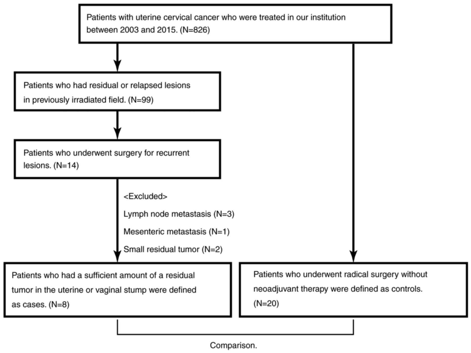
Ninety-seven patients had PRRCC, and 14 of them
underwent surgery for a recurrent lesion. After excluding six
patients because of lymph node metastasis or small residual tumors,
eight patients with uterine or vaginal stump recurrence were
investigated. Twenty patients who underwent radical surgery without
neoadjuvant therapy were extracted as a control (Fig. 1).
Immunohistochemical (IHC) staining and
its evaluation
Archival formalin-fixed, paraffin-embedded tumor
tissue obtained at surgery was used in this study. Sections of 4-µm
thickness were prepared using a microtome. The sections were
deparaffinized and rehydrated, subjected to antigen retrieval in 10
mM sodium citrate (pH 6.0) for 20 min at 95°C in a microwave, and
treated with 0.3% hydrogen peroxide in methanol for 20 min after
being washed with phosphate-buffered saline (PBS). Then, the
sections were blocked with appropriate serum using Histofine
SAB-PO(R) kit or Histofine SAB-PO(M) kit according to the
manufacturer's protocol (Nichirei, Tokyo, Japan), and incubated
with an appropriate first antibody diluted by PBS at 4°C overnight.
After rinsing with PBS, the sections were incubated with an
appropriate second antibody, and then peroxidase labeled
streptavidin using the kit. Then, the sections were rinsed with PBS
and developed by the 3, 3′-diaminobenzidine (DAB)
substrate-chromogen. After rinsing in water, the sections were
incubated with hematoxylin, dehydrated, and mounted. Details about
the reagents are presented in Table
I.
 | Table I.Details about antibodies and
immunohistochemistry kits. |
Table I.
Details about antibodies and
immunohistochemistry kits.
| A, Primary
antibodies |
|---|
|
|---|
| Antibody name | Manufacturer | Product no. | Host | Dilution rate |
|---|
| Anti-VEGF
antibody | Abcam | ab46154 | Rabbit | 1:100 |
| Anti-VEGF receptor 1
antibody | Abcam | ab2350 | Rabbit | 1:100 |
| Anti-HIF-1α
antibody | Abcam | ab1 | Rabbit | 1:100 |
|
| B,
Immunohistochemistry kits |
|
| Kit name |
Manufacturer | Product
no. |
|
|
|
| Histofine SAB-PO (R)
kit | Nichirei | 424032 |
|
|
| 10%
Normal goat serum |
|
|
|
|
| Biotin
labeled anti-rabbit IgG antibody |
|
|
|
|
|
Peroxidase labeled
streptavidin |
|
|
|
|
| Histofine SAB-PO (M)
kit | Nichirei | 424022 |
|
|
| 10%
Normal rabbit serum |
|
|
|
|
| Biotin
labeled anti-mouse IgG + IgA + IgM antibody |
|
|
|
|
|
Peroxidase labeled
streptavidin |
|
|
|
|
| Liquid DAB+ Substrate
Chromogen system | Dako | K3468 |
|
|
Based on the IHC activity, a four-tiered
semi-quantitative score was assigned according to the intensity and
area of stained cells, as follows: For the evaluation of IHC
expression, the staining intensity was scored as: 0, negative; 1,
weak; 2, medium; or 3, strong. The percentage of the staining area
was scored as ‘focal’ (1–10%), ‘sporadic’ (11–50%), and diffuse
(>51%) relative to the total tumor area. Carcinoma cells and
stroma were separately evaluated by two researchers, and the final
score was decided according to Table
II (‘negative’, ‘weak’, ‘moderate’, and ‘strong’,
respectively).
| Table II.The evaluation of
immunohistochemistry. |
Table II.
The evaluation of
immunohistochemistry.
| A, Cancer area |
|---|
|
|---|
|
| Intensity |
|---|
|
|
|
|---|
| Cancer area | Negative | Weak | Medium | Strong |
|---|
| Focal | 0 | 1 | 1 | 2 |
| Sporadic | 0 | 1 | 2 | 3 |
| Diffuse | 0 | 2 | 2 | 3 |
|
| B, Stroma
area |
|
|
|
Intensity |
|
|
|
| Stroma
area |
Negative | Weak | Medium | Strong |
|
|
|
| Focal | 0 | 1 | 1 |
|
| Sporadic | 0 | 1 | 2 |
|
| Diffuse | 0 | 2 | 3 |
|
All photographs were taken using Zeiss Axio
Imager.A1 (Carl Zeiss, Tokyo, Japan).
Statistics
All statistical analyses were performed with EZR
(Saitama Medical Center, Jichi Medical University, Saitama, Japan),
which is a graphical user interface for R (The R Foundation for
Statistical Computing, Vienna, Austria). More precisely, it is a
modified version of R commander designed to add statistical
functions frequently used in biostatistics. Differences between
recurrent cancer and control patients were assessed by the
Mann-Whitney U test and t-test, and the correlation of each
expression was assessed by Spearman's correlation coefficient.
Differences at P<0.05 were considered significant.
Results
We first compared clinical backgrounds of eight
patients with PRRCC and those of 20 patients with primary uterine
cervical cancer. Distributions of the age, tumor size, and
lymphovascular space invasion between the two groups were not
significantly different. All patients had squamous cell carcinoma
(SCC), and the serum SCC level at surgery was not significantly
different (Table III). Detailed
characteristics of PRRCC patients are presented in Table IV. All of the patients had previously
received radiotherapy, and six of them had received CCRT. Salvage
hysterectomy was performed for seven patients, and pelvic
exenteration was performed for case 2, who had vaginal stump
recurrence after vaginal total hysterectomy for carcinoma in
situ (CIS).
| Table III.Patients' characteristics. |
Table III.
Patients' characteristics.
|
| Cases (N=8) |
|
|
|---|
|
|
|
|
|
|---|
| Characteristic | #1 | #2 | Controls
(N=20) | P-value |
|---|
| Age |
|
|
| 0.321 |
|
Median | 43 | 44 | 39 |
|
|
(Range) | (34–73) | (34–74) | (20–68) |
|
| Stage |
|
|
| 0.795 |
|
CIS | 1 |
| 0 |
|
| Stage
I | 3 |
| 10 |
|
| Stage
II | 4 |
| 10 |
|
| Tumor size |
|
|
| 0.100 |
| <4
cm | 5 | 8 | 18 |
|
| ≥4
cm | 3 | 0 | 2 |
|
| LVSI |
|
|
| 0.591 |
|
Yes |
| 6 | 15 |
|
| No |
| 1 | 5 |
|
|
Unknown |
| 1 | 0 |
|
| Nodal
metastasis |
|
|
| 0.576 |
|
Yes | 3 | 1 | 10 |
|
| No | 5 | 0 | 10 |
|
| Unknown |
| 7 |
|
|
| Serum SCC
level |
| <2.0
ng/ml | 3 | 4 | 8 | 0.660 |
| ≥2.0
ng/ml | 4 | 4 | 12 |
|
|
Unknown | 1 |
|
|
|
| Previous
treatment |
|
|
| <0.0001 |
|
CCRT |
| 5 | 0 |
|
| RT |
| 3 | 0 |
|
| Table IV.Characteristics and oncologic
outcomes of patients with recurrence. |
Table IV.
Characteristics and oncologic
outcomes of patients with recurrence.
| No. | Agea | TNM | Ageb | PFS (months) | Previous
treatments | OS (months) | Outcome |
|---|
| 1 | 34 | cT1b2N1M0 | 34 | 3 | CCRT (PFx4 kur + WP
56.4 Gy, RALS 15 Gy) | 20 | DOD |
| 2 | 35 | pTisN0M0 | 38 | 16 | VTH → CCRT (PF ×5
kur + WP 50.4 Gy, RALS 13 Gy) | 47 | NED |
| 3 | 37 | cT2bN0M0 | 40 | 4 | CCRT (PFx5 kur + WP
50.4 Gy, RALS 16 Gy) → TC ×6 kur → CPT-11 ×3 kur | 44 | NED |
| 4 | 42 | cT1b2N0M0 | 43 | 7 | CCRT (PFx2 kur + WP
50.4 Gy, RALS 24 Gy) | 13 | DOD |
| 5 | 44 | cT2bN1M0 | 45 | 16 | CCRT (PFx1 kur,
CBDCA ×1 kur, NDP ×3 kur + WP 50.4 Gy, RALS 18 Gy) | 41 | NED |
| 6 | 53 | cT1bN0M0 | 54 | 5 | RT (WP 50.4 Gy,
RALS 24 Gy) → PF ×2 kur → TP ×4 kur | 24 | DOD |
| 7 | 73 | cT2bN0M0 | 74 | 5 | RT (WP 50.4 Gy,
RALS 13 Gy) | 35 | DOD |
| 8 | 73 | cT2aN0M0 | 74 | 10 | RT (WP 50.4 Gy,
RALS 9 Gy) | 14 | NED |
Representative images of IHC are shown in Fig. 2. In both carcinoma and stroma cells,
the expressions of VEGF-A were significantly higher in the PRRCC
group than in controls [PRRCC vs. control: P=0.0003 (carcinoma) and
P=0.0014 (stroma), respectively] (Fig.
2A and Table VA). Similarly, the
expressions of VEGFR-1 were also significantly stronger [PRRCC vs.
control: P=0.0003 (carcinoma) and P<0.0001 (stroma),
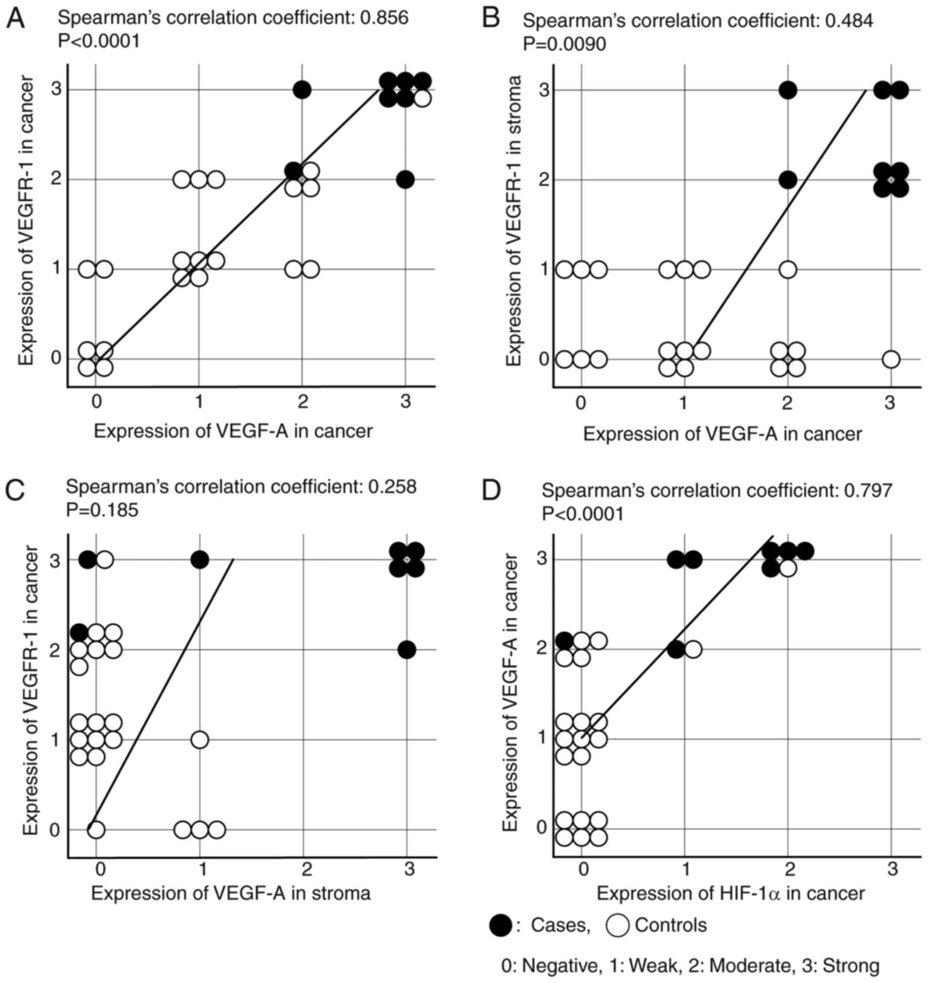
respectively] (Fig. 2B and Table VB). Of note, the expressions of VEGF-A
and VEGFR-1 in carcinoma cells were significantly correlated with
each other (Spearman's correlation coefficient: 0.856; P<0.0001)
(Fig. 3A). In addition, the
correlation of the VEGF-A expression in carcinoma cells and the
VEGFR-1 expression in stroma cells was moderate (Spearman's
correlation coefficient: 0.484; P=0.0090) (Fig. 3B), and that of the VEGF-A expression
in stroma cells and the VEGFR-1 expression in carcinoma cells was
weak (Spearman's correlation coefficient: 0.258; P=0.185) (Fig. 3C).
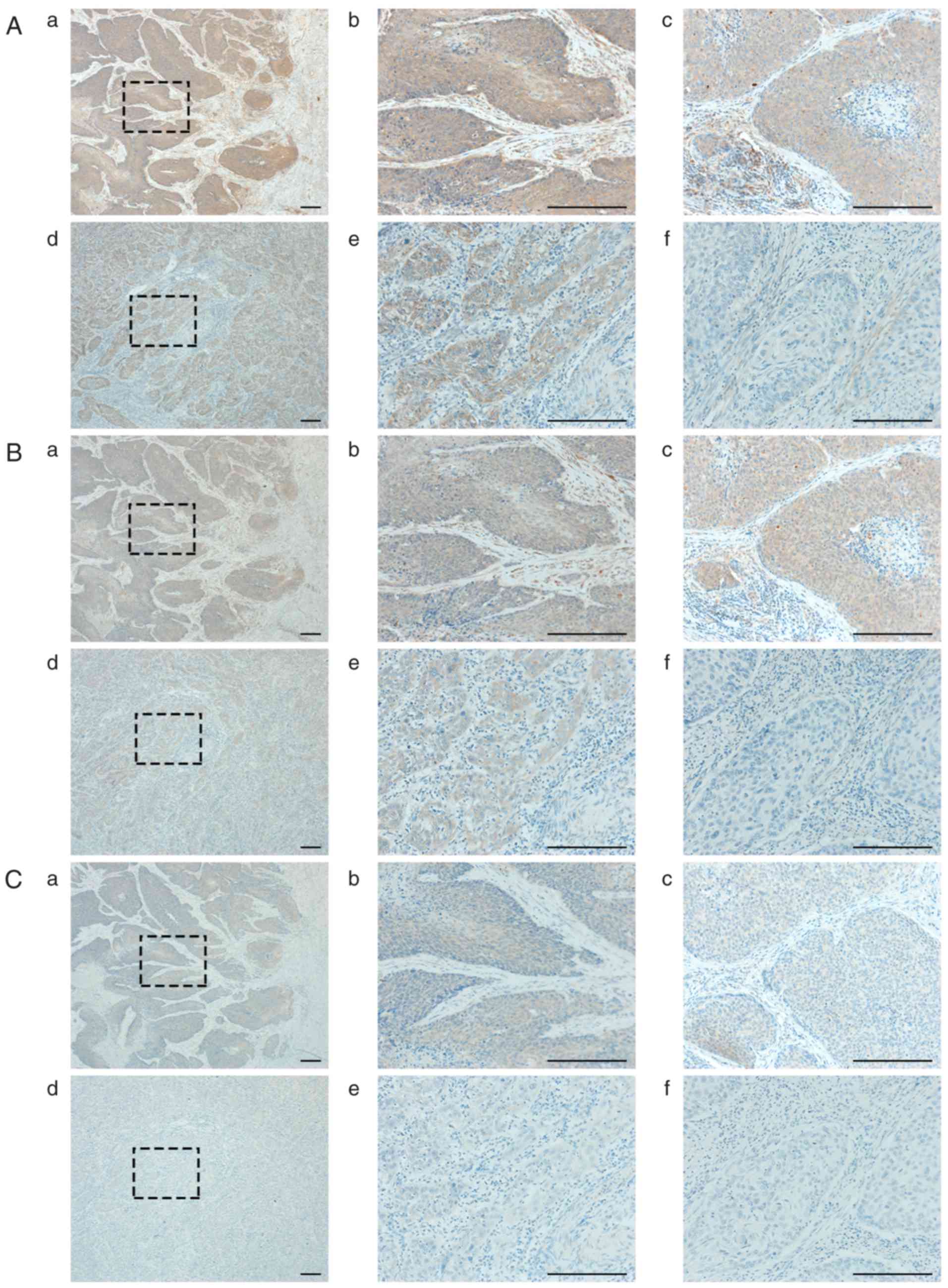 | Figure 2.Representative images of
immunohistochemistry. (A, B, and C) Representative images of
VEGF-A, VEGFR-1, and HIF-1α, respectively. a and b show those of
case 3, c shows that of case 2, d and e show those of control 1,
and f shows that of control 2. All scale bars, 200 µm. A-b shows
strong expression in cancer (cancer-3), A-c: cancer-3, A-e:
cancer-2, and A-f: cancer-0. A-b shows strong expression in stroma
(stroma-3), A-c: stroma-3, A-e: stroma-0, and A-f: stroma-1. B-b:
cancer-3, B-c: cancer-3, B-e: cancer-2, and A-f: cancer-0. B-b:
stroma-3, B-c: stroma-2, B-e: stroma-0, and A-f: stroma-1. C-b:
cancer-2, C-c: cancer-2, C-e: cancer-1, and A-f: cancer-0. C-b:
stroma-1, C-c: stroma-1, C-e: stroma-0, and A-f: stroma-0. |
| Table V.The expressions of VEGF-A, VEGFR-1,
and HIF-1α. |
Table V.
The expressions of VEGF-A, VEGFR-1,
and HIF-1α.
| A, VEGF-A
expression |
|---|
|
|---|
| VEGF-A | Negative | Weak | Moderate | Strong | P-value |
|---|
| Cancer |
|
Cases |
|
| 2 | 6 | 0.0003 |
|
Controls | 6 | 8 | 5 | 1 |
|
| Stroma |
|
Cases | 2 | 1 |
| 5 | 0.0014 |
|
Controls | 16 | 4 |
|
|
|
|
| B, VEGFR-1
expression |
|
| VEGFR-1 |
Negative | Weak |
Moderate | Strong | P-value |
|
| Cancer |
|
Cases |
|
| 2 | 6 | 0.0003 |
|
Controls | 4 | 9 | 6 | 1 |
|
| Stroma |
|
Cases |
|
| 5 | 3 | <0.0001 |
|
Controls | 13 | 7 |
|
|
|
|
| C, HIF-1α
expression |
|
| HIF-1α |
Negative | Weak |
Moderate | Strong | P-value |
|
| Cancer |
|
Cases | 1 | 3 | 4 |
| 0.0001 |
|
Controls | 18 | 1 | 1 |
|
|
| Stroma |
|
Cases | 4 | 4 |
|
| 0.343 |
|
Controls | 14 | 6 |
|
|
|
The expression of HIF-1α in carcinoma cells was also
significantly stronger in the PRRCC group than control group,
although that in stroma cells was weak and showed no significant
difference (P=0.343) (Fig. 2C and
Table VC). Moreover, the expression
of HIF-1α was significantly correlated with that of VEGF-A in
carcinoma cells, but not in stroma cells [Spearman's correlation
coefficient: 0.797; P<0.0001 (carcinoma); P=0.343 (stroma)]
(Fig. 3D).
Discussion
VEGF is an important factor for tumor angiogenesis,
and there have been a number of reports evaluating the VEGF
expression of primary surgery specimens or that of serum in uterine
cervical cancer (12–16). However, to the best of our knowledge,
there have been no reports concerning VEGF expression in PRRCC. In
this study, we investigated the expression of VEGF and related
molecules using tumor samples from patients with PRRCC. The
expressions of both VEGF-A and VEGFR-1 were significantly higher in
PRRCC sections than in controls. These results led us to
hypothesize two possible mechanisms: ‘natural selection’ and
‘evolution’. Intra-tumor genetic heterogeneity is also known in
cervical cancer, and subpopulations of each tumor showed
differential responses to chemoradiotherapy (20). In addition, patients with high VEGF
expression in cancer tissue or serum were associated with a poor
response to radiotherapy and poor survival (12–15).
Therefore, the ‘natural selection’ hypothesis suggests that
subpopulations of cervical cancer with high VEGF expression survive
through chemoradiation, and then these selected subpopulations
develop, leading to recurrence. On the other hand, in addition to
its therapeutic effects, ionizing radiation is known to promote the
malignant behaviors of surviving cancer cells. Radiation induced
HIF-1α and VEGF, and those factors were related to radioresistance
(21,22). Therefore, the ‘evolution’ hypothesis
suggests that some cervical cancer cells are evolutionarily induced
to acquire VEGF expression by radiation while most of them are
killed, and then cancer with acquired resistance develops, leading
to recurrence. Regardless of the two independent hypotheses, if
VEGF and its receptor are upregulated in PRRCC, VEGF-targeting
therapy is expected as an effective therapeutic strategy for this
tumor.
Hypoxia is an important cancer microenvironment, and
most solid human cancers including cervical cancer are known to
induce such an environment (23). It
is possible that PRRCC tissue is exposed to hypoxic conditions by
tissue fibrosis after radiotherapy. HIF-1α has been reported to
mediate essential homeostatic responses by activating the
transcription of multiple genes including VEGF (24). Indeed, in the present study, we showed
that the expression of HIF-1α in carcinoma cells was also
significantly higher in the PRRCC than control group consistent
with our current findings. According to earlier studies, hypoxic
conditions enhanced the radiation resistance dependent on HIF-1α by
elevating the expression of VEGF and inhibiting the expression of
p53 (25). In addition, high-level
expression of HIF-1α is associated with treatment-resistance, and,
conversely, the inhibition of HIF-1α transactivation enhances
radiotherapy responses (23,26). Burri et al reported that
multivariate analyses revealed HIF-1α expression to be an
independent factor for overall survival based on an
immunohistochemical analysis of 78 patients with uterine cervix
carcinoma treated with external beam radiotherapy (27). This evidence prompted us to
hypothesize that HIF-1α plays a crucial role in VEGF upregulation
and the treatment refractoriness of PRRCC.
In the current study, high-level expressions of
VEGF-A and VEGFR-1 were observed in the stroma as well as in
carcinoma cells. Cancer-associated fibroblasts (CAFs) are major
components of the tumor stroma and involved in tumor progression. A
previous report demonstrated the effects to protect against
radiation of CAF-cancer cell crosstalk through multiple growth
factors including VEGF in vitro (28). In order to inhibit VEGF-VEGFR
interactions between carcinoma and stroma cells, VEGF inhibitors
such as bevacizumab have been widely used. In cervical cancer, VEGF
inhibitors also showed clinical benefits for patients with
advanced, persistent, or recurrent lesions (18,19,29).
Especially, according to Tewari's sub-group analysis, bevacizumab
was more favorable in patients with recurrent or persistent lesions
than those with advanced lesions, and also in those who previously
received chemoradiotherapy (18).
These results suggest that recurrent or persistent cancer after
chemoradiotherapy expressed VEGF-A and VEGFR-1 more strongly than
advanced cancer, being consistent with our results.
The main limitation of this study was the fact that
only eight patients with PRRCC were available despite the
enrollment of over 800 patients with cervical cancer. This limited
patient number is consistent with the actual clinical situation
whereby the selection of surgical treatment for PRRCC is extremely
rare. Second, we did not evaluate the association between the VEGF
expression and efficacy of VEGF inhibitors in patients with PRRCC.
Moreover, we could not directly compare the immunohistochemical
expressions between pre- and post-treatment sample sets in the same
patient. Actually, it was difficult to obtain enough specimens from
patients with primary CCRT before treatment. As a result, we used
specimens of primary surgery as a control. An additional
large-scale study to confirm our current findings is desirable by
accumulating more patients with PRRCC from multiple institutions.
Therefore, in the present study, we could not verify the direct
effect of the radiation-induced expression of VEGF in tumor tissues
of patients with PRRCC. We would like to verify the
radiation-induced upregulation of VEGF expression effects in
vitro and using an animal model in a future study.
In conclusion, the expressions of VEGF-A and VEGFR-1
were significantly upregulated in PRRCC. These results are
important and valuable because there has been no evidence of VEGF
expression in PRRCC. For further evaluation, a large-scale study of
VEGF in advanced, residual, and recurrent cervical cancer is
desired and the efficacy of VEGF inhibitors must be investigated.
The prognoses of these patients are expected to improve in the
future. We believe that our results will help clarify the efficacy
of bevacizumab.
Acknowledgements
We sincerely thank members of the Deptartment of
OBGYN, Nagoya University for collaborating in data collection.
Glossary
Abbreviation
Abbreviations:
|
PRRCC
|
post-radiotherapy relapsed/persistent
cervical cancer
|
References
|
1
|
Siegel RL, Miller KD and Jemal A: Cancer
statistics, 2016. CA Cancer J Clin. 66:7–30. 2016. View Article : Google Scholar : PubMed/NCBI
|
|
2
|
Morris M, Eifel PJ, Lu J, Grigsby PW,
Levenback C, Stevens RE, Rotman M, Gershenson DM and Mutch DG:
Pelvic radiation with concurrent chemotherapy compared with pelvic
and para-aortic radiation for high-risk cervical cancer. N Engl J
Med. 340:1137–1143. 1999. View Article : Google Scholar : PubMed/NCBI
|
|
3
|
Green JA, Kirwan JM, Tierney JF, Symonds
P, Fresco L, Collingwood M and Williams CJ: Survival and recurrence
after concomitant chemotherapy and radiotherapy for cancer of the
uterine cervix: A systematic review and meta-analysis. Lancet.
358:781–786. 2001. View Article : Google Scholar : PubMed/NCBI
|
|
4
|
Benedet JL, Odicino F, Maisonneuve P,
Beller U, Creasman WT, Heintz AP, Ngan HY and Pecorelli S:
Carcinoma of the cervix uteri. Int J Gynaecol Obstet. 83 Suppl
1:S41–S78. 2003. View Article : Google Scholar
|
|
5
|
Mabuchi S, Isohashi F, Yoshioka Y, Temma
K, Takeda T, Yamamoto T, Enomoto T, Morishige K, Inoue T and Kimura
T: Prognostic factors for survival in patients with recurrent
cervical cancer previously treated with radiotherapy. Int J Gynecol
Cancer. 20:834–840. 2010. View Article : Google Scholar : PubMed/NCBI
|
|
6
|
Waggoner SE: Cervical cancer. Lancet.
361:2217–2225. 2003. View Article : Google Scholar : PubMed/NCBI
|
|
7
|
Legge F, Chiantera V, Macchia G, Fagotti
A, Fanfani F, Ercoli A, Gallotta V, Morganti AG, Valentini V,
Scambia G and Ferrandina G: Clinical outcome of recurrent locally
advanced cervical cancer (LACC) submitted to primary multimodality
therapies. Gynecol Oncol. 138:83–88. 2015. View Article : Google Scholar : PubMed/NCBI
|
|
8
|
Kosmas C, Mylonakis N, Tsakonas G, Vorgias
G, Karvounis N, Tsavaris N, Daladimos T, Kalinoglou N, Malamos N,
Akrivos T and Karabelis A: Evaluation of the
paclitaxel-ifosfamide-cisplatin (TIP) combination in relapsed
and/or metastatic cervical cancer. Br J Cancer. 101:1059–1065.
2009. View Article : Google Scholar : PubMed/NCBI
|
|
9
|
Benjapibal M, Thirapakawong C,
Leelaphatanadit C, Therasakvichya S and Inthasorn P: A pilot phase
II study of capecitabine plus cisplatin in the treatment of
recurrent carcinoma of the uterine cervix. Oncology. 72:33–38.
2007. View Article : Google Scholar : PubMed/NCBI
|
|
10
|
Borcoman E and Le Tourneau C:
Pembrolizumab in cervical cancer: Latest evidence and clinical
usefulness. Ther Adv Med Oncol. 9:431–439. 2017. View Article : Google Scholar : PubMed/NCBI
|
|
11
|
Dallas NA, Fan F, Gray MJ, Van Buren G II,
Lim SJ, Xia L and Ellis LM: Functional significance of vascular
endothelial growth factor receptors on gastrointestinal cancer
cells. Cancer Metastasis Rev. 26:433–441. 2007. View Article : Google Scholar : PubMed/NCBI
|
|
12
|
Loncaster JA, Cooper RA, Logue JP,
Davidson SE, Hunter RD and West CM: Vascular endothelial growth
factor (VEGF) expression is a prognostic factor for radiotherapy
outcome in advanced carcinoma of the cervix. Br J Cancer.
83:620–625. 2000. View Article : Google Scholar : PubMed/NCBI
|
|
13
|
Lee IJ, Park KR, Lee KK, Song JS, Lee KG,
Lee JY, Cha DS, Choi HI, Kim DH and Deung YK: Prognostic value of
vascular endothelial growth factor in Stage IB carcinoma of the
uterine cervix. Int J Radiat Oncol Biol Phys. 54:768–779. 2002.
View Article : Google Scholar : PubMed/NCBI
|
|
14
|
Bachtiary B, Selzer E, Knocke TH, Pötter R
and Obermair A: Serum VEGF levels in patients undergoing primary
radiotherapy for cervical cancer: Impact on progression-free
survival. Cancer Lett. 179:197–203. 2002. View Article : Google Scholar : PubMed/NCBI
|
|
15
|
Cheng WF, Chen CA, Lee CN, Wei LH, Hsieh
FJ and Hsieh CY: Vascular endothelial growth factor and prognosis
of cervical carcinoma. Obstet Gynecol. 96:721–726. 2000. View Article : Google Scholar : PubMed/NCBI
|
|
16
|
Iwasaki K, Yabushita H, Ueno T and
Wakatsuki A: Role of hypoxia-inducible factor-1α, carbonic
anhydrase-IX, glucose transporter-1 and vascular endothelial growth
factor associated with lymph node metastasis and recurrence in
patients with locally advanced cervical cancer. Oncol Lett.
10:1970–1978. 2015. View Article : Google Scholar : PubMed/NCBI
|
|
17
|
Zhang J, Liu J, Zhu C, He J, Chen J, Liang
Y, Yang F, Wu X and Ma X: Prognostic role of vascular endothelial
growth factor in cervical cancer: A meta-analysis. Oncotarget.
8:24797–24803. 2017.PubMed/NCBI
|
|
18
|
Tewari KS, Sill MW, Long HJ III, Penson
RT, Huang H, Ramondetta LM, Landrum LM, Oaknin A, Reid TJ, Leitao
MM, et al: Improved survival with bevacizumab in advanced cervical
cancer. N Engl J Med. 370:734–743. 2014. View Article : Google Scholar : PubMed/NCBI
|
|
19
|
Schefter T, Winter K, Kwon JS, Stuhr K,
Balaraj K, Yaremko BP, Small W Jr, Sause W and Gaffney D: Radiation
Therapy Oncology Group (RTOG): RTOG 0417: efficacy of bevacizumab
in combination with definitive radiation therapy and cisplatin
chemotherapy in untreated patients with locally advanced cervical
carcinoma. Int J Radiat Oncol Biol Phys. 88:101–105. 2014.
View Article : Google Scholar : PubMed/NCBI
|
|
20
|
Cooke SL, Temple J, Macarthur S, Zahra MA,
Tan LT, Crawford RA, Ng CK, Jimenez-Linan M, Sala E and Brenton JD:
Intra-tumour genetic heterogeneity and poor chemoradiotherapy
response in cervical cancer. Br J Cancer. 104:361–368. 2011.
View Article : Google Scholar : PubMed/NCBI
|
|
21
|
Gorski DH, Beckett MA, Jaskowiak NT,
Calvin DP, Mauceri HJ, Salloum RM, Seetharam S, Koons A, Hari DM,
Kufe DW and Weichselbaum RR: Blockage of the vascular endothelial
growth factor stress response increases the antitumor effects of
ionizing radiation. Cancer Res. 59:3374–3378. 1999.PubMed/NCBI
|
|
22
|
Moeller BJ, Cao Y, Li CY and Dewhirst MW:
Radiation activates HIF-1 to regulate vascular radiosensitivity in
tumors: Role of reoxygenation, free radicals, and stress granules.
Cancer Cell. 5:429–441. 2004. View Article : Google Scholar : PubMed/NCBI
|
|
23
|
Rey S, Schito L, Koritzinsky M and Wouters
BG: Molecular targeting of hypoxia in radiotherapy. Adv Drug Deliv
Rev. 15:45–62. 2017. View Article : Google Scholar
|
|
24
|
Jiang BH, Agani F, Passaniti A and Semenza
GL: V-SRC induces expression of hypoxia-inducible factor 1 (HIF-1)
and transcription of genes encoding vascular endothelial growth
factor and enolase 1: Involvement of HIF-1 in tumor progression.
Cancer Res. 57:5328–5335. 1997.PubMed/NCBI
|
|
25
|
Fu Z, Chen D, Cheng H and Wang F:
Hypoxia-inducible factor-1α protects cervical carcinoma cells from
apoptosis induced by radiation via modulation of vascular
endothelial growth factor and p53 under hypoxia. Med Sci Monit.
21:318–325. 2015. View Article : Google Scholar : PubMed/NCBI
|
|
26
|
Dewhirst MW, Cao Y and Moeller B: Cycling
hypoxia and free radicals regulate angiogenesis and radiotherapy
response. Nat Rev Cancer. 8:425–437. 2008. View Article : Google Scholar : PubMed/NCBI
|
|
27
|
Burri P, Djonov V, Aebersold DM, Lindel K,
Studer U, Altermatt HJ, Mazzucchelli L, Greiner RH and Gruber G:
Significant correlation of hypoxia-inducible factor-1alpha with
treatment outcome in cervical cancer treated with radical
radiotherapy. Int J Radiat Oncol Biol Phys. 56:494–501. 2003.
View Article : Google Scholar : PubMed/NCBI
|
|
28
|
Chu TY, Yang JT, Huang TH and Liu HW:
Crosstalk with cancer-associated fibroblasts increases the growth
and radiation survival of cervical cancer cells. Radiat Res.
181:540–547. 2014. View Article : Google Scholar : PubMed/NCBI
|
|
29
|
Symonds RP, Gourley C, Davidson S, Carty
K, McCartney E, Rai D, Banerjee S, Jackson D, Lord R, McCormack M,
et al: Cediranib combined with carboplatin and paclitaxel in
patients with metastatic or recurrent cervical cancer (CIRCCa): A
randomised, double-blind, placebo-controlled phase 2 trial. Lancet
Oncol. 16:1515–1524. 2015. View Article : Google Scholar : PubMed/NCBI
|