Introduction
Breast cancer is the second-leading cause of
cancer-associated mortality among women worldwide (1) and constitutes 22.9% of cancer cases in
women (2). In Egypt, it was reported
that breast cancer represented ~38.2% of female malignancies
(3). Despite advancements in
treatment strategies, metastatic breast cancer remains incurable,
with available therapies predominantly aiming to provide
symptomatic relief and extend the overall survival time of the
patient (4).
The development of effective vaccines for specific
tumor cell antigens is uncomplicated; however, this approach has
achieved limited success in solid tumors, particularly in the case
of breast cancer (5). This is due to
the number of breast cancer cell subgroups, which vary in
morphology, biology, behavior and response to therapy (5). Constructing an effective, reliable,
tolerable and safe cancer vaccine is the core principle of cancer
immunotherapy. Dendritic cells (DCs) are critical players in
producing an antitumor immune response and have been employed as a
cellular cancer vaccine in a number of clinical trials (6,7). Using DCs
provides a unique immune response against tumor-associated antigens
(TAAgs); therefore, DC-based vaccines are capable of regulating and
maintaining T cell functions that generate an effective cellular
immune response (8,9).
The present study was designed to construct a
potential breast cancer vaccine in which DCs were primed by intact
viable cancer cells. This strategy is advantageous in that the
cancer cells provide a full complement of TAAgs, including major
histocompatibility complex (MHC) class I and class II-restricted
epitopes, and can provoke an immune response against numerous
unknown tumor antigens and may therefore overcome the hazards of
immunological escape by antigen loss variants.
Materials and methods
Patient selection
The current study was conducted using 30 patients
with breast cancer lesions and 15 age-matched healthy controls. The
patients presented to the Surgical Oncology Unit of the National
Cancer Institute (NCI), Cairo University (Cairo, Egypt), between
March 2014 and June 2016. The ethics committee of the NCI of Egypt
approved the study, and written informed consent was obtained from
patients prior to enrollment.
MCF-7 breast cancer cell line
MCF-7 cells were obtained frozen in liquid nitrogen
(−180°C) from the American Type Culture Collection (Manassas, VA,
USA). The tumor cell line was maintained via serial sub-culturing
in RPMI-1640 medium supplemented with 10% heat-inactivated fetal
bovine serum (FBS), 100 U/ml penicillin and 100 µg/ml streptomycin
(Biowest LLC, Kansas City, MO, USA) in a humidified incubator at
37°C with 5% CO2, at the Cancer Biology Lab of the
NCI.
Isolation of mononuclear cells and
production of DCs and lymphocytes
Mononuclear cells were isolated from peripheral
blood by Ficoll-Hypaque density gradient centrifugation (Biowest
LLC, Riverside, MO, USA). Centrifuging was performed at 60–100 × g
for 30–40 min at 18–20°C (10,11). The
mononuclear cells were cultured in RPMI-1640 medium supplemented
with 10% heat-inactivated FBS, 100 U/ml penicillin and 100 µg/ml
streptomycin for 24 h in a humidified incubator at 37°C with 5%
CO2. The non-adherent cells (lymphocytes) were removed
by gently washing with pre-warmed (37°C) tissue culture medium
containing recombinant human interleukin (IL)-2 (2 ng/ml). The
adherent cells (monocytes) were cultured in RPMI-1640 medium
containing recombinant human cytokines, granulocyte monocyte-colony
stimulating factor (GM-CSF) and IL-4 (700 and 500 IU/ml,
respectively; Koma Biotech Inc., Seoul, South Korea), thereby
producing immature DCs.
Immature DC (IDC) loading with MCF-7
breast cancer cells
On day 5 of culture, IDCs were co-cultured with
MCF-7 breast cancer cells for 24 h at a ratio of 10:1 in RPMI-1640
medium with 10% FBS, 100 U/ml penicillin and 100 µg/ml streptomycin
in a humidified incubator at 37°C with 5% CO2 to
generate mature DCs (MDCs).
T cell priming by autologous MDCs
Lymphocytes were counted and added to the flask of
co cultured MDCs and MCF-7 cell line at ratio of (lymphocytes: DCs;
10:1) for 2 days. The count was estimated upon characterization of
DCs using a flow cytometer and confirmed using a hemocytometer.
Note that mature DCs are not adherent to the flask compared with
the tumor cell line. A number of time periods were investigated
(not shown) and the time period of least duration with favorable
results [increased expression of cluster of differentiation
(CD)4+, CD8+ and decreased expression of
CD4+CD25+brightFoxp3+] was
selected, as the present study aimed to make a simple
immunotherapeutic model in the shortest time possible.
Cytokine detection via ELISA
Media was obtained prior to and following co-culture
of DCs and T cells for estimation of interferon-γ (IFN-γ), IL-12
(cat nos. K0331121 and K0331124, respectively; Koma Biotech Inc.)
and Foxp3 (cat no. 201702; Glory Science Co., Ltd, Shanghai, China)
via an ELISA, according to the manufacturer's protocol. Cytokine
release was reported as the mean ± standard error of the mean
(SEM).
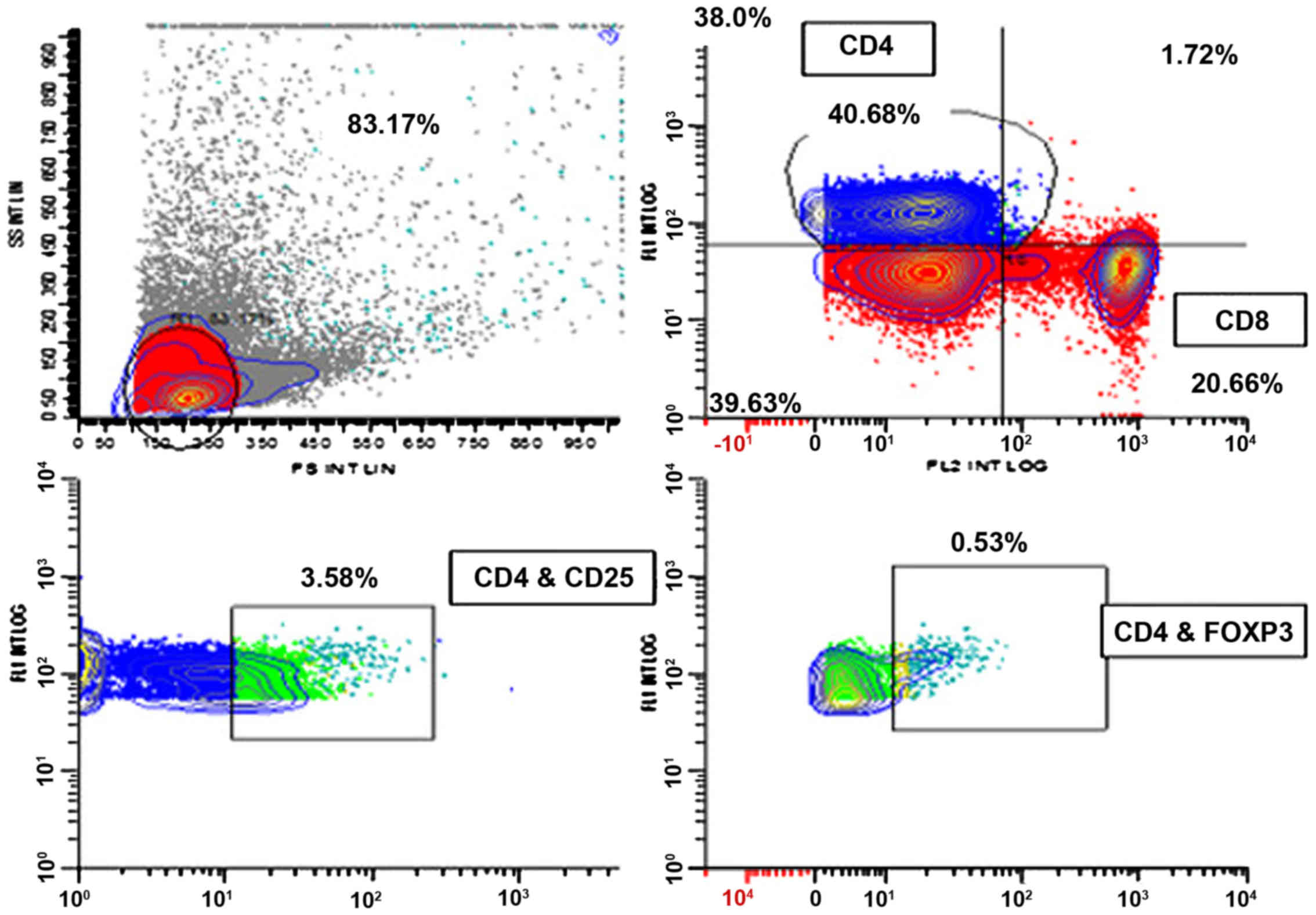
Flow cytometric analysis
DC phenotypes were determined using monoclonal
anti-human CD86-FITC, CD83-PE and MHC-II-APC antibodies (cat nos.
FAB141F, FAB1774P and IC7169A, respectively; R&D System, Inc.,
Minneapolis, MN, USA) prior to and following loading with tumor
cells. T-cell immunophenotype characterization was conducted via
CD3-APC, CD4-FITC, CD8-PE conjugate (cat no. 15080668, eBioscience,
Inc.), CD25-ECD and Foxp3 PE-CY7 (cat nos. 4238109 and 560046,
respectively; Beckman Coulter, Inc., USA). For surface markers,
samples were stained according to the manufacturer's protocol.
Cells were lysed and washed twice with PBS, centrifuged at 1800 rpm
for 3 min and incubated for 30 min in darkness at room temperature.
Foxp3 was prepared using an IntaPrep permeabilization kit (Beckman
Coulter, Inc., Brea, CA, USA). Intracellular staining was performed
using IntaPrep permeabilization reagent, by which cells were fixed
with reagent 1 (fixation reagent using formaldehyde). Following
washing, cells were permeabilized using reagent 2 (using Saponine
for permeability). Buffer was supplied from a Foxp3/Transcription
Factor Staining Buffer Set (00–5523; eBioscience; Thermo Fisher
Scientific. Inc., Waltham, MA, USA). Samples were analyzed using a
flow cytometer (NAVIOS; Beckman Coulter, Inc., Figs. 1 and 2).
Detection of cytotoxicity produced by
activated CTLs
Cytotoxicity was assessed by measuring LDH release
from the MCF-7 breast cancer cell line prior to and following co
culture with CTL for 2 days, using an ELISA (cat no. SEB864Hu,
Cloud-Clone Corp., Katy, TX, USA) according to the manufacturer's
protocol. T cells were added to the population of tumor cells and
DCs in the same flask to ensure that antigen presentation, T cell
activation and exposure to tumor cells occurred simultaneously in
order to mimic what occurs in the tumor microenvironment, and allow
interactions with different cytokines. This was a preliminary step
for immunotherapy preparation.
Reverse transcription-quantitative
polymerase chain reaction (RT-qPCR) assay of FOXP3 gene expression
in CTLs
RNA was purified from samples using the SV Total RNA
Isolation System (Promega Corporation, Madison, WI, USA), and cDNA
was produced using a High-Capacity cDNA Reverse Transcription kit
(Applied Biosystems; Thermo Fisher Scientific, Inc.), conducted
according to the manufacturer's protocols. Two primer sets were
designed to eliminate the possibility of primer dimer formation and
nonspecific annealing using Primer3 (version 4.0; http://bioinfo.ut.ee/primer3-0.4.0/) and
oligoanalyzer 3.1 (https://eu.idtdna.com/calc/analyzer) software: One for
the gene of interest, FOXP3 (forward, 5′-ACTGACCAAGGCTTCATCTGTG-3′;
and reverse, 5′-GGAACTCTGGGAATGTGCTGT-3′); and one for the
housekeeping gene β-actin (forward, 5′-ATGATATCGCCGCGCTCA-3′; and
reverse, 5′-CGCTCGGTGAGGATCTTCA-3′). Expression levels of β-actin
were measured as a reference for the target gene expression. Each
PCR was performed in a final volume of 10 µl, including 1 µg of the
cDNA product, 1 µl (50 nM) of each primer and 2X reaction mixtures
containing Fast Start DNA polymerase, reaction buffer, dNTPs, and
SYBR-Green (Applied Biosystems; Thermo Fisher Scientific, Inc.)
according to the manufacturer's protocol (12). The thermal cycling conditions
comprised a temperature profile of 95°C for 10 min for
denaturation, followed by 40 cycles (95°C for 15 sec and 60°C for
60 min) in a ViiA™ 7 Real-Time PCR System (Applied
Biosystems; Thermo Fisher Scientific, Inc.). The RT-qPCR
amplification products were analyzed via melting curve
analysis.
The relative expression of gene transcripts was
calculated according to the ΔCq and 2−ΔΔCq formulas
according to Schmittgen and Livak (13). Finally, the ratios of target to
reference gene were determined with the Pfaffl method (14).
Statistical analysis
Statistical analysis was conducted using SPSS
version 24 (IBM Corp., Armonk, NY, USA). Data are expressed as the
mean ± SEM. Comparison between groups was performed using paired
Student's t-test for comparison between variables prior to and
following activation. Pearson correlation analysis was applied for
quantitative variables. P<0.05 was considered to indicate a
statistically significant difference.
Results
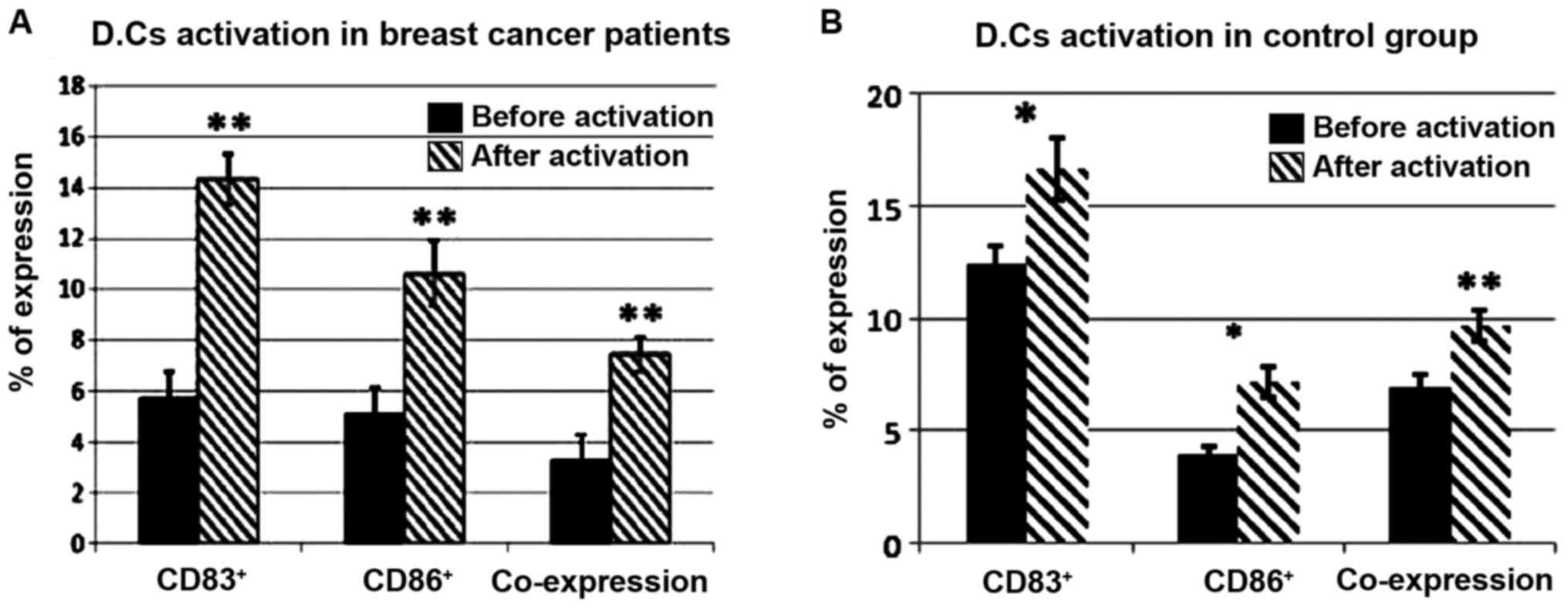
DC activation and maturation ex vivo
by exposure to tumor antigens of viable breast cancer cells
The present study was conducted on 30 patients with
breast cancer and 15 control healthy females with mean ages of
53.9±10.9 and 50±9.4 years, respectively (Table I). CD83 and CD86 were significantly
increased on DCs isolated from breast cancer patients and healthy
controls (P<0.001) following loading with viable MCF breast
cancer cells for 24 h (Fig. 3).
 | Table I.Clinicopathological characteristics
of 30 patients with breast cancer. |
Table I.
Clinicopathological characteristics
of 30 patients with breast cancer.
| Characteristic | Number |
|---|
| Diagnosis |
|
|
Invasive ductal carcinoma | 23 |
|
Invasive lobular
carcinoma | 4 |
| Tubular
carcinoma | 2 |
|
Papillary carcinoma | 1 |
| Grade |
|
| I | 3 |
| II | 20 |
|
III | 3 |
| IV | 4 |
| Axillary
lymphadenopathy |
|
|
Yes | 23 |
| No | 7 |
| Distant
metastasis |
|
|
Yes | 4 |
| No | 26 |
| Stage |
|
| I | 6 |
| II | 4 |
|
III | 16 |
| IV | 4 |
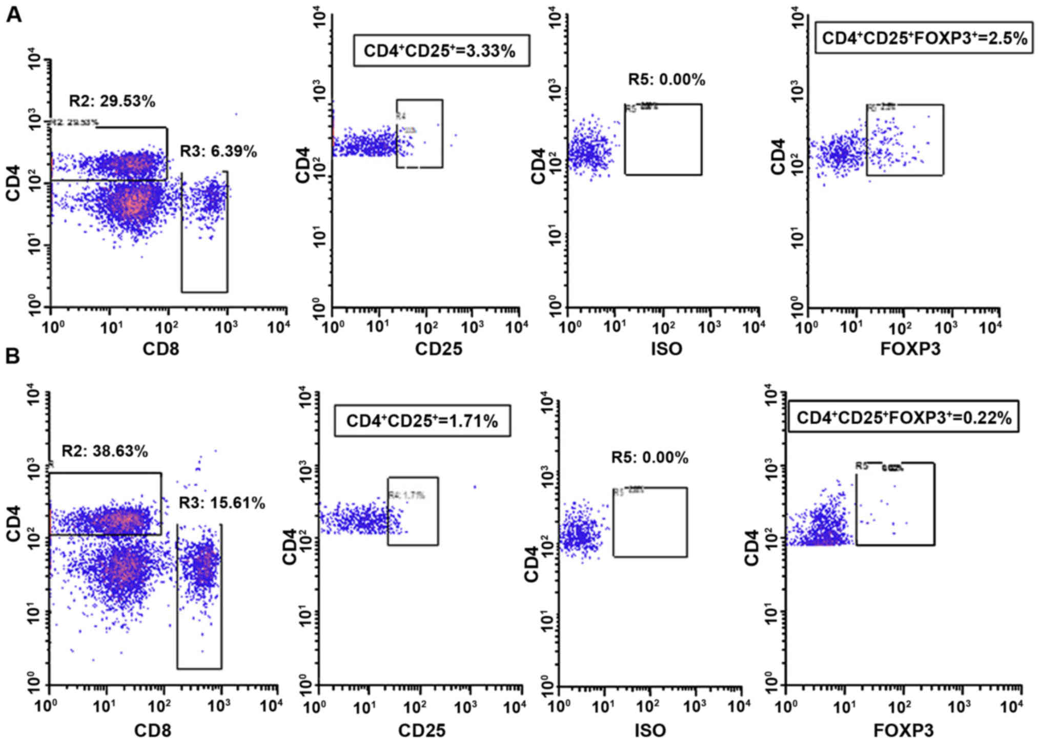
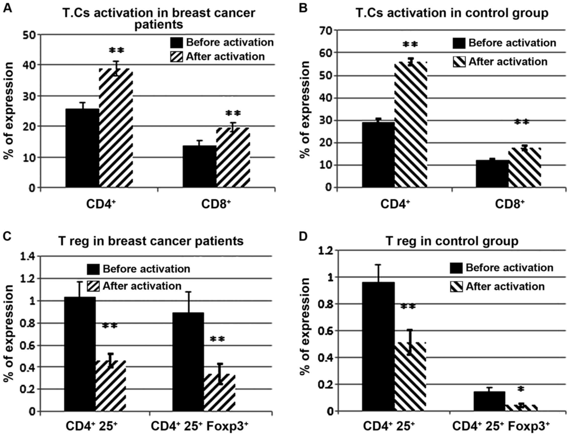
Activation of CTLs
The activation of T lymphocytes was confirmed by the
upregulation of CD4, CD8 and CD3, detected by flow cytometric
analysis. Significant increases in the levels of CD4+
T-helper (Th) cells and CD8+ CTLs were observed
(P<0.001; Fig. 4A and B). However,
there was a significant decrease in the
CD4+CD25+brightFoxp3+ regulatory T
cell (Treg) subpopulation (P<0.001) isolated from breast cancer
patients and healthy controls following activation with MDCs
previously loaded with whole viable MCF-7 cells (Fig. 4C and D).
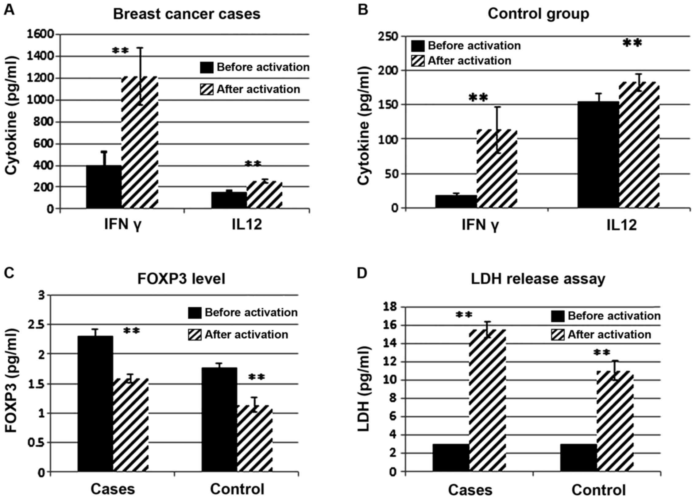
Cytokine detection by ELISA
The effect of priming DCs with viable MCF-7 breast
cancer cells resulted in a significant increase in IL-12 secretion
in the media of DCs from breast cancer patients and healthy
controls (P<0.001). Furthermore, activation of Th cells by the
vaccine resulted in a significant elevation in the IFN-γ level in
the media of T cells isolated from breast cancer patients and
healthy controls (P=0.001; Fig. 5A and
B). This was confirmed by the significant association between
the IL-12 and IFN-γ levels (P<0.05; r=0.399). IL-12 also
positively correlated with LDH (P<0.05; r=0.418). There was a
significant decrease in Foxp3 released in the media of T cells
isolated from breast cancer patients and healthy controls
(P<0.001) following activation with MDCs previously loaded with
whole viable cancer cells (Fig.
5C).
Detection of CTL cytotoxicity
There was a significant elevation in LDH release in
the media of MCF-7 cells following mixing with CTLs induced by the
whole viable cancer cell-primed DCs (P<0.001; Fig. 5D).
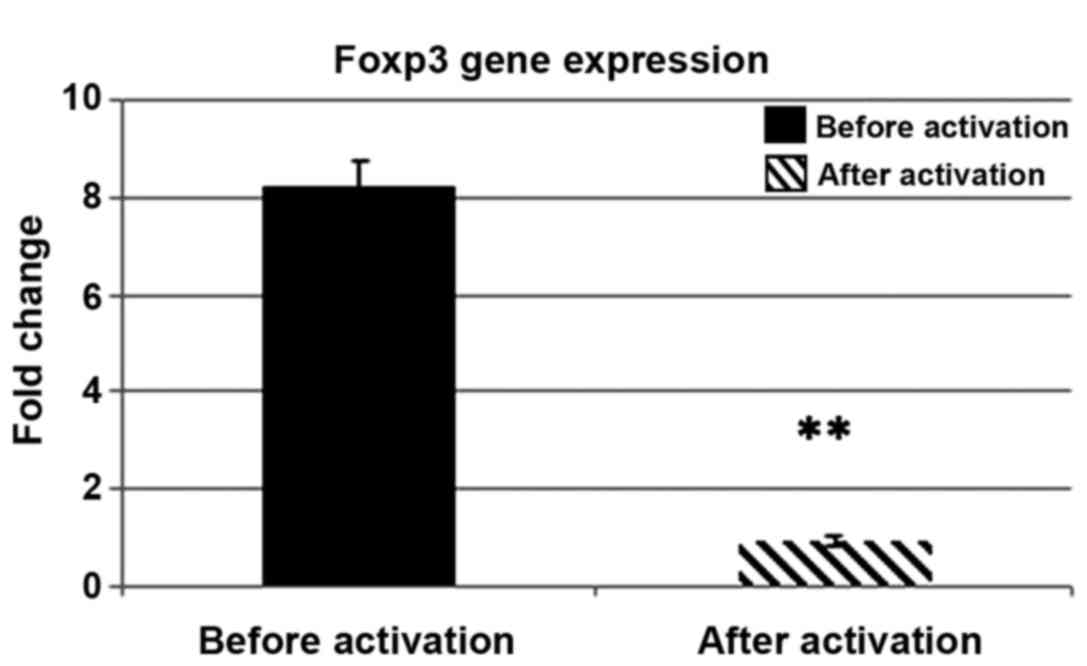
RT-qPCR for Foxp3 gene expression
There was a significant decrease in FOXP3 gene
expression in CTLs induced by the DC-based vaccine (P<0.001).
The fold change was 8.26±0.77 prior to activation, and 0.92±0.09
following activation (Fig. 6). In
breast cancer patients, there was a significant inverse correlation
between Foxp3 gene expression and the level of CD4+ Th
cells (P<0.05; r=−0.389).
Discussion
A number of studies have been conducted to enhance
the antitumor immune response in breast cancer through priming DCs;
however, selecting and optimizing the antigen-loading strategy has
been demonstrated to be a challenging undertaking (6,8,15). The most common method is by using
freeze-thawed ordinary lysates that are obtained by subjecting
cancer cells to several freeze and thaw cycles using liquid
nitrogen and water baths (16,17).
However, DCs loaded with such lysates have not provided complete
protection against tumor responses in differential animal models
(16,18,19).
Furthermore, in clinical trials, numerous studies have demonstrated
that freeze-thawed lysates are ineffective for therapy as they
suppress DC maturation and function (17,20,21).
The present study attempted to optimize tumor
antigen loading with DCs by a novel strategy. This was obtained by
subjecting DCs isolated from breast cancer patients, or healthy
controls as a comparison, to intact viable MCF-7 cells in complete
conditioned RPMI-1640 medium. The activation of DCs was
demonstrated by significantly increased levels of the stimulatory
molecule CD86, maturation molecule CD83 and MHC-II. The elevation
of such molecules may enhance the capacity of DCs to prime T cell
responses. By contrast, other studies have used DCs loaded with
cell culture supernatants that may not have contained sufficient or
adequate antigens secreted in the media (22). Additionally, a previous study
demonstrated reduced levels of CD80, CD86 and CD40 in DCs loaded
with irradiated tumor cells (23).
Another important feature of mature active DCs is
the production of IL-12. In the present study, there was a
significant increase in its secretion, from 160.03±10.27 pg/ml
prior to activation to 254.60±12.67 pg/ml following activation of
DCs with viable tumor antigens. IL-12 has a crucial importance in
the differentiation of naive T cells into Th cells and subsequently
in initiating an active specific immune response through the
induction of IFN-γ and tumor necrosis factor-α from T cells and
natural killer cells, as demonstrated by Vieira et al
(24).
The results of the present study revealed that the
interaction of viable cancer cells and DCs resulted in stimulation
of CD4+ Th cells and CD8+ CTLs against a wide
range of tumor antigens. This was confirmed by the increased
expression of CD4+, CD8+ and CD3+
cells by flow cytometry, as well the increased secretion of IFN-γ
by reactive tumor antigen-specific CD4+ Th cells. An
explanation for these findings is that these cells were capable of
inducing IFN-γ and TNF-α, and serve a role in priming
tumor-specific CTLs through the release of IL-2 (25). These results suggested that DC priming
by whole, intact tumor cells induced a differential MHC class I and
II cross-presentation of tumor antigen to T cells, as reported by
Kini Bailur et al (26), and
therefore induced a potent antitumor immune response.
Furthermore, this vaccine type resulted in a
significant decrease in an important subset of T cells,
CD4+CD25+Foxp3+ Tregs, which are
increased in the blood and tumor microenvironment of patients with
breast cancer compared with healthy subjects (27,28) and
its level is correlated with advanced clinical stages (29). Previous studies have emphasized the
role of Tregs in the suppression of antitumor immune responses, as
they are considered to exhibit critical functions in the
progression and modulation of immunological escape mechanisms in
malignancies. These cells express FOXP3 and CTL-associated
protein-4 (CTLA-4), as negative regulatory molecules of active
immune cells, and are increased in breast cancer patients (30,31).
Increased expression of Foxp3 and subsequently Tregs are considered
obstacles that may hinder the desired response of potential immune
therapeutic strategies (30–32). Therefore, in the present study the
level of Foxp3 protein secreted in the media of cultured T
lymphocytes from breast cancer patients was assessed, which
demonstrated a significant decrease in Foxp3 following the
subjection of T cells to tumor cell-primed DCs (P<0.001). This
was confirmed by a significant downregulation of Foxp3 gene
expression in CTLs as detected by RT-qPCR. Furthermore a
significant upregulation of Foxp3 gene expression (~12-fold higher)
was observed in peripheral blood of patients compared with normal
healthy controls, which was consistent with the results of
Hamidinia et al (33). This
was also confirmed by the inverse correlation between FOXP3 gene
expression and CD4+ Th cell levels identified in the
peripheral blood of the patients enrolled in the present study.
These findings suggested that the immune system was suppressed in
breast cancer patients, which may be due to an augmentation in the
Treg population and suppression of effector Th cells.
An alternative way to assess the efficacy of the
viable cancer cell-DC based vaccine was through the detection of
cytotoxicity exerted by activated CTLs on MCF-7 cells through the
measurement of LDH release. The results demonstrated a significant
increase in LDH level, indicting a highly active and potent immune
response against these cancer cells. As described by Faloppi et
al (34), LDH is typically
released from necrotic cells. Therefore, the higher the degree of
necrosis, which is related to tumor volume, the higher the level of
LDH. It is important to clarify that a significant positive
correlation was identified between IL-12 produced by this type of
viable cancer cell-based vaccine and the increased level of IFN-γ,
also, IL-12 was positively correlated with increased cytotoxicity
and necrosis of tumor cells via the increased level of LDH.
Therefore, the current study has identified an avenue for further
studying the development of novel effective immunotherapy for
patients suffering from breast cancer using DCs.
In summary, the current study demonstrated that a
viable cancer cells are an effective source of TAAs for pulsing
DCs, which may be utilized in the immunotherapeutic treatment of
breast cancer. The matured DCs induced expansion of TAA-specific T
cells, namely CTLs and Th cells. A strong cytokine response was
provoked through increased IFN-γ and IL-12 levels. Additionally, an
augmentation of the immune response was indicated by decreased
Tregs and Foxp3 expression, and an elevation of CTL activity, as
indicated by an increase in LDH release.
Acknowledgements
The present study was supported by the National
Cancer Institute, Cairo University (Cairo, Egypt).
References
|
1
|
Siegel RL, Miller KD and Jemal A: Cancer
statistics, 2016. CA Cancer J Clin. 66:7–30. 2016. View Article : Google Scholar : PubMed/NCBI
|
|
2
|
Lee J, Park S, Kim S, Kim J, Ryu J, Park
HS, Kim SI and Park BW: Characteristics and survival of breast
cancer patients with multiple synchronous or metachronous primary
cancers. Yonsei Med J. 56:1213–1220. 2015. View Article : Google Scholar : PubMed/NCBI
|
|
3
|
Ibrahim AS, Khaled HM, Mikhail NN, Baraka
H and Kamel H: Cancer incidence in Egypt: Results of the national
population-based cancer registry program. J Cancer Epidemiol.
2014:4379712014. View Article : Google Scholar : PubMed/NCBI
|
|
4
|
Caffarel MM, Andradas C, Pérez-Gómez E,
Guzmán M and Sánchez C: Cannabinoids: A new hope for breast cancer
therapy? Cancer Treat Rev. 38:911–918. 2012. View Article : Google Scholar : PubMed/NCBI
|
|
5
|
Zhang P, Yi S, Li X, Liu R, Jiang H, Huang
Z, Liu Y, Wu J and Huang Y: Preparation of triple-negative breast
cancer vaccine through electrofusion with day-3 dendritic cells.
PLoS One. 9:e1021972014. View Article : Google Scholar : PubMed/NCBI
|
|
6
|
Berneman Z, Van de Velde A, Anguille S,
Willemen Y, Huizing M, Germonpré P, Saevels K, Nijs G, Cools N, Van
Driessche A, et al: Vaccination with Wilms' Tumor Antigen (WT1)
mRNA-electroporated dendritic cells as an adjuvant treatment in 60
cancer patients: Report of clinical effects and increased survival
in acute myeloid leukemia, metastatic breast cancer, glioblastoma
and mesothelioma. Cytotherapy. 18:S13–S14. 2016. View Article : Google Scholar
|
|
7
|
Bigalke I, Honnashagen K, Lundby M, Solum
G, Skoge L, Inderberg EMS, Kasten J, Saboe-Larssen S, Schendel DJ
and Kvalheim G: Abstract 2516: A new generation of dendritic cells
to improve cancer therapy shows prolonged progression-free survival
in patients with solid tumors. Cancer Res. 75 15 Suppl:S25162015.
View Article : Google Scholar
|
|
8
|
Song QK, Ren J, Zhou XN, Wang XL, Song GH,
Di LJ, Yu J, Hobeika A, Morse MA, Yuan YH, et al: The prognostic
value of peripheral CD4+CD25+ T lymphocytes
among early stage and triple negative breast cancer patients
receiving dendritic cells-cytokine induced killer cells infusion.
Oncotarget. 6:41350–41359. 2015. View Article : Google Scholar : PubMed/NCBI
|
|
9
|
Iranpour S, Nejati V, Delirezh N, Biparva
P and Shirian S: Enhanced stimulation of anti-breast cancer T cells
responses by dendritic cells loaded with poly lactic-co-glycolic
acid (PLGA) nanoparticle encapsulated tumor antigens. J Exp Clin
Cancer Res. 35:1682016. View Article : Google Scholar : PubMed/NCBI
|
|
10
|
Ciccocioppo R, Ricci G, Rovati B, Pesce I,
Mazzocchi S, Piancatelli D, Cagnoni A, Millimaggi D, Danova M and
Corazza GR: Reduced number and function of peripheral dendritic
cells in coeliac disease. Clin Exp Immunol. 149:487–496. 2007.
View Article : Google Scholar : PubMed/NCBI
|
|
11
|
Bøyum A: Isolation of lymphocytes,
granulocytes and macrophages. Scand J Immunol. Suppl 5:S9–S15.
1976. View Article : Google Scholar
|
|
12
|
Kwok S: Procedures to minimize PCR-product
carry-over. PCR protocols: A Guide Met Appl. 142–145. 1990.
|
|
13
|
Schmittgen TD and Livak KJ: Analyzing
real-time PCR data by the comparative C(T) method. Nat Protoc.
3:1101–1108. 2008. View Article : Google Scholar : PubMed/NCBI
|
|
14
|
Pfaffl MW: A new mathematical model for
relative quantification in real-time RT-PCR. Nucleic Acids Res.
29:e452001. View Article : Google Scholar : PubMed/NCBI
|
|
15
|
Wei FQ, Sun W, Wong TS, Gao W, Wen YH, Wei
JW, Wei Y and Wen WP: Eliciting cytotoxic T lymphocytes against
human laryngeal cancer-derived antigens: Evaluation of dendritic
cells pulsed with a heat-treated tumor lysate and other
antigen-loading strategies for dendritic-cell-based vaccination. J
Exp Clin Cancer Res. 35:182016. View Article : Google Scholar : PubMed/NCBI
|
|
16
|
Fields R, Shimizu K and Mulé JJ: Murine
dendritic cells pulsed with whole tumor lysates mediate potent
antitumor immune responses in vitro and in vivo. Proc Natl Acad Sci
USA. 95:9482–9487. 1998. View Article : Google Scholar : PubMed/NCBI
|
|
17
|
Nesrua FO, Atuaolcl S, Gilliet M, Sun Y,
Grabbe S, Dummer R, Burg G and Schadendorf D: Vaccination of
melanoma patients with peptide- or tumor lysate-pulsed dendritic
cells. Nat Med. 4:328–332. 1998. View Article : Google Scholar : PubMed/NCBI
|
|
18
|
Jouanneau E, Poujol D, Gulia S, Le Mercier
I, Blay J, Belin MF and Puisieux I: Dendritic cells are essential
for priming but inefficient for boosting antitumour immune response
in an orthotopic murine glioma model. Cancer Immunol Immunother.
55:254–267. 2006. View Article : Google Scholar : PubMed/NCBI
|
|
19
|
Zhang Y, Yoneyama H, Wang Y, Ishikawa S,
Hashimoto S, Gao JL, Murphy P and Matsushima K: Mobilization of
dendritic cell precursors into the circulation by administration of
MIP-1alpha in mice. J Natl Cancer Inst. 96:201–209. 2004.
View Article : Google Scholar : PubMed/NCBI
|
|
20
|
Hersey P, Menzies SW, Halliday GM, Nguyen
T, Farrelly ML, DeSilva C and Lett M: Phase I/II study of treatment
with dendritic cell vaccines in patients with disseminated
melanoma. Cancer Immunol Immunother. 53:125–134. 2004. View Article : Google Scholar : PubMed/NCBI
|
|
21
|
Höltl L, Zelle-Rieser C, Gander H, Papesh
C, Ramoner R, Bartsch G, Rogatsch H, Barsoum AL, Coggin JH Jr and
Thurnher M: Immunotherapy of metastatic renal cell carcinoma with
tumor lysate-pulsed autologous dendritic cells. Clin Cancer Res.
8:3369–3376. 2002.PubMed/NCBI
|
|
22
|
Ascierto ML, Idowu MO, Zhao Y, Khalak H,
Payne KK, Wang XY, Dumur CI, Bedognetti D, Tomei S, Ascierto PA, et
al: Molecular signatures mostly associated with NK cells are
predictive of relapse free survival in breast cancer patients. J
Transl Med. 11:1452013. View Article : Google Scholar : PubMed/NCBI
|
|
23
|
Idoyaga J, Moreno J and Bonifaz L: Tumor
cells prevent mouse dendritic cell maturation induced by TLR
ligands. Cancer Immunol Immunother. 56:1237–1250. 2007. View Article : Google Scholar : PubMed/NCBI
|
|
24
|
Vieira PL, de Jong EC, Wierenga EA,
Kapsenberg ML and Kaliński P: Development of Th1-inducing capacity
in myeloid dendritic cells requires environmental instruction. J
Immunol. 164:4507–4512. 2000. View Article : Google Scholar : PubMed/NCBI
|
|
25
|
Benchetrit F, Gazagne A, Adotevi O,
Haicheur N, Godard B, Badoual C, Fridman WH and Tartour E:
Cytotoxic T lymphocytes: Role in immunosurveillance and in
immunotherapy. Bull Cancer. 90:677–685. 2003.PubMed/NCBI
|
|
26
|
Bailur Kini J, Gueckel B and Pawelec G:
Prognostic impact of high levels of circulating plasmacytoid
dendritic cells in breast cancer. J Transl Med. 14:1512016.
View Article : Google Scholar : PubMed/NCBI
|
|
27
|
Morse MA, Secord AA, Blackwell K, Hobeika
AC, Sinnathamby G, Osada T, Hafner J, Philip M, Clay TM, Lyerly HK
and Philip R: MHC class I-presented tumor antigens identified in
ovarian cancer by immunoproteomic analysis are targets for T-cell
responses against breast and ovarian cancer. Clin Cancer Res.
17:3408–3419. 2011. View Article : Google Scholar : PubMed/NCBI
|
|
28
|
Woo EY, Yeh H, Chu CS, Schlienger K,
Carroll RG, Riley JL, Kaiser LR and June CH: Cutting edge:
Regulatory T cells from lung cancer patients directly inhibit
autologous T cell proliferation. J Immunol. 168:4272–4276. 2002.
View Article : Google Scholar : PubMed/NCBI
|
|
29
|
Wang ZK, Yang B, Liu H, Hu Y, Yang JL, Wu
LL, Zhou ZH and Jiao SC: Regulatory T cells increase in breast
cancer and in stage IV breast cancer. Cancer Immunol Immunother.
61:911–916. 2012. View Article : Google Scholar : PubMed/NCBI
|
|
30
|
Jaberipour M, Habibagahi M, Hosseini A,
Habibabad SR, Talei A and Ghaderi A: Increased CTLA-4 and FOXP3
transcripts in peripheral blood mononuclear cells of patients with
breast cancer. Pathol Oncol Res. 16:547–551. 2010. View Article : Google Scholar : PubMed/NCBI
|
|
31
|
Suzuki S, Ishida T, Yoshikawa K and Ueda
R: Progress in clinical use of CC chemokine receptor 4 antibody for
regulatory T cell suppression. Inflam Immun Cancer: Springer.
207–227. 2015.
|
|
32
|
Liu F, Lang R, Zhao J, Zhang X, Pringle
GA, Fan Y, Yin D, Gu F, Yao Z and Fu L: CD8+ cytotoxic T
cell and FOXP3+ regulatory T cell infiltration in
relation to breast cancer survival and molecular subtypes. Breast
Cancer Res Treat. 130:645–655. 2011. View Article : Google Scholar : PubMed/NCBI
|
|
33
|
Hamidinia M, Boroujerdnia Ghafourian M,
Talaiezadeh A, Solgi G, Roshani R, Iranprast S and Khodadadi A:
Increased P-35, EBI3 transcripts and other treg markers in
peripheral blood mononuclear cells of breast cancer patients with
different clinical stages. Adv Pharm Bull. 5:261–267. 2015.
View Article : Google Scholar : PubMed/NCBI
|
|
34
|
Faloppi L, Bianconi M, Memeo R, Gardini
Casadei A, Giampieri R, Bittoni A, Andrikou K, Del Prete M, Cascinu
S and Scartozzi M: Lactate dehydrogenase in hepatocellular
carcinoma: Something old, something new. Biomed Res Int.
2016:71962802016. View Article : Google Scholar : PubMed/NCBI
|